R E S E A R C H
Open Access
Resistin and visfatin concentrations are
related to central obesity and inflammation
in Brazilian children
Natalia Figuerôa Simões
1, Ana Luiza Gomes Domingos
2*, Fernando Luiz Pereira De Oliveira
3, Ivo Santana Caldas
4,
Mariana Reis Guedes
5, Virgínia Capistrano Fajardo
6and Silvia Nascimento De Freitas
1Abstract
Background:The evidence that cardiovascular disease begins in childhood and adolescence, especially in the presence of excess weight, is associated with dysfunction on adipokine pro-inflammatory secretion. These affect glucose metabolism and lead to other complications related to insulin resistance and cardiovascular disease. This study assessed the association of anthropometric and metabolic parameters related to obesity, cardiovascular risk, and insulin resistance with concentrations of resistin and visfatin, in children.
Methods:A cross-sectional study was developed with 178 children of 6–10 years old enrolled in public city schools. Anthropometric data, composition body, clinical, and biochemical were measured according to standard procedures. We used multiple regression models by stepwise method to evaluate the associations of resistin and visfatin with variables of interest.
Results:In healthy weight children, resistin was associated with LDL cholesterol, visfatin, atherogenic index, and waist-to-height ratio, whereas in obese children resistin was associated with visfatin and interaction between conicity index and HOMA-AD. Furthermore, in healthy weight children, visfatin was associated to resistin and triceps skinfold thickness and negatively associated to HOMA-AD, while in obese ones visfatin was associated with waist-to-height ratio, atherogenic index, resistin, and interaction between trunk adiposity index and adiponectin and was negatively associated with the HOMA-IR index.
Conclusions:Our study shows an association between anthropometric and biochemical variables related to
visceral fat and inflammation. These results suggest the resistin and visfatin as good pro-inflammatory markers. In addition, both adipokines are strongly related to central obesity, in children.
Keywords:Adipokines, Adipose tissue, Insulin resistance, Waist-height ratio, Adiposity, Body composition, Metabolic diseases
Background
Obesity is a condition of chronic inflammation of low intensity resulted primarily from excess fat [1, 2]. Several anthropometric and body composition indexes have been used to measure obesity and to assess the risk of metabolic and cardiovascular diseases [3, 4].
Excess body fat is a risk factor for cardiovascular disease (CVD) and type 2 diabetes (DM2) [1, 4–6], especially the
visceral fat. Thus, it is essential to investigate measures that not only reflect body proportions but also body fat distribution. Waist circumference, trunk adiposity index (TAI), and waist-to-height ratio (WHtR) have a good correlation with central adiposity and the low cost make these available and easily applicable indicators in clinical practice and research [6–9]. Some studies have shown that waist circumference in children tends to increase with growth, requiring specific references for sex and age [10]. Waist-to-height ratio has the advantage of not requiring such references or points of cut-offs, which facilitates its use [10–12].
* Correspondence:al-domingos@hotmail.com
2Graduate Program in Nutrition Science, Federal University of Viçosa, Viçosa, Minas Gerais, Brazil
Full list of author information is available at the end of the article
Studying anthropometric indicators that can predict accumulation of visceral fat as a risk factor is relevant. The visceral fat increased is related with macrophage population, which is majorly responsible for production of inflammatory cytokines. The inflammatory cytokines have a paracrine action that compromise vascular homeostasis and lead to systemic inflammation and endothelial dysfunction, such as changes in the concen-tration of resistin and visfatin [3, 13, 14].
Studies have shown that resistin has direct effect on endothelial cells of human coronary arteries, which con-tributes to arterial inflammation, accumulation of chol-esterol, triglycerides, and insulin resistance [15, 16]. Other studies have shown the important role of resistin in controlling the cascade of inflammatory cytokines, which makes it a pro-inflammatory marker next to visfa-tin [2, 13, 17]. In addition to pro-inflammatory action, visfatin is essential forβ-pancreatic cells in insulin secre-tion [18, 19] and also stands as a marker of visceral adipose tissue [4, 20].
Furthermore, accumulation of adipose tissue, dyslipid-emia, and inflammation are risk factors for atherosclerosis and insulin resistance [21]. Studying these factors in obese children is an opportunity to examine the consequences of early stages of obesity, free of other pro-inflammatory conditions such as smoking, cardiovascular disease, and kidney and liver disease. Our study will help to determine the association between anthropometric and metabolic parameters related to obesity, cardiovascular risk, and insulin resistance with concentrations of resistin and visfatin in children.
Methods
Study design and population
A cross-sectional study was developed in 2009 with 178 children of 6–10 years old, 104 girls and 74 boys, enrolled in public city schools of Nova Era, in the state of Minas Gerais, Brazil.
The city of Nova Era had 1024 children aged 6–10 years enrolled in public schools; of those, 6.4% (n= 65) are with obesity [22]. We included two healthy weight children (n= 130) for each obese child (n= 65), resulting in a 195 student sample. We excluded students with acute and chronic diseases, changes in the gastrointestinal tract, weight loss in the last 6 months, special diets, using medicines that alter metabolism and/or affect inflammation pathways, and C-reactive protein (CRP) levels above 10 mg/L. In the end, we included 57 obese and 121 healthy weight children.
The study followed the principles of the Declaration of Helsinki and was approved by the Research Ethics Com-mittee of the Federal University of Ouro Preto under the number 2007/93. A written consent was signed in dupli-cate and obtained from those responsible.
Anthropometric, clinical, and body composition variables The students were instructed to attend school with light clothes, fast for at least 4 h, and not to do vigorous exer-cise before the anthropometric and body composition assessment. Body mass index (BMI) was calculated by dividing weight (Tanita®BC554 Ironman, Illinois, USA) and the square of height (AlturExata®, Belo Horizonte, Brazil), which were measured according to the protocol [23]. These measures were also used for calculating the ponderal index (PI) = (weight, kg)/(height, m)3[24].
Circumferences were measured using a tape measure with 0.1 cm accuracy. Arm circumference (AC) was measured in duplicate at the midpoint between the acro-mion process of the scapula and the olecranon. Waist circumference (WC) was measured in triplicate at the midpoint between the anterior superior iliac crest and the last rib, then used to calculate the conicity index (C index), C index = (WC, cm)/(0.019 √((weight, kg)/ (height, m))) [25]. All measurements were performed in the right side of the body.
Triceps skinfold (TSF) and subscapular skinfold (SSF) were also measured in triplicate and not consecutively using the caliper Cescorf® (Cescorf Equipment Ltd., Porto Alegre, Brazil). TSF was measured at the midpoint between the acromion and the olecranon on the back of the arm and SSF was measured in the marked point at the 45° diagonal, 2 cm below the lower angle of the scapula. Trunk adiposity index (TAI) was calculated based on these measurements, TAI = SSF (mm)/TSF (mm) [26].
Blood pressure (BP) was measured by vascular Doppler ultrasound using Doppler DV 610 after 5-min rest. The procedure was performed in triplicate with 2-min inter-vals between each measurement. All values were replaced by the corresponding arithmetic means.
Body fat percentage (BF%) was assessed through tetrapo-lar bioelectrical impedance (TBI) using Maltron® (Maltron® Bioscan BF-916, Maltron International Ltd., Essex, UK). We also used an equation to estimate body fat percentage by the sum of both triceps and subscapular skinfolds, as proposed by Slaughter et al. [27].
Biochemical variables
After 12 h fasting, a 10-mL blood sample was withdrawn by puncture of the cubital vein into 10-mL disposable tubes and then fractionated into vials containing sodium fluoride for glucose analysis and without anticoagulant for dosing cholesterol and ratios. After being centrifuged (Excelsa Baby® model 206–2; FANEM, São Paulo, Brazil), the serum was divided into three amber microtubes and stored at−80 °C for further analysis.
calculated homeostatic model assessment for insulin resistance (HOMA-IR) using the equation HOMA-IR = (fasting insulin (μUI/mL) × fasting glucose (mmol/mL)/ 22.5) [28] and homeostatic mode assessment-adiponectin (HOMA-AD) using the equation HOMA-AD = fasting insu-lin (mU/L) × fasting glucose (mg/dL)/adiponectin (μg/mL) [29]. The quantitative insulin sensitivity check index (QUICKI) was obtained by the equation QUICKI = 1/log (fasting insulin) + log (fasting glucose) [30].
Triacylglycerol concentrations and total cholesterol (TC) were determined using the CM 200 analyzer (WIENER LAB, Rosario, Argentina) by the enzymatic colorimetric method using Triglycerides Liquicolor mono kits and Cholesterol Liquicolor (Human of Brazil, Itabira, Brazil). The high-density lipoprotein cholesterol (HDL-C) fraction was measured by direct enzymatic colorimetric method HDL-PP (Analisa, Gold Analisa Diagnostics Ltd., Belo Horizonte) while the concentrations of low-density lipoprotein cholesterol (LDL-C) were calculated using the Friedewald equation [31]. Based on these concentrations, the atherogenic index (AI) was calculated by the ratio of total cholesterol/HDL-C [32].
Concentrations of adiponectin (Linco Research Kit—St Charles, Missouri, USA), resistin (PeproTech® Kit, Rocky Hill, NJ, USA), and visfatin (USCN Kit Life Science Inc., Wuhan, China) were obtained by ELISA (enzyme-linked immunosorbent assay). C-reactive protein concentra-tions were processed in the IMMAGE® 800 analyzer (Beckman Coulter, Fullerton, California, USA) by nephe-lometry, with detection over 0.1 mg/dL.
Statistical analysis
Data were presented as mean and standard deviation for normally distributed variables or as mean and interquartile range for not-normal distributed, previ-ously tested by Kolmogorov-Smirnov test. Comparing groups, samples with normal distribution were ana-lyzed using a t-test of two independent samples, and non-parametric samples were tested by Wilcoxon-Mann-Whitney test.
To determine which variables were associated with adipokines and the magnitude of these associations, multiple regression models were constructed by a stepwise method with resistin and visfatin as dependent variables. Initially, all independent variables were included in the model, leaving only significant variables in the subsequent models. Some interactions were also included at this stage. If a significant interaction is present in the model, the effect of a variable x1 on the average response depends on the second variable x2and vice versa. In this case, we considered a multiple regres-sion model with two regresregres-sion variables, as shown in the formula:
Y¼β0þβ1x1þβ2x2þβ3x1x2
|{z}
Interaction
Analysis of residuals of each model was performed to assess validity of assumptions of normality, homoscedas-ticity, and independence between observations. The sta-tistics Cook’s distance and variance inflation factor (VIF) were used to identify outliers and to check for possible multicollinearity between independent variables [33, 34].
This study considered α of 0.05 as significant for all statistical tests. For analysis, we used the statistical software R.2.13.1.
Results
The characteristics of the students are shown in Table 1. The sample consisted of 104 girls (58.4%) and 74 boys (41.6%). The obese group showed mean/median an-thropometric values higher than the healthy weight group. In addition, all variables analyzed were signifi-cantly different between groups, except age, HDL-C, adiponectin, and resistin.
Regression models in Tables 2 and 3 showed β -coefficients ± SE,R2, and VIF. The VIF is responsible for quantifying multicollinearity between the independent variables, since it measures how much the variance of an estimated regression coefficient is greater because of collinearity [35]. For all adjusted models, VIF was lower than 2.0, indicating no multicollinearity between the independent variables, as all adjusted models were significant at the 0.05 significance level. Adjustments for age and sex were not performed, as the groups did not differ for these parameters.
Resistin was associated with LDL-C, visfatin, AI, and WHtR among healthy weight children (Table 2). While in obese ones, resistin was associated with visfatin with inter-action between conicity index (CI) and HOMA-AD consid-ering a significance level of 10% (Table 2). Visfatin levels were positively associated with concentration of resistin and triceps skinfold and negatively with HOMA-AD index in healthy weight children (Table 3), whereas in obese ones visfatin was positively associated with WHtR, AI, resistin, and interaction of TAI vs. adiponectin, and a negative asso-ciation with HOMA-IR index (Table 3).
Discussion
In this study, obese children had higher concentrations of visfatin (p< 0.05) and similar concentrations of resistin (p= 0.479) compared to healthy weight children. These results confirm earlier studies [36–40]. Further, both adi-pokines were associated with central obesity markers, lipid metabolism, and insulin resistance.
with BMI and body fat percentage. This suggests that these adipokine concentrations are related not only with total fat but also with fat distribution. Scientific evidence shows that WHtR was strongly associated with abdominal fat measured by advanced imaging techniques such as com-puted tomography and magnetic resonance imaging [18]. Several studies of sensitivity and specificity demonstrated the WHtR as a method to use for identifying childhood overweight, obesity, and tracking of inflammatory and cardiometabolic risk in 6- to 16-year-old children [4, 6, 7, 10–12].
Another relevant factor in central obesity is the evi-dence that increase in body fat, especially visceral, re-sults in cellular hypoxia process, which lead to adipocyte death by necrosis or apoptosis, and increased macro-phage population. This inflammatory process is primar-ily responsible for production of adipokines, resistin and visfatin [13, 41]. A study by Li et al. showed that pubes-cent girls with acute inflammation showed no associ-ation between resistin and anthropometric variables, implying that pro-inflammatory effects of this adipokine were not dependent on obesity only. This can be seen in
Table 1Anthropometric, clinical, and metabolic characteristics and adipokine levels of schoolchildren 6–10 years of Nova Era, Minas
Gerais, Brazil, 2009
Normal weight Obese pvalue
n= 121 n= 57
Sex (nM/F) 49/72 25/32 –
Age (years) 8.0 (7.0–9.0) 8.0 (7.0–9.0) 0.618
Weight (kg) 27.7 (24.0–30.6) 40.9 (36.1–48.3) < 0.001*
Height (cm) 130.83 ± 9.52a 135.11 ± 9.45a
0.006*
BMI (kg/m2) 15.7 (15.1–16.53) 22.4 (20.7–25.13)
< 0.001*
Arm circumference (cm) 19.3 (18.0–20.2) 25.7 (24.05–27.65) < 0.001*
Waist circumference (cm) 58.5 (55.7–61.0) 76.5 (70.87–83.0) < 0.001*
Triceps skinfold (mm) 8.66 (7.18–10.60) 21.13 (16.23–24.11) < 0.001*
Subscapular skinfold (mm) 6.03 (5.23–7.6) 19.46 (12.83–24.86) < 0.001*
BF from skinfolds (%) 14.22 (11.96–17.33) 32.08 (27.11–36.94) < 0.001*
BF tetrapolar (%) 15.78 ± 5.50a 27.66 ± 5.83a
< 0.001*
Systolic BP Doppler (mmHg) 92.6 (87.3–98.3) 103.3 (94.8–111.6) < 0.001*
Fasting glucose (mg/dL) 83.48 ± 7.45a 86.14 ± 7.08a
0.023*
Fasting insulin (mg/dL) 4.72 (3.25–6.42) 7.61 (5.34–13.13) < 0.001*
Total cholesterol (mg/dL) 147.75 ± 27.08a 161.84 ± 30.68a
0.004*
HDL cholesterol (mg/dL) 57.0 (50.0–66.0) 54.0 (48.0–64.0) 0.160
LDL cholesterol (mg/dL) 74.82 ± 23.00a 87.38 ± 23.86a
0.001*
VLDL cholesterol (mg/dL) 12.59 (9.68–19.79) 18.82 (13.05–23.68) < 0.001*
Triacylglycerols (mg/dL) 63.0 (48.5–99.0) 94.0 (65.0–118.5) < 0.001*
Waist/height ratio 0.44 (0.43–0.46) 0.56 (0.53–0.60) < 0.001*
Atherogenic index 2.58 ± 0.50a 2.94 ± 0.49a
< 0.001*
Conicity index 1.17 (1.14–1.20) 1.26 (1.22–1.29) < 0.001*
Trunk adiposity index 0.76 (0.64–0.84) 0.92 (0.77–1.06) < 0.001*
HOMA-AD index 14.01 (8.42–24.21) 25.21 (16.37–51.0) < 0.001*
HOMA-IR index 0.96 (0.69–1.32) 1.64 (1.13–2.76) < 0.001*
QUICKI 0.38 (0.36–0.40) 0.35 (0.32–0.37) < 0.001*
Adiponectin (μg/mL) 26.89 (18.23–39.66) 26.30 (19.74–35.11) 0.473
C-reactive protein (mg/mL) ND (ND–0.051) 0.148 (ND–0.449) < 0.001*
Resistin (ng/mL) 4.02 ± 1.86a 4.27 ± 2.00a 0.479
Visfatin (ng/mL) 2.19 (1.40–3.26) 2.66 (2.10–3.71) 0.037*
our study, since there were significant associations in healthy weight children opposing a low explanatory power in the model for resistin in obese children (R= 10.6%).
In the same context, high concentrations of visfatin are associated with the area of visceral adipose tissue, obesity, and metabolic syndrome in children [18, 40, 42–44]. In this study, analysis confirmed the relationship between visfatin concentrations and central obesity through associ-ation with WHtR and TAI in obese children. This model reveals a significant interaction between trunk adiposity index and adiponectin in the variation of average visfatin concentration. Currently, it is recognized that adiponectin concentrations are inversely proportional to body fat [45, 46]. A possible explanation for this interaction may be
attributed to reduction of adiponectin levels by increasing trunk fat in obese children, causing the variation of visfa-tin concentrations.
In addition to reducing adiponectin concentration, childhood obesity is associated with increased C-reactive protein concentration and visceral fat. The C-reactive protein is a non-specific marker of inflammation, and visceral fat has been increasingly demonstrated as an important endocrine organ involved in the relationship between obesity and inflammation through accumula-tion of macrophages, which in turn promotes synthesis of these pro-inflammatory cytokines [47]. In our sample, previously published data showed important relation-ships between C-reactive protein (CRP) and adiponectin with anthropometric variables, and metabolic and body composition which highlighted the role of adipose tissue in the inflammatory process [48].
Authors have shown that resistin controls the pro-inflammatory cytokine cascade [2, 17] and that increased visfatin concentration was related to inflammation in over-weight individuals [40, 44, 49]. Such inflammatory signaling significantly alters lipid metabolism in adipose tissue, liver, and skeletal muscle, and the interaction between dyslipid-emia and inflammation accelerates the development of atherosclerosis and insulin resistance [50, 51].
Analysis of the adjusted models strengthened the role of adipokines in lipid metabolism in our study. Both resistin and visfatin were associated with atherogenic index and resistin also was associated with cholesterol concentration LDL-C. In contrast with results obtained in children, some studies have shown visfatin positively associated with HOMA-IR [43, 52] but in our study showed negative association between these two variables. Araki et al. found no difference in visfatin concentra-tions in a group of obese children stratified by HOMA-IR and hyperinsulinemia, suggesting that visfatin is a marker for visceral adipose tissue (VAT) accumulation but not a marker for insulin metabolism [18].
Although some studies have shown an insulin-mimetic property of visfatin, other reports consider visfatin to be a molecule homologous to nicotinamide phosphoribosyl transferase (NAMPT). The NAMPT is an enzyme that triggers the synthesis of nicotinamide adenine dinucleo-tide (NAD), which is essential for the functioning of cells
β-pancreaticin insulin secretion [2]. As in both cases visfatin may promote reduction of blood glucose, a nega-tive association is suggested between visfatin and insulin resistance [53, 54], as observed in our results.
In the case of resistin, it was observed that our model presents an adipokine association with the interaction between conicity and HOMA-AD in-dexes. This result demonstrates the importance of resistin in insulin resistance and this is obesity-dependent, since the conicity index estimates central
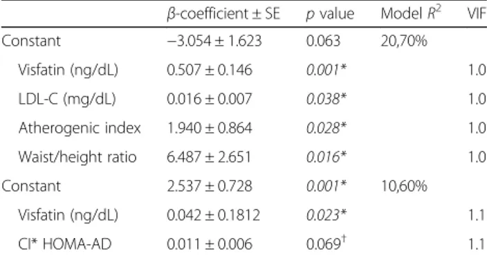
Table 2Multiple regression analyses between resistin level with anthropometric and biochemical variables in schoolchildren
6–10 years of Nova Era, Minas Gerais, Brazil, 2009
β-coefficient ± SE pvalue ModelR2 VIF
Constant −3.054 ± 1.623 0.063 20,70%
Visfatin (ng/dL) 0.507 ± 0.146 0.001* 1.0
LDL-C (mg/dL) 0.016 ± 0.007 0.038* 1.0
Atherogenic index 1.940 ± 0.864 0.028* 1.0
Waist/height ratio 6.487 ± 2.651 0.016* 1.0
Constant 2.537 ± 0.728 0.001* 10,60%
Visfatin (ng/dL) 0.042 ± 0.1812 0.023* 1.1
CI* HOMA-AD 0.011 ± 0.006 0.069†
1.1
Model normal weight: resistin = visfatin + LDL-C + atherogenic index + waist/ height ratio. Model obese: resistin =β0+ visfatin + conicity index × HOMA-AD Abbreviations:CIconicity index,HOMA-ADhomeostatic
model assessment-adiponectin *p< 0.05;†
p< 0.10. Significant values are in italic
Table 3Multiple regression analyses between visfatin level with anthropometric and biochemical variables in schoolchildren
6–10 years of Nova Era, Minas Gerais, Brazil, 2009
β-coefficient ± SE pvalue ModelR2 VIF
Constant 0.871 ± 0.409 0.036* 20,00%
Resistin (ng/mL) 0.186 ± 0.060 0.003* 1.0
Triceps skinfold (mm) 0.116 ± 0.037 0.002* 1.1
HOMA-AD −0.018 ± 0.006 0.008* 1.1
Constant −2.691 ± 1.356 0.054 37,70%
Resistin (ng/mL) 0.241 ± 0.075 0.003* 1.1
Waist/height ratio 4.078 ± 1.913 0.039* 1.1 Atherogenic index 0.834 ± 0.2854 0.006* 1.1
HOMA-IR −0.334 ± 0.112 0.005* 1.1
TAI*adiponectin (μg/mL) 0.029 ± 0.014 0.046* 1.1
Model normal weight: visfatin =β0+ resistin + triceps skinfold−HOMA-AD. Model obese: resistin + waist/height ratio + atherogenic index−HOMA-IR + trunk adiposity index × adiponectin
Abbreviations:HOMA-ADhomeostatic model assessment-adiponectin,HOMA-IR
obesity [55, 56] and the HOMA-AD index was posi-tively associated with levels of resistin in obese children.
However, the fact that both resistin and visfatin are as-sociated with insulin resistance should be analyzed with caution, since no association was found between concen-trations of resistin and visfatin with fasting glucose or QUICKI. According to a study by Keskin et al., HOMA-IR index is more reliable than QUICKI to measure insulin resistance in children [57], and this is a likely explanation for the absence of association with QUICKI in our sample. In addition, several discrepancies between the-ories described in various studies maybe due to different characteristics of the studied populations or confounding factors such as sex and age, and use of different laboratory methods in the assessment of adipokines.
Thus, these findings add evidence to underscore the importance of early identification of risk factors for cardiometabolic diseases and insulin resistance in order to promote early intervention and improved quality of life in this population. In addition, these results reinforce the importance of conducting studies that infer causality to endorse the use of anthropometric parameters and body composition in the estimation of resistin and visfa-tin concentrations, thus allowing a more applicable diag-nosis in clinical practice.
Conclusion
Our study shows a significant association of anthropomet-ric and metabolic variables with resistin and visfatin concentrations in children. These results suggesting that both adipokines are pro-inflammatory markers and strongly related to central obesity, especially waist-height ratio.
Furthermore, studies are needed to investigate the adipokine receptors, especially their regulation, whereas this could help in understanding the metabolic pathways linking obesity with atherosclerosis and other cardiovascular diseases, and it could determine causal assumptions.
Abbreviations
AC:Arm circumference; AI: Atherogenic index; BF: Body fat; BMI: Body mass index; BMI/A: Body mass index for age; BP: Blood pressure; CI: Conicity index; CRP: C-reactive protein; CVD: Cardiovascular disease; DM2: Type 2 diabetes; HDL-C: High-density lipoprotein cholesterol; HOMA-AD: Homeostatic model assessment-adiponectin; HOMA-IR: Homeostatic model assessment for insulin resistance; LDL-C: Low-density lipoprotein cholesterol; NAD: Nicotinamide adenine dinucleotide; NAMPT: Nicotinamide phosphoribosyl transferase; PI: Ponderal index; QUICKI: Quantitative insulin sensitivity check index; SSF: Subscapular skinfold; TAI: Trunk adiposity index; TBI: Tetrapolar bioelectrical impedance; TC: Total cholesterol; TSF: Triceps skinfold; VAT: Visceral adipose tissue; VIF: Variance inflation factor; VLDL-C: Very-low-density lipoprotein cholesterol; WC: Waist circumference; WHtR: Waist-to-height ratio
Acknowledgements
The authors thank the Pilot Laboratory of Clinical Analysis of Federal University of Ouro Preto for biochemical analysis.
Funding
This project was funded by Fundação de Amparo à Pesquisa do Estado de Minas Gerais (APQ-02851-10) and Federal University of Ouro Preto. The funders had no role in study design, data collection and analysis, decision to publish, or preparation of the manuscript.
Availability of data and materials
The datasets used and/or analyzed during the current study are available from the corresponding author on reasonable request.
Authors’contributions
NFS wrote and edited the manuscript and is responsible for the data analysis and interpretation; ALGD wrote and edited the manuscript and is
responsible for the interpretation; MRG and VCF wrote and edited the manuscript; FLPO wrote and edited the manuscript and is responsible for the statistical analysis and data interpretation; SNF wrote and edited the manuscript and the conducted field work and data analysis and interpretation. All authors read and approved the final manuscript.
Ethics approval and consent to participate
The study followed the principles of the Declaration of Helsinki and was approved by the Research Ethics Committee of the Federal University of Ouro Preto under the number 2007/93. A written consent was signed in duplicate and obtained from those responsible.
Consent for publication
Not applicable
Competing interests
The authors declare that they have no competing interests.
Publisher’s Note
Springer Nature remains neutral with regard to jurisdictional claims in published maps and institutional affiliations.
Author details
1Graduate Program in Health and Nutrition, School of Nutrition, Federal University of Ouro Preto, Ouro Preto, Minas Gerais, Brazil.2Graduate Program in Nutrition Science, Federal University of Viçosa, Viçosa, Minas Gerais, Brazil. 3Department of Statistics, Federal University of Ouro Preto, Ouro Preto, Minas Gerais, Brazil.4Department of Pathology and Parasitology, Federal University of Alfenas, Alfenas, Minas Gerais, Brazil.5Research Center in Biological Sciences, Federal University of Ouro Preto, Ouro Preto, Minas Gerais, Brazil. 6School of Medicine, Federal University of Ouro Preto, Ouro Preto, Minas Gerais, Brazil.
Received: 2 June 2017 Accepted: 12 January 2018
References
1. Stastny J, Bienertova-Vasku J, Vasku A. Visfatin and its role in obesity development. Diabetes & metabolic syndrome. 2012;6:120–4.
2. AL-Suhaimi EA, Shehzad A. Lepitin, resistin and visfatin: the missing link between endocrine metabolic disorders and immunity. Eur J Med Res. 2013;18:12.
3. Iantorno M, Campia U, Di Daniele N, Nistico S, Forleo GB, Cardillo C, et al. Obesity, inflammation and endothelial dysfunction. J Biol Regul Homeost Agents. 2014;28:169–76.
4. Olza J, Aguilera CM, Gil-Campos M, Leis R, Bueno G, Vall M, et al. Waist-to-height ratio, inflammation and CVD risk in obese children. Public Health Nutr. 2014;17:2378–85.
5. Pagano C, Pilon C, Olivieri M, Mason P, Fabris R, Serra R, et al. Reduced plasma visfatin/pre-B cell colony-enhancing factor in obesity is not related to insulin resistance in humans. J Clin Endocrinol Metab. 2006;91:3165–70. 6. Maffeis C, Banzato C, Talamini G. Waist-to-height ratio, a useful index
to identify high metabolic risk in overweight children. J Pediatr. 2008; 152:207–13.
7. Kuba VM, Leone C, Damiani D. Is waist-to-height ratio a useful indicator of cardio-metabolic risk in 6-10-year-old children? BMC Pediatr. 2013;13:91. 8. Alvarez MM, Vieira ACRE, Sichieri R, Veiga GVD. Associação das medidas
síndrome metabólica em uma amostra probabilística de adolescentes de escolas públicas. Arq Bras Endocrinol Metabol. 2008;52:649–57. 9. Magalhães EIS, Sant’Ana LFR, Priore SE, Franceschini SDCC. Waist
circumference, waist/height ratio, and neck circumference as parameters of central obesity assessment in children. Rev Paul Pediatr. 2014;32:273–81.
10. Marrodán MD, Álvarez JRM, Espinosa MGM, López-Ejeda N, Cabañas MD, Prado C. Precisión diagnóstica del índice cintura-talla para la identificación del sobrepeso y de la obesidad infantil. Med Clin (Barc). 2012;140:296–301. 11. Nambiar S, Hughes I, Davies PS. Developing waist-to-height ratio cut-offs to
define overweight and obesity in children and adolescents. Public Health Nutr. 2010;13:1566–74.
12. Petroff D, Kromeyer-Hauschild K, Wiegand S, Binder G, Schwab K, Stachow R, et al. Introducing excess body weight in childhood and adolescence and comparison with body mass index and waist-to-height ratio. Int J Obes. 2015;39:52–60.
13. Curat CA, Wegner V, Sengenès C, Miranville A, Tonus C, Busse R, et al. Macrophages in human visceral adipose tissue: increased accumulation in obesity and a source of resistin and visfatin. Diabetologia. 2006;49:744–7. 14. Fontana L, Eagon JC, Trujillo ME, Scherer PE, Klein S. Visceral fat adipokine
secretion is associated with systemic inflammation in obese humans. Diabetes. 2007;56:1010–3.
15. Rae C, Graham A. Human resistin promotes macrophage lipid accumulation. Diabetologia. 2006;49:1112–4.
16. Calabro P, Cirillo P, Limongelli G, Maddaloni V, Riegler L, Palmieri R, et al. Tissue factor is induced by resistin in human coronary artery endothelial cells by the NF-kB-dependent pathway. J Vasc Res. 2011;48:59–66. 17. Maggio ABR, Wacker J, Montecucco F, Galan K, Pelli G, Mach F, et al. Serum
resistin and inflammatory and endothelial activation markers in obese adolescents. J Pediatr. 2012;161:1022–7.
18. Maury E, Brichard SM. Adipokine dysregulation, adipose tissue inflammation and metabolic syndrome. Mol Cell Endocrinol. 2010;314:1–16.
19. Martos-Moreno GA, Kopchick JJ, Argente J. Adipoquinas en el niño sano y con obesidad. An Pediatr (Barc). 2013;78:189.
20. Araki S, Dobashi K, Kubo K, Kawagoe R, Yamamoto Y, Kawada Y, et al. Plasma visfatin concentration as a surrogate marker for visceral fat accumulation in obese children. Obesity. 2008;16:384–8.
21. van Diepen J, Berbée J, Havekes L, Rensen PC. Interactions between inflammation and lipid metabolism: relevance for efficacy of anti-inflammatory drugs in the treatment of atherosclerosis. Atherosclerosis. 2013;228:306–15.
22. Md O, Onyango AW, Borghi E, Siyam A, Nishida C, Siekmann J. Development of a WHO growth reference for school-aged children and adolescents. Bull World Health Organ. 2007;85:660–7.
23. Lohman TG, Roche AF, Martorell R. Anthropometric standardization reference manual. Illinois: Human Kinetics Books; 1991.
24. Cook D, Mendall M, Whincup PH, Carey IM, Ballam L, Morris JE, et al. C-reactive protein concentration in children: relationship to adiposity and other cardiovascular risk factors. Atherosclerosis. 2000;149:139–50. 25. Valdez R. A simple model-based index of abdominal adiposity. J Clin
Epidemiol. 1991;44:955–6.
26. Haffner S, Stern M, Hazuda HP, Pugh J, Patterson JK. Do upper-body and centralized adiposity measure different aspects of regional body-fat distribution? Relationship to non-insulin-dependent diabetes mellitus, lipids, and lipoproteins. Diabetes. 1987;36:43–51.
27. Slaughter M, Lohman T, Boileau R, Horswill CA, Stillman RJ, Van Loan MD, et al. Skinfold equations for estimation of body fatness in children and youth. Hum Biol. 1988;60:709–23.
28. Matthews D, Hosker J, Rudenski A, Naylor BA, Treacher DF, Turner RC. Homeostasis model assessment: insulin resistance and beta-cell function from fasting plasma glucose and insulin concentrations in man. Diabetologia. 1985;28:412–9.
29. Matsuhisa M, Yamasaki Y, Emoto M, Shimabukuro M, Funahashi T, Matsuzawa Y. A novel index of insulin resistance determined from the homeostasis model assessment index and adiponectin levels in Japanese subjects. Diabetes Res Clin Pract. 2007;77:151–4.
30. Katz A, Nambi S, Mather K, Baron AD, Follmann DA, Sullivan G, et al. Quantitative insulin sensitivity check index: a simple, accurate method for assessing insulin sensitivity in humans. J Clin Endocrinol Metab. 2000;85:2402–10.
31. Friedewald W, Levy R, Fredrickson D. Estimation of the concentration of low-density lipoprotein cholesterol in plasma, without use of the preparative ultracentrifuge. Clin Chem. 1972;18:499–502.
32. Castelli W. Cholesterol and lipids in the risk of coronary artery disease—the Framingham heart study. The Canadian journal of cardiology. 1988; 4 Suppl A: 5A-10A.
33. Charnet R, Freire CAL, Charnet EMR, BONVINO H, CHARNET R. Análise de Modelos de Regressão Linear–com aplicações. 2ed. Campinas: UNICAMP; 2008. 34. Montgomery DC, Peck EA, Vining GG. Introduction to linear regression
analysis 5a ed. New York: John Wiley John Wiley & Sons; 2012. 35. O’brien RM. A caution regarding rules of thumb for variance inflation
factors. Quality & Quantity. 2007;41:673–90.
36. Jaleel A, Aheed B, Jaleel S, Majeed R, Zuberi A, Khan S, et al. Association of adipokines with obesity in children and adolescents. Biomark Med. 2013;7:731–5.
37. Ko B-J, Lee M, Park HS, Han K, Cho GJ, Hwang TG, et al. Elevated vaspin and leptin levels are associated with obesity in prepubertal Korean children. Endocr J. 2013;60:609–16.
38. Rubin DA, McMurray RG, Hackney AC, Harrell JS. Relationship between cardiovascular risk factors and adipokines in adolescents. Hormone research in paediatrics. 2011;76:123–9.
39. Ortega L, Riestra P, Navarro P, Gavela-Pérez T, Soriano-Guillén L, Garcés C. Resistin levels are related to fat mass, but not to body mass index in children. Peptides. 2013;49:49–52.
40. Martos-Moreno G, Kratzsch J, Körner A, Barrios V, Hawkins F, Kiess W, et al. Serum visfatin and vaspin levels in prepubertal children: effect of obesity and weight loss after behavior modifications on their secretion and relationship with glucose metabolism. Int J Obes. 2011;35:1–8.
41. Sun K, Kusminski CM, Scherer PE. Adipose tissue remodeling and obesity. J Clin Invest. 2011;121:2094–101.
42. Haider DG, Holzer G, Schaller G, Weghuber D, Widhalm K, Wagner O, et al. The adipokine visfatin is markedly elevated in obese children. J Pediatr Gastroenterol Nutr. 2006;43:548–9.
43. Davutoglu M, Ozkaya M, Guler E, Garipardic M, Gursoy H, Karabiber H, et al. Plasma visfatin concentrations in childhood obesity relationships with insulin resistance and anthropometric indices. Swiss Med Wkly. 2009;139:22–7.
44. Kolsgaard ML, Wangensteen T, Brunborg C, Joner G, Holven KB, Halvorsen B, et al. Elevated visfatin levels in overweight and obese children and adolescents with metabolic syndrome. Scand J Clin Lab Invest. 2009;69:858–64.
45. Savino F, Petrucci E, Nanni G. Adiponectin: an intriguing hormone for paediatricians. Acta Paediatr. 2008;97:701–5.
46. Riestra P, Garcia-Anguita A, Lasuncion MA, Cano B, de Oya M, Garcés C. Relationship of adiponectin with metabolic syndrome components in pubertal children. Atherosclerosis. 2011;216:467–70.
47. Bastien M, Poirier P, Lemieux I, Després JP. Overview of epidemiology and contribution of obesity to cardiovascular disease. Prog Cardiovasc Dis. 2014; 56:369–81.
48. Domingos ALG, Coelho GLLM, Volp ACP, Oliveira FLPD, Caldas IS, Freitas SN. Association between nutritional status, C-reactive protein, adiponectin and HOMA-AD in Brazilian children. Nutr Hosp. 2014;30:66–74.
49. Chen X-Y, Zhang J-H, Liu F, Liu HM, Song YY, Liu YL. Association of serum resistin levels with metabolic syndrome and early atherosclerosis in obese Chinese children. J Pediatr Endocrinol Metab. 2013;26:855–60.
50. Glass C, Olefsky J. Inflammation and lipid signaling in the etiology of insulin resistance. Cell Metab. 2012;15:635–45.
51. Diepen JAv, Berbée JFP, Havekes LM, Rensen PC. Interactions between inflammation and lipid metabolism: relevance for efficacy of anti-inflammatory drugs in the treatment of atherosclerosis. Atherosclerosis 2013;228:306–315.
52. Taşçılar ME, Çekmez F, Meral C, Pirgon Ö, Tanju IA, Canpolat FE. Evaluation
of adipocytokines in obese children with insulin resistance. Turk J Pediatr. 2011;53:269–73.
53. Kim HJ, Park SY, Choi YJ, Han SJ, Lee KW, Kim DJ. Differential significance of plasma visfatin concentrations according to adiposity in children and adolescents. Horm Res Paediatr. 2013;79:208–13.
55. Sant'Ana MSL, Tinôco ALA, Sant'Ana LFDR, Rosado LEFPD, Santos TF, Mello AD, et al. Eficácia do índice de conicidade e da relação cintura/ estatura em predizer o percentual de gordura corporal em crianças. Nutrire. 2010;35:67–80.
56. Taylor RW, Jones IE, Williams SM, Goulding A. Evaluation of waist circumference, waist-to-hip ratio, and the conicity index as screening tools for high trunk fat mass, as measured by dual-energy X-ray absorptiometry, in children aged 3-19 y. Am J Clin Nutr. 2000;72:490–5.
57. Keskin M, Kurtoglu S, Kendirci M, Atabek ME, Yazici C. Homeostasis model assessment is more reliable than the fasting glucose/insulin ratio and quantitative insulin sensitivity check index for assessing insulin resistance among obese children and adolescents. Pediatrics. 2005;115:e500–3.
• We accept pre-submission inquiries
• Our selector tool helps you to find the most relevant journal
• We provide round the clock customer support
• Convenient online submission
• Thorough peer review
• Inclusion in PubMed and all major indexing services
• Maximum visibility for your research
Submit your manuscript at www.biomedcentral.com/submit