International Journal of Pharmaceutics 349 (2008) 152–160
Release profiles and morphological characterization by atomic
force microscopy and photon correlation spectroscopy of
99m
Technetium-fluconazole nanocapsules
Danielle Nogueira de Assis
a, Vanessa Carla Furtado Mosqueira
b,∗, Jos´e M´ario Carneiro Vilela
c,
Margareth Spangler Andrade
c, Valbert Nascimento Cardoso
aaDepartamento de An´alises Cl´ınicas, Faculdade de Farm´acia, Universidade Federal de Minas Gerais, Av. Antˆonio Carlos,
6627 Belo Horizonte, MG 31270-901, Brazil
bDepartamento de Farm´acia, Escola de Farm´acia, Universidade Federal de Ouro Preto,
Rua Costa Sena, 171 Centro, Ouro Preto, MG 35400-000, Brazil
cFunda¸c˜ao Centro Tecnol´ogico de Minas Gerais – CETEC, Avenida Jos´e Cˆandido da Silveira,
2000 Belo Horizonte, MG 31170-000, Brazil
Received 5 May 2007; received in revised form 3 August 2007; accepted 7 August 2007 Available online 11 August 2007
Abstract
Several classes of antifungal have been employed in candidiasis treatment, but patients with advanced immunodeficiency can present unsatis-factory results after therapy. In these cases, high doses of drugs or the use of multiple agents are sometimes used, and hence increasing the risk of serious side effects. Considering theses difficulties, the encapsulation of antifungal agents in nanoparticulate carriers has been used with the objective of modifying the pharmacokinetic of drugs resulting in more efficient treatments with less side effects. The purpose of this work was the preparation, characterization and the investigation of the release profiles of radiolabeled fluconazole nanocapsules. The size, homogeneity and zeta potential of NC preparations were determined with a Zetasizer 3000HS. The morphology and the structural organization were evaluated by atomic force microscopy (AFM). The release studyin vitroof NC was evaluated in physiologic solution with or without 70% mouse plasma. The labeling yield of fluconazole with99mTc was 94% and the radiolabeled drug was stable within 24 h period. The encapsulation percentage of 99mTc-fluconazole in PLA-POLOX NC and PLA-PEG NC was approximately of 30%. The average diameter calculated by photon correlation
spectroscopy (PCS) varied from 236 to 356 nm, while the average diameter determined by AFM varied from 238 to 411 nm. The diameter/height relation decreased significantly when 25% glutaraldehyde was used for NC fixation on mica. The zeta potential varied from−55 to−69 nm and surface-modified NC showed lower absolute values than conventional NC. Thein vitrorelease of99mTc-fluconazole in plasma medium of the
conventional and surface-modified NC was greater than in saline. The drug release in plasma medium from conventional NC was faster than for surface-modified NC. The results obtained in this work suggest that the nanocapsules containing fluconazole could be used to identify infectious foci, due to the properties, such as size, zeta potential and controlled release of 99mTc-fluconazole. The surface-modified nanocapsules could
constitute a long-circulating intravenous formulation of fluconazole for treatingsepsiscaused by disseminated form of candidiasis. However,in vivostudies should be considered and are under investigation.
© 2007 Elsevier B.V. All rights reserved.
Keywords: Nanocapsules; Atomic force microscopy;99mTechnetium-fluconazole; Photon correlation spectroscopy; Morphological characterization; Radioactive
labeling
∗Corresponding author. Tel.: +55 31 35591638; fax: +55 31 35591628. E-mail address:[email protected](V.C.F. Mosqueira).
1. Introduction
Candida albicansis the most common fungal pathogen, and is the organism responsible for the majority of localized fun-gal infections in humans (Martin, 1999). Patients with impaired immunity, such as those who have AIDS or are neutropenic as a result of cancer therapy, are at particular risk of developing
C. albicansinfections, which may become systemic. Dissem-inated infection leading to candidemia can be devastating and cause up to a 60 percent mortality rate in medical or post-surgical intensive care wards (Appleton, 2000).
Several category of antifungal has been used in candidiasis treatment, but the efficacy of therapy depends on precocious diagnosis and immunity of patient. Patients with advanced immunodeficiency can present clinical failure after therapy. In these cases are necessary high doses of drugs and the use of mul-tiple agents, thereby increasing the risk of serious side effects. Despite of these difficulties, the encapsulation of antifungal agents in nanoparticulate carriers can be used with several advan-tages, such as the possibilities of to restrict access of the drug to the chosen sites and of drug delivering it at a controlled and sustained rate to the desired site, increasing the drug therapeutic efficacy and reducing the side effects. Some antifungal agents have been encapsulated in nanoparticles and promising results has been observed in animal models and in clinical therapy. These studies include mainly the polyenes drugs, represented by amphotericin B and nystatin (Maesaki, 2002; Mehta et al., 1987a,b; Otsubo et al., 1999). There are some few studies about association of azole antifungal with nanoparticles (Chasteigner et al., 1998; Gupta et al., 2000; Agarwal and Katare, 2002).
Previous works have studied and characterized azole antifun-gal loaded in nanoparticles. The physico-chemical properties of nanoparticles are very important to evaluate the mechanisms of drug-carrier association, the stability, homogeneity and mor-phology of the formulations.Chasteigner et al. (1998)showed that the itraconazole, a triazole antifungal, yielded greater asso-ciation efficiencies with chemically modified -cyclodextrin nanospheres composed of poly--caprolactone (PCL), and a negatively charged steroidal surfactant, sodium deoxycholate, than with other lipid-based drug carriers, such as liposomes and cholesterol complexes.Molina et al. (2001)studied three azoles antifungals (itraconazole, ketoconazole and DO870) loaded in PLA-PEG nanospheres that showed a monodisperse size distribution between 100 and 200 nm and efficiency of drug entrapment of 90, 87 and 92%, respectively. Moreover, the itraconazole was also encapsulated in PLGA nanocapsules, and the concentration of polymer, oil and drug had significant effect on nanocapsules particle size and on the amount of itra-conazole entrapped in the nanoparticles (Prakobvaitayakit and Nimmannit, 2003).
A special attention was devoted to the nanocapsules (NC) that are a special type of nanometric colloidal carrier that pos-sesses an oily core surrounded by a polymeric wall, where lipophilic and/or hydrophilic surfactants can be placed at the interface (Legrand et al., 1999). The ability of NC to mod-ify the biopharmaceutical properties of lipophilic substances has been demonstrated before by several authors (Barratt, 2000). The NC generally presents high entrapment efficiency for lipophilic drugs and low polymer content compared to nanospheres in total formulation (Mosqueira et al., 2006). However, conventional nanoparticles are rapidly cleared from the circulation by phagocytes of the mononuclear phagocyte system (MPS) (Gref et al., 1994). Surface modification of nanocapsules with hydrophilic polymers, such as
polyethylene-glycol (PEG), resulted in decreased recognition by the MPS, thereby increasing the half-life of their circulation in the blood (Mosqueira et al., 2001b). The fluconazole is a can-didate to be entrapped within the oily core of nanocapsules because this drug has low water solubility and a favorable par-tition coefficient octanol/PBS (McEvoy, 2003). In this way, the aim of the present work was the preparation, characterization and the investigation of the release profiles of nanocapsules prepared with poly-d,l-lactide (PLA) and with the
copoly-mer of monomethoxy-polyethyleneglycol-co-poly-d,l-lactide
(PLA-PEG), containing radiolabeled fluconazole, which could further be employed in biodistribution studies.
2. Materials and methods
2.1. Materials
Soy lecithin (Epikuron® 170) was purchased from Lucas Meyer (France). Poly(d,l-lactic acid) PLA50 with an average molecular mass (MW) of 75,000 Da and the surfactant polox-amer 188 (Synperonic®F 68) were provided by Sigma–Aldrich (USA). Miglyol 810N (caprylic/capric triglyceride) was gift from Hulls (Germany). PLA-PEG (PLA of 49,000 Da containing approximately 10% PEG with a MW of 5000 Da) was provided by Alkermes (USA) and was used without further purification. The fluconazole was provided by Galena (Brazil) and 99mTc (pertechnetate form) was obtained as a sodium solution from a 99Mo generator (IPEN/Brazil). All the solvents used were ana-lytical grade, and other chemicals were commercially available reagent grade and used without further purification. Water was purified by reverse osmosis (Simplicity 185, Millipore, Bedford, USA).
2.2. Methods
2.2.1. Procedure and efficiency of fluconazole labeling with
99mTechnetium
Fluconazole was labeled according to the method described byLupetti et al. (2002). In a labeling vial were mixed 50L of a fluconazole solution (2 mg/mL), 4L of a solution containing 1 mg/mL of SnCl2·2H2O and 2 mg/mL of sodium pyrophos-phate and 4L of a solution containing 10 mg/mL of KBH4 in 0.1 mol/L NaOH. After addition of 0.1 mL of99mTc-sodium pertechnetate solution (74 MBq), the mixture was gently stirred at room temperature for 120 min and diluted with 0.01 mol/L acetic acid to a final volume of 1 mL. The mixture was applied to a Maxi-Clean C18cartridge (Alltech Associations, EUA) pre-viously flushed with 20 mL 0.01 mol/L acetic acid, to remove the radioactive impurities, free pertechnetate and colloids. After being rinsed with 20 mL acetic acid, 99mTc-fluconazole was eluted with 2 mL methanol which was evaporated using hot air. This procedure was repeated twice. The colloidal radioactivity was entrapped in the cartridge.
Tc-fluconazole remained at the site of origin. The stability of 99mTc-fluconazole was also determined after incubation of 500L of the solution in 500L of mouse serum at 37◦C. After 15, 30, 60, 90, 120, 360 and 1440 min, the amounts of free/released pertechnetate were assessed by ITLC as described above. The radioactivity of chromatography strips was deter-minate in a gamma radiation counter equipped with a NaI (TI) crystal (ANSR, ABBOT, USA).
2.2.2. Size-exclusion chromatography analysis
A 2 mL volume of radiolabeled fluconazole was applied into a column of Sephadex G150 (20 cm×0.6 cm) previously flushed with 20 mL of phosphate-buffered saline (PBS, pH 7.3). Aliquots of 1 mL were eluted and the radioactivity was deter-mined in a gamma radiation counter equipped with a NaI (TI) crystal (ANSR, ABBOT, USA). The same procedure was per-formed with unlabeled fluconazole (2 mg/mL) and the eluted aliquots were evaluated by absorbance measurements at 220 nm. The profiles of the two formulations were compared.
2.2.3. Preparation of nanocapsules
Conventional nanocapsules (PLA NC) were prepared accord-ing to the method described by Fessi et al. (1989)and those sterically stabilized with PEG were prepared using the method reported byMosqueira et al. (2001a). Poly(d,l-lactic acid) (MW
75,000 Da) was used together with Poloxamer 188, a surfac-tant that contains polyethyleneglycol chains in its structure, to prepare conventional NC. In this case, the poloxamer is only adsorbed at the NC surface. Surface-modified NC was prepared using the diblock polymer PLA-PEG (PLA, 49,000 Da and PEG, 5000 Da) in the absence of poloxamer. Briefly, the polymer (0.6%, w/v) was dissolved in a mixture of methanol acetone (1:3) containing 0.75% (w/v) of soy lecithin (Epikuron®170) and 2.5% (v/v) of Miglyol®810N. In the case of conventional NC, this organic solution was poured into the external aque-ous phase containing 0.75% (w/v) of Synperonic® F68 with stirring. The solvents were evaporated to 10 mL under reduced pressure (Fisatom Rotary Evaporator, Brazil). The NC contain-ing the drug was prepared by the same method with purified 99mTc-fluconazole suspended in a mixture methanol acetone (1:3).
2.2.4. Determination of99mTc-fluconazole encapsulated in nanocapsules
The percentage of encapsulation of 99mTc-fluconazole in the nanocapsules was calculated by the difference between the total drug radioactivity in the colloidal suspension and the free 99mTc-fluconazole in the external aqueous phase. The frac-tion of colloidal suspension in the external phase was assessed by the ultrafiltration/centrifugation method in an AMICON device (Microcon, MWCO 10,000 Da, Millipore®) centrifuging 200L of NC suspension at 1800×g for 15 min. The non-encapsulated 99mTc-fluconazole was found in the ultrafiltrate, and the99mTc-fluconazole associated with the NC was retained in the upper compartment of the device. The percentage of radioactivity associated with the NC was determined by the
following Eq.(1):
(% drug loading)= A
Total−Aultrafiltrate
ATotal ×100 (1)
where A: activity determined in a gamma radiation counter equipped with an NaI (TI) crystal (ANSR, ABBOT, USA).
To obtain purified NC, free of unloaded99mTc-fluconazole, the method described above was used, and the 99m Tc-fluconazole-loaded NC were then diluted in saline solution. These purified NC were used in further characterization and release experiments.
2.2.5. 99mTc-fluconazole release from nanocapsules
The 99mTc-fluconazole released from the NC formula-tions was determined in saline solution and in 70% mouse plasma/saline at different times at 37◦C under sink
condi-tions. In the release experiments, 125L of NC suspension was incubated with 500L of each medium at 37◦C with
moderate stirring. At each point of time chosen, the exter-nal phase was separated from the total NC suspension by the ultrafiltration/centrifugation technique in Ultrafree®units with a 0.1m pore size (Millipore®) that allowed the filtration of serum proteins. In this method, 400L of medium with NC suspension was added to the upper compartment of the device, and it was centrifuged at 1800×gfor 15 min. The separation membrane used in this study allowed the elution of the free 99mTc-fluconazole or that associated with plasmatic protein, while the NC remained in the compartment above the ultrafil-tration membrane. This analysis was evaluated in time periods of up to 240 min. The percentage of radioactivity released from NC was determined from following Eq. (2), where A: activ-ity determined in a gamma radiation counter (ANSR, ABBOT, USA).
(% drug release)= A ultrafiltrate
ATotal ×100 (2)
2.2.6. Characterization of the nanocapsules
2.2.6.1. Photon correlation spectroscopy analysis. The mean size of the nanocapsules was determined by photon correla-tion spectroscopy. This method allows the determinacorrela-tion of the mean diameter of the particle and the polydispersity index (PI), which is a dimensionless measure of the broadness of the par-ticle size distribution. Parpar-ticle size was evaluated in a Zetasizer HS3000 (Malvern Instruments, Malvern, UK). Samples were analyzed after appropriate dilution in ultra-pure Milli-Q water. Reported values were expressed as mean±standard deviation for ten different batches of each nanocapsule formulation.
glu-Table 1
Composition of NC preparations
NC formulation Polymer (molar ratio) Polymer (%, w/v) POLOX (%, w/v) Lecithin (%, w/v) Miglyol (%, w/v) FLU (mg/mL)
PLA-POLOX PLA50 0.6 0.75 0.75 2.5 −
PLA-PEG PLA50-PEG 49-5a 0.6 − 0.75 2.5 −
99mTc-FLU-PLA-POLOX PLA
50 0.6 0.75 0.75 2.5 +
99mTc-FLU-PLA-PEG PLA
50-PEG 49-5a 0.6 − 0.75 2.5 +
FLU-PLA-POLOX PLA50 0.6 0.75 0.75 2.5 2
FLU-PLA-PEG PLA50-PEG 49-5a 0.6 − 0.75 2.5 2
FLU: fluconazole; PEG: polyethyleneglycol; PLA: poly(d,l-lactic acid); POLOX: poloxamer 188; Lecithin: soy lecithin with approximately 70% phosphatidylcholine. aFormulations containing approximately 10% PEG.
taraldehyde was deposited on a freshly cleaved mica surface before deposition of the NC sample. After applying the sam-ple, the liquids were mixed and dried with a stream of argon. The images were obtained in tapping mode, using commercial silicon probes from NanosensorsTM, with cantilevers having a length of 228m, resonance frequencies of 75–98 kHz, spring constants of 29–61 N/m and a nominal tip curvature radius of 5–10 nm. The scan rate used was 1 Hz. Dimensional analyses were performed using the “section of analyses” program of the system. A minimum of ten images from each sample was analyzed to assure reproducible results. The values represent an average±standard deviation of approximately 40 particle measurements.
2.2.6.3. Zeta potential analyses. The zeta potential was evalu-ated by Laser Doppler Electrophoresis (LDE) measurements in a Zetasizer HS3000 (Malvern Instruments, Malvern, UK). The samples were analyzed following dilution by 1:1000 in 1 mM NaCl at a conductivity of approximately 120±20 mS/cm2. Val-ues reported are the mean±standard deviation of at least three different batches of each nanocapsule formulation.
2.2.7. Statistics
All experiments were performed in triplicate and were expressed as mean values±standard deviations, unless other-wise stated. Mean sizes, zeta potential and drug release data at each time point were compared by an ANOVA test using the Prism 4.0 program while considering a probability of 5% to be significant.
3. Results and discussion
NC containing99mTc-fluconazole were prepared by the inter-facial deposition of the preformed polymer followed by solvent evaporation, a simple and rapid method of obtaining nanocap-sules suitable for intravenous administration. Two types of nanocapsules containing99mTc-fluconazole were developed in this work: conventional PLA NC and surface-modified PLA-PEG NC. The composition of NC is summarized inTable 1. Surface-modified nanocapsules (PLA-PEG NC) were included in this study because their uptake by the mononuclear phago-cyte system is substantially delayed, thereby increasing the time during which they circulate in the blood stream (Mosqueira et al., 2001b).
Table 2
99mTcO
4−released after incubation of99mTc-fluconazole with mouse serum
Time (min) %99mTcO
4−
0 5.54±1.20a
15 5.83±1.17a
30 5.72±0.70a
60 6.62±1.36a
90 6.07±1.23a
120 7.15±1.64a
360 6.28±1.26a
1440 5.91±1.42a
The results represent the mean±standard deviation (n= 4) for each determina-tion.
aIdentical letters indicate that there is no significant difference between the
data presented (P> 0.05).
3.1. Labeling yield of fluconazole with99mTechnetium
After the purification process, 94% of the radioactivity was associated with 99mTc-fluconazole. The results of stabil-ity studies of 99mTc-fluconazole in mouse serum for 24 h at 37◦C did not show a significant increase in 99mTcO
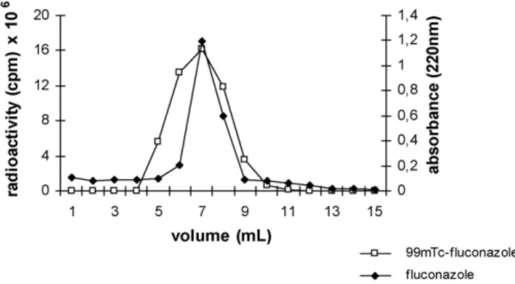
4− release (P> 0.05) as determined by ITLC (Table 2). These results showed that 99mTc-fluconazole is a stable molecule in the presence of serum components, and it proved to be suit-able for use in biodistribution studies. Furthermore, the elution profile of 99mTc-fluconazole by size-exclusion chromatogra-phy in Sephadex G150 showed a radioactivity peak coincident with unlabeled fluconazole, between 5 and 9 mL, detected by absorbance measurement at 220 nm (Fig. 1). These results
con-Fig. 1. Chromatography profile of99mTc-fluconazole and unlabeled fluconazole
Table 3
Characteristics of the NC preparations
NC formulation Mean size±S.D.a
(nm)
Mean size±S.D.b (nm)
Mean size±S.D.b (nm)c
Polydispersity index
Potential±S.D.d (mV)e
PCS AFM AFM (glut.)
PLA-POLOX 282±24 411±128 193±26 0.340f −69.6±8.4
PLA-PEG 236±40 238±124 212±39 0.340f −57.9±3.7
99mTc-Fluconazole-PLA-POLOX 356±45 298±151 200±40 0.375f −66.2±6.7
99mTc-Fluconazole-PLA-PEG 351±24 264±95 202±45 0.418f −55.4±7.8
Fluconazole-PLA-POLOX 347±15 nd nd 0.116g −62.3±1.6
Fluconazole-PLA-PEG 330±20 nd nd 0.272g −58.1±6.2
nd: not determined.
aStandard deviation of measurements from 10 different batches. b Standard deviation of size measurements of 40 nanocapsules. cSamples fixed with 25% glutaraldehyde.
d Standard deviation of at least three measurements obtained from different batches. eMeasurement after 1:1000 dilution in 1 mM NaCl (conductivity, 120±20S/cm). fHomogeneous samples (≤0.5).
g Monodispersed samples (≤0.3).
firmed the chemical integrity of fluconazole molecule after the radiolabeling process. Free pertechnetate and colloids repre-sented less than 6% of the total radioactivity in the labeling solution. The fluconazole labeling yield obtained in the present work are in agreement with those previously published by
Lupetti et al. (2002).
3.2. Nanocapsule characterization
The percentage of encapsulation was 29.3±3.5 and 32.2±1.5% for PLA NC and PLA-PEG NC preparations, respectively. The nature of the polymer, PLA or PLA-PEG, did not modify the percentage of 99mTc-fluconazole encapsulated (P> 0.05).
The particle size, polydispersity index and zeta potential of prepared conventional and surface-modified NC containing 99mTc-fluconazole are presented inTable 3. The mean size and size distribution of the nanoparticles have a key role in determin-ing their fate followdetermin-ing administration, their interaction with the cell membrane and their penetration across the physiological drug barriers (Feng et al., 2004). Some authors have demon-strated that the mean diameter of nanoparticles has an influence on the biodistribution studies because large particles are rapidly taken up by MPS, thereby reducing their half-lives in the circula-tory system (Owens and Peppas, 2006). The polydispersity index is indicative of the size distribution, and it showed that the differ-ent NC formulations prepared by the nanoprecipitation method were homogeneous (PI < 0.5). However only fluconazole-loaded NC were monodisperse (PI≤0.3). The particle size of this for-mulation was the most homogeneous even when compared with the unloaded PLA and PLA-PEG NC. The mean size of NC obtained in this study (236–356 nm) are in agreement with data obtained by others authors that observed NC diameters between 200 and 300 nm (Couvreur et al., 2002; Mosqueira et al., 2006).Torchilin (2000)showed that nanoparticles ranging from 10 to 500 nm in size can extravasate and accumulate in areas with increased vascular permeability. Therefore, the NC sizes obtained in this study are suitable for reaching infectious
and inflammatory foci. It seems that the introduction of flu-conazole in the NC composition increases the particle size. The mean diameters of unloaded NC were smaller than NC contain-ing99mTc-fluconazole or unlabeled fluconazole (P< 0.05). The interaction of fluconazole with phospholipid bilayers has previ-ously been investigated (Pascual et al., 1993; Ambrosini et al., 1998), and it could be one of the reasons for the effect observed in this work because all NC formulations contained a high per-centage of phosphatidylcholine. Ambrosini and co-workers have suggested that fluconazole interacts with the polar head group region without penetration into the apolar hydrocarbon region of bilayers.
Zeta potential results (Table 3) showed that the unloaded NC and NC containing radiolabeled and unlabeled fluconazole exhibited a negative charge with values ranging from−55.4 to −69.6 mV, as is typically observed for these types of systems composed of PLA polymer (Mosqueira et al., 2001a). The pres-ence of PEG resulted in a reduction in absolute values of the zeta potential for unloaded NC and NC containing99mTc-fluconazole (P< 0.05). Previous studies showed that formulations with PEG chains have a reduced zeta potential when compared with con-ventional nanoparticles (Fresta et al., 2001; Mosqueira et al., 2001b; Ameller et al., 2004; Faria et al., 2005). According to
Mosqueira et al. (2006), the reduction of the zeta potential is a result of the presence of the PEG layer at NC surface that shifts the plane of shear to the outer boundary of the layer, thereby masking the real zeta potential value. Although the PLA-PEG NC showed reduced zeta potential, the absolute values are still high compared with previously reported zeta potential for PLA-PEG nanospheres (Gref et al., 1994). It was demonstrated that lecithin in the nanocapsules formulation contributed to the main-tenance of the high negative surface charge masking the effects of PEG chains in the zeta potential measurements (Mosqueira et al., 2001a). However, in vivo the same amount of PEG at the NC surface increases eight fold the AUC (plasma) in mice (Mosqueira et al., 2001b).
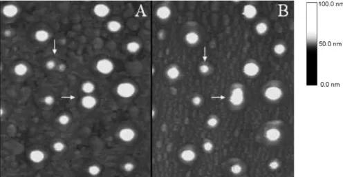
Fig. 2. AFM images of height (A and C) and phase (B and D) of PLA-POLOX NC containing99mTc-fluconazole (upper images) and of PLA-PEG NC containing 99mTc-fluconazole (lower images) showing spherical nanostructures. Scan size: 5m×5m.
(Neves et al., 1998). In this technique, the preparation of the sample is very simple. It is deposited in a partially dried state onto freshly cleaved mica plates, a fact that allows simultane-ous characterization of particle shape, structure and interparticle organization. All nanocapsule preparations presented a spherical form in the AFM images (Figs. 2 and 3). The NC presented a het-erogeneous distribution in size and height in three-dimensional images (Fig. 3). The PLA-POLOX nanocapsules (Fig. 2A and B) presented more complex images than those obtained with
Fig. 3. Three-dimensional AFM image of height of PLA-POLOX NC.
PLA-PEG (Fig. 2C and D) because they had few nanometer layers (2–10 nm) with different heights, probably because the presence of lecithin and poloxamer. On the other hand, PLA-PEG NC (Fig. 2C and D), without poloxamer, presented more homogeneous particles that were less contaminate by layers. The average size of unloaded conventional NC obtained by AFM were 411±128 nm, while those containing99mTc-fluconazole were 298±151 nm (n= 40). However, these differences were not significant, in contrast to the results observed by PCS. The average diameter calculated was 238±124 nm (n= 40) for unloaded PLA-PEG NC and 264±95 nm (n= 40) for PLA-PEG NC containing99mTc-fluconazole. The mean sizes were not significantly different (P> 0.05). However, the variation in diameters represented by the standard deviation of measure-ments obtained by AFM was larger than those measured by PCS for both NC types. This observation can be explained as a possi-ble NC flattening and aggregation process that takes place after drying on a mica surface. In fact, the flattening phenomenon was probably related to variations in polymeric wall thickness and in its homogeneity (Leite et al., 2005).
The diameter/height ratios were calculated from the topo-graphical profile (Table 4). The values showed a ratio of approximately 11 for both types of unloaded NC. These results confirmed the existence of flattened forms that were also suggested by Montasser et al. (2002)who worked with nanocapsules prepared with the co-polymer
con-Table 4
Diameter/height ratios of nanocapsules analyzed by AFM
NC formulation Diameter/height ratio±S.D.a Not fixed with
glutaraldehyde
Fixed with glutaraldehyde
PLA-POLOX 11.1±1.1 1.7±0.5
PLA-PEG 11.0±2.6 6.1±2.3
99mTc-Fluconazole-PLA-POLOX 16.6±3.6 2.2±0.6 99mTc-Fluconazole-PLA-PEG 15.4±3.0 6.0±1.6
aStandard deviation of 40 measurement.
firming the flattening process during AFM analysis. When the 99mTc-fluconazole was associated with the NC, the diam-eter/height ratios increased to values of 15.4 and 16.6 for 99mTc-fluconazole-PLA-POLOX and99m Tc-fluconazole-PLA-PEG NC, respectively (Table 4). These NC were evaluated by AFM, 24 and 48 h after deposition on the mica surface, as is shown in detail inFig. 4. The observations demonstrated the occurrence of an aggregation process and flattening phenom-ena on mica that caused a fusion of NC and, consequently, an increase in NC sizes after 48 h. NC containing 99m
Tc-fluconazole showed more pronounced flattening phenomena in the samples when compared with unloaded NC, probably due to increased complexity on NC wall formation in the presence of the radiolabeled drug. On the other hand, this aggrega-tion process was not observed when 25% glutaraldehyde was used on the mica surface (Fig. 5), suggesting that nanocapsules maintained their structures unaltered 48 h after the deposition. Furthermore, the diameter/height ratio obtained for NC fixed with 25% glutaraldehyde (Table 4) decreased significantly to 1.7 and 6.1 for unloaded PLA-POLOX NC and PLA-PEG NC, respectively. The same process occurred with NC containing 99mTc-fluconazole, where the values were reduced to 2.2 and 6 for PLA-POLOX NC and PLA-PEG NC, respectively (Table 4). The greater ratio founded for PLA-PEG NC, despite of drug association, indicate that PEG located on NC surface probably induces a lower degree of NC wall interaction with glutaralde-hyde. Thus, these observations suggest that the fixation of NC containing radiolabeled fluconazole on the mica surface by glu-taraldehyde could be an important method in sample preparation for AFM analysis. The glutaraldehyde seems to reduce the flattening process by hardening the wall of the NC and thus preserving its shape and morphology. This sample treatment
Fig. 4. AFM images of height of PLA-POLOX NC containing99mTc-fluconazole, 24 h (A) and 48 h (B) after deposition of formulation on the mica surface. The
white arrows indicate the aggregation of NC. Scan size 5m×5m.
Fig. 5. AFM images of height of PLA-POLOX NC containing99mTc-fluconazole previously fixed with 25% glutaraldehyde, 24 h (A) and 48 h (B) after deposition
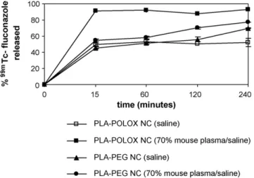
Fig. 6. Release profile of99mTc-fluconazole from PLA and PLA-PEG NC in
saline or saline with 70% of mouse plasma at 37◦C.
furnished information that more closely approximated the real NC size and morphology.
3.3. 99mTc-fluconazole release from nanocapsules
The profiles forin vitrorelease of99mTc-fluconazole from PLA and PLA-PEG nanocapsules are shown in Fig. 6. The experiments were preformed under sink conditions to avoid the interference of the solubility of the drug with thein vitrorelease. A fast release of radioactivity for all NC types was observed in the first 15 min. A fast release of drug in the first few minutes after dilution in release media has been interpreted by several authors as drug adsorbed at the NC surface (Schaffazick et al., 2003; Mosqueira et al., 2006). However, plasma protein prob-ably acts as a driving force that promotes 99mTc-fluconazole desorption from the NC surface as a result of its binding capac-ity, which removes drug from bulk solution and thus maintains the concentration gradient (Mosqueira et al., 2006). Therefore, the release of 99mTc-fluconazole by POLOX and PLA-PEG NC was greater in the plasmatic medium. In this medium, it was possible to distinguish differences between the two for-mulations. The drug release by PLA-PEG NC was significantly smaller at all times studied (P< 0.05). The PEG layer around the NC probably reduced the amount of drug released by impairing protein binding at NC surface, such as also observed byGref et al. (1995)andMosqueira et al. (2006).
After the immediate release of99mTc-fluconazole by PLA-POLOX NC, a plateau was observed, indicating no further additional release of effectively encapsulated drug. The poly-meric wall of PLA was not able to control release of the drug, which would explain the fast, although incomplete release from PLA-POLOX NC. Furthermore, previous studies suggest that the poloxamer could increase the amount of drug release from NC (Mosqueira et al., 2006). On the other hand, the PLA-PEG NC showed different biphasic release profile, with the initial burst and after a gradual and incomplete drug release up to 240 min, in plasma and saline media. Those release profiles of 99mTc-fluconazole from NC indicate that PLA-PEG NC show different properties of drug release control compared with
PLA-POLOX NC. These differences could be attributed to the form of PEG or poloxamer association to the NC surface, by covalent grafting (PLA-PEG) or by adsorption (PLA-POLOX).
Finally, the results indicate that PLA-PEG NC offered advan-tageous properties as a delivery system of 99mTc-fluconazole over PLA-POLOX formulation, because they show a reduc-tion in drug release as a result of the minor interacreduc-tion with plasma proteins. These data are in agreement with the stud-ies ofMosqueira et al. (2006), where a better ability to retain and release a lipophilic antimalarial drug was observed with PLA-PEG NC when compared with PLA NC stabilized with poloxamer in media containing plasma.
4. Conclusion
Both types of nanocapsule preparations studied in this work were able to encapsulate the99mTc-fluconazole with the same yield. The homogeneity, the adequate sizes and the negative zeta potential of both types of nanocapsules containing99m Tc-fluconazole indicate that these formulations could be used for reaching infectious foci, such as those induced by Can-dida albicans. Moreover, the presence of covalently grafted PEG chains at the surface provides a hydrophilic steric bar-rier, limiting and controlling the release of99mTc-fluconazole. Therefore, it is expected that in furtherin vivostudies, the PLA-PEG nanocapsules containing fluconazole would constitute in a long-circulating intravenous formulation for treatingCandida
infections.
Acknowledgments
The authors thank the financial support of FAPEMIG/ NANOBIOMG Network for this study (EDT-1806/02 grant). The authors would like to thank Maira Alves Pereira for her important collaboration.
References
Agarwal, R., Katare, O.P., 2002. Preparation andin vitroevaluation of micona-zole nitrate–loaded topical liposomes. Pharm. Technol. 26, 48–60. Ambrosini, A., Bossi, G., Dante, S., Dubini, B., Gobbi, L., Leone, L., Bossi,
M.G.P., Zolese, G., 1998. Lipid-drug interaction: thermodynamic and struc-tural effects of antimicotic fluconazole on DPPC liposomes. Chem. Phys. Lipids 95, 37–47.
Ameller, T., Marsaud, V., Legrand, P., Gref, R., Renoir, J.-M., 2004. Pure antiestrogen RU 58668-loaded nanospheres: morphology, cell activity and toxicity studies. Eur. J. Pharm. Sci. 21, 361–370.
Appleton, S.S., 2000. Candidiasis: pathogenesis, clinical characterisitics, and treatment. J. Can. Dent. Assoc. 28, 942–948.
Barratt, G., 2000. Therapeutic applications of colloidal drug carriers. Pharm. Sci. Technol. Today 3, 163–171.
Chasteigner, S., Fessi, H., Devissaguet, J.-P., Puisieux, F., 1998. Comparative study of the association of itraconazole with colloidal drug carriers. Drug Dev. Res. 38, 125–133.
Couvreur, P., Barratt, G., Fattal, E., Legrand, P., Vauthier, C., 2002. Nanocapsule technology: a review. Crit. Rev. Drug Carrier Syst. 19, 99–134.
Feng, S.S., Li, M., Khin, W., Guofeng, H., 2004. Nanoparticles of biodegradable polymers for clinical administration of paclitaxel. Curr. Med. Chem. 11, 413–424.
Fessi, H., Puisieux, F., Devissaguet, J.-P., Ammoury, N., Benita, S., 1989. Nanocapsule formation by interfacial polymer deposition following solvent displacement. Int. J. Pharm. 55, R1–R4.
Fresta, M., Fontana, G., Bucolo, C., Cavallaro, G., Giammona, G., Puglisi, G., 2001. Ocular tolerability andin vivobioavailability of poly(ethylene glycol) (PEG)-coated polyethyl-2-cyanoacrylate nanosphere-encapsulated acyclovir. J. Pharm. Sci. 90, 288–297.
Gref, R., Domb, A., Quellec, P., Blunk, T., Muller, R.H., Verbavatz, J.M., Langer, R., 1995. The controlled intravenous delivery of drugs using PEG-coated sterically stabilized nanospheres. Adv. Drug Deliv. Rev. 16, 215–233. Gref, R., Minamitake, Y., Peracchia, M.T., Trubetskoy, V., Torchilin, V., Langer,
R., 1994. Biodegradable long-circulating polymeric nanospheres. Science 263, 1600–1603.
Gupta, S.K., Dhingra, N., Velpandian, T., Jaiswal, J., 2000. Efficacy of flu-conazole and liposome entrapped fluflu-conazole for C. albicans induced experimental mycotic endophthalmitis in rabbit eyes. Acta Ophthalmol. Scand. 78, 448.
Legrand, P., Barratt, G., Mosqueira, V., Fessi, H., Devissaguet, J.P., 1999. Poly-meric nanocapsules as drug delivery systems: a review. S.T.P. Pharm. Sci. 9, 411–418.
Leite, E.A., Vilela, J.M.C., Mosqueira, V.C.F., Andrade, M.S., 2005. Poly-caprolactone nanocapsules morphological features by atomic force microscopy. Microsc. Microanal. 11, 48–51.
Lupetti, A., Welling, M.M., Mazzi, U., Nibbering, P.H., Pauwels, E.K.J., 2002. Technetium-99m labeled fluconazole and antimicrobial peptides for imaging ofCandida albicansandAspergillus fumigatusinfections. Eur. J. Nucl. Med. 29, 674–679.
Maesaki, S., 2002. Drug delivery system of anti-fungal and parasitic agents. Curr. Pharm. Des. 8, 433–440.
Martin, M.V., 1999. The use of fluconazole and itraconazole in the treatment ofCandida albicansinfections: a review. J. Antimicrob. Chemother. 44, 429–437.
McEvoy, G.K., 2003. Antifungal antibiotics—fluconazole. In: McEvoy, G.K. (Ed.), AHFS Drug Information. American Society of Health System Phar-macists Inc., Bethesda, MD, p. 101.
Mehta, R.T., Hopfer, R.L., Gunner, L.A., Juliano, R.L., Lopez-Berestein, G., 1987a. Formulation, toxicity, and antifungal activity in vitro of liposome-encapsulated nystatin as therapeutic agent for systemic candidia-sis. Antimicrob. Agents Chemother. 31, 1897–1900.
Mehta, R.T., Hopfer, R.L., McQueen, T., Juliano, R.L., Lopez-Berestein, G., 1987b. Toxicity and therapeutic effects in mice of liposome-encapsulated nystatin for systemic fungal infections. Antimicrob. Agents Chemother. 31, 1901–1903.
Molina, J., Urbina, J., Gref, R., Brener, Z., Junior, J.M.R., 2001. Cure of experimental Chagas’s disease by the bis-triazole DO870 incorporated into stealth polyethyleneglycol-polylactide nanospheres. Antimicrob. Agents Chemother. 47, 101–104.
Montasser, I., Fessi, H., Coleman, A.W., 2002. Atomic force microscopy imag-ing of novel type of polymeric colloidal nanostructures. Eur. J. Pharm. Biopharm. 54, 281–284.
Mosqueira, V.C.F., Legrand, P., Barratt, G., 2006. Surface-modified and con-ventional nanocapsules as novel formulation for parenteral delivery of halofantrine. J. Nanosci. Nanotechnol. 9–10, 3193–3202.
Mosqueira, V.C.F., Legrand, P., Gulik, A., Bourdon, O., Gref, R., Labarre, D., Barratt, G., 2001a. Relationship between complement activation, cellular uptake and physicochemical aspects of novel PEG-modified nanocapsules. Biomaterials 22, 2967–2979.
Mosqueira, V.C.F., Legrand, P., Morgat, J., Vert, M., Mysiakine, E., Gref, R., Devissaguet, J.-P., Barratt, G., 2001b. Biodistribution of long-circulating PEG-grafted nanocapsules in mice: effects of PEG chain length and density. Pharm. Res. 18, 1411–1419.
Neves, B.R.A., Vilela, J.M.C., Andrade, M.S., 1998. Microscopia de varredura por sonda mecˆanica: uma introduc¸˜ao. Cerˆamica 44, 212–219.
Otsubo, T., Maesaki, S., Hossain, M.A., Yamamoto, Y., Tomono, K., Tashiro, T., Seki, J., Tomii, Y., Sonoke, S., Kohno, S., 1999.In vitroandin vivo
activities of NS-718, a new lipid nanosphere incorporating amphotericin B, againstAspergillus fumigatus. Antimicrob. Agents Chemother. 43, 471– 475.
Owens III, D.E., Peppas, N.A., 2006. Opsonization, biodistribution, and phar-macokinetics of polymeric nanoparticles. Int. J. Pharm. 307, 93–102. Pascual, A., Garcia, I., Conejo, C., Perea, E.J., 1993. Uptake and intracellular
activity of fluconazole in human polymorphonuclear leukocytes. Antimi-crob. Agents Chemother. 37, 187–190.
Prakobvaitayakit, M., Nimmannit, U., 2003. Optimization of polylactic-co-glycolic acid nanoparticles containing itraconazole using 23factorial design.
AAPS Pharm. Sci. Technol. 4, 1–9.
Schaffazick, S.R., Guterres, S.S., Freitas, L.L., Pohlmann, A.R., 2003. Caracterizac¸˜ao e estabilidade f´ısico-qu´ımica de sistemas polim´ericos nanoparticulados para administrac¸˜ao de f´armacos. Qu´ımica Nova 26, 726–737.