Review Article
Biological Effects of Medicinal Plants on Induced Periodontitis:
A Systematic Review
Jefferson Soares de Oliveira,
1Moara e Silva Conceição Pinto,
2Lucas de Araújo de Bastos Santana,
1Antonione Santos Bezerra Pinto,
3David di Lenardo,
1and Daniel Fernando Pereira Vasconcelos
21Laboratory of Biology and Biochemistry Plants (BIOqPLANT), Federal University of Piaui, Parnaiba, PI, Brazil 2Laboratory of Histological Analysis and Prepare (LAPHIS), Federal University of Piaui, Parnaiba, PI, Brazil 3Department of Morphology, LABICONTE, Federal University of Ceara, Fortaleza, CE, Brazil
Correspondence should be addressed to Daniel Fernando Pereira Vasconcelos; vasconcelos@ufpi.edu.br
Received 1 June 2016; Revised 22 June 2016; Accepted 23 June 2016
Academic Editor: Gul Atilla
Copyright © 2016 Jeferson Soares de Oliveira et al. his is an open access article distributed under the Creative Commons Attribution License, which permits unrestricted use, distribution, and reproduction in any medium, provided the original work is properly cited.
Objective. he aim of this systematic review was to investigate the advances in the study of medicinal plants and their biologic efects on periodontitis in animal models.Study Design. A systematic search was conducted by three independent researchers, who screened articles published up to March/2016, to identify the studies that contained suicient and clear information on the association of the medicinal plants and periodontitis in murine models. he searches were performed using PubMed, Cochrane, and Science Direct databases. Results. Ater a critical analysis of titles and abstracts, 30 studies were inally eligible for analysis. he studies presented a great diversity of the experiment designed regarding the methods of induced periodontitis and the evaluation of the medicinal plants eicacy. None of the studies described the possible toxic efects associated with the administration of the plant material to animals and whether they could prevent damage to organs caused by systemic efect of induced periodontitis. Gel-based formulations containing plant substances are seen as an interesting strategy to treat periodontitis.Conclusions. In this systematic review, the state-of-the-art knowledge on the medicinal plants and the induced periodontitis was critically evaluated and discussed from the experiment designed to the possible clinical application.
1. Introduction
Periodontitis is one of the most extensive oral problems that afect human population, resulting from an inlammatory response against microorganisms involved with plaque accu-mulation on the subgingival dental surface. he development of the process depends on the interaction between the bacteria in the site of infection. he presence of an oral bioilm composed by bacteria and their products includes also lipopolysaccharides and proteinases that are responsible for the progression of periodontitis. Bacteria stimulate host immunopathological and inlammatory mechanisms that result in the destruction of the periodontal tissue [1].
Diferent strategies have been used to treat periodon-tal diseases. Mechanical therapy and surgical procedures reduce microbial burden, being efective in the control of
the periodontitis progression. Nevertheless, this regulation is not always satisfactory, possibly due to the prominent role of immunogenetic response on periodontal destruction. In some cases, adjunctive therapies may be required [2]. hus, the discovery and development of potential therapeutic drugs with the ability to regulate the host immune and bacteria-mediated inlammatory interactions are a valuable approach for the prevention and treatment of the peri-odontal disease [2–4]. In this context, plants can represent an interesting source of molecules with a potential activ-ity against periodontal disease progression. he growing incidence of periodontitis, the increased resistance of oral bacteria to antibiotics, and the adverse efects of some drugs used in dentistry all motivate the search for safe and efective molecules to treat and prevent the disease [5].
2 International Journal of Dentistry
Medicinal plants have been fundamental for thousands of years to provide bioactive molecules used to treat diferent types of human inirmities, such as inlammation, pain, and tumors. Also, they can be a source of compounds to be tested in the treatment of periodontal diseases. Nowadays, there is an increasing number of scientiic investigation exploring plant extracts or puriied molecules in periodontal diseases [3]. Several of these studies were performed using animal models since biochemical, histological, and anatomical fea-tures are similar to humans [3, 4, 6, 7].
he aim of this systematic review was to investigate the advances in the study of medicinal plants and the develop-ment of induced periodontitis in animal models. Data were described and discussed in order to evaluate the limitation and also the perspectives of the application of these agents in the treatment of the periodontal disease.
2. Material and Methods
A systematic search was conducted by three indepen-dent researchers who screened articles published up to March/2016 in order to identify the studies that contained suicient and clear information on the association of the medicinal plants and periodontitis in murine models. he searches were performed using PubMed (http://www.ncbi .nlm.nih.gov/pubmed), Cochrane (http://www.cochraneli-brary.com/), and Science Direct (http://www.sciencedirect
.com/) databases using the key wordsplant,periodontitis,and
ratsand search details “plant” [All Fields] AND
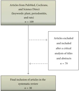
“periodonti-tis” [All Fields] AND “rats” [All Fields]. he articles selected by each researcher were compared to remove duplicate records and 109 diferent articles were initially evaluated. Ater a critical analysis of titles and abstracts, 79 articles were excluded such as articles that were not in English, articles that were not fully available, or those that did not report an association between medicinal products and murine model of periodontitis. Hence, 30 studies were inally eligible for a qualitative analysis for this review (Figure 1).
3. Results and Discussion
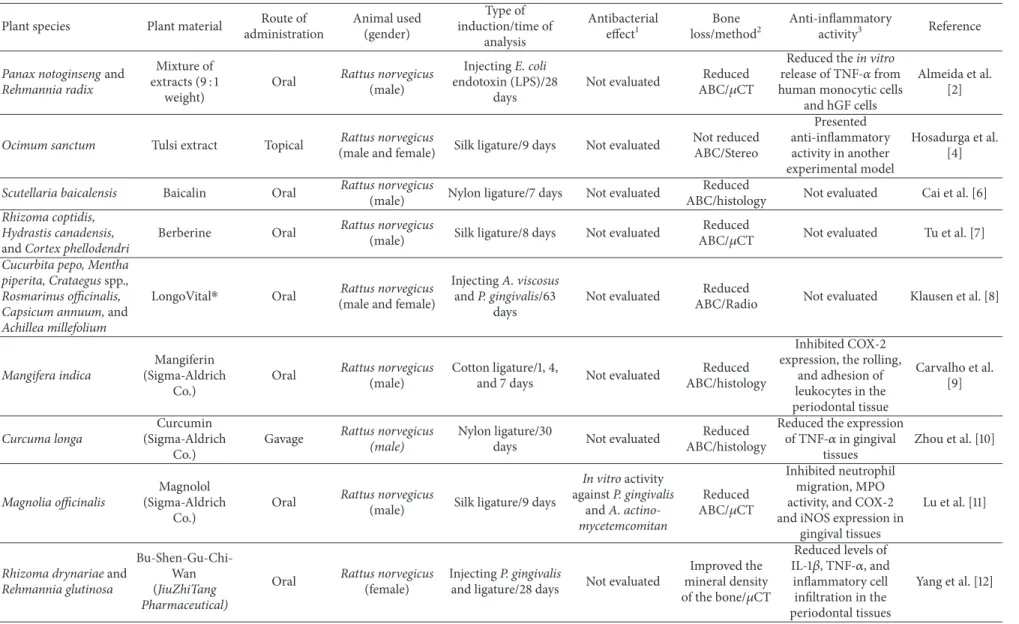
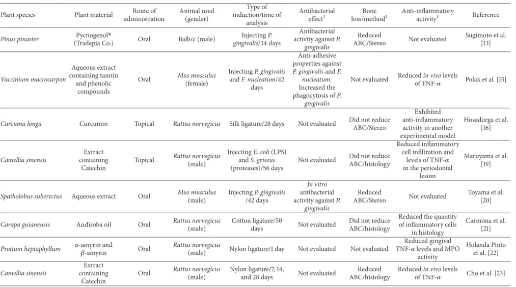
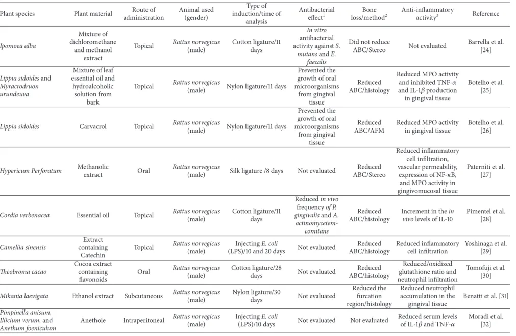
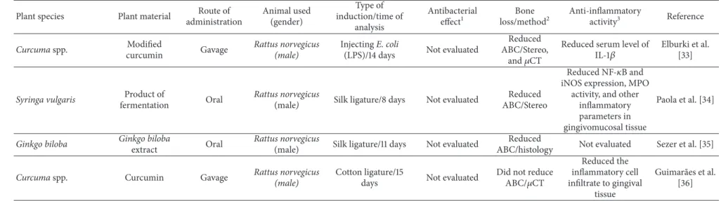
Table 1 summarize plant species, plant material, route of administration, animal used, type of induction and time of analysis, and the ability of the medicinal plant to reduce alveolar bone loss related to 30 selected articles.
he majority of articles (43.3%) described studies con-ducted with plant extracts, 40% with puriied compounds and 16.6% with a mixture of two or more plant extracts. Among them, six articles described experiments developed using a commercial product [8–13], containing puriied compounds or a mixture of plant extracts. None of authors mentioned the process of obtaining selected plant material as an important limitation for development of the research.
In general, studies were preferentially conducted using
Rattus norvegicus(90%) of diferent strains Wistar, Lewis, or Sprague-Dawley. his specie is one of the mostly used for
in vivoexperimental models, including the pathogenesis of periodontal disease because they ofer some advantages such as price, easy handling, and the possibility of microbiological,
Articles from PubMed, Cochrane, and Science Direct (keywords: plant, periodontitis,
and rats)
Articles excluded
and included
after a critical
analysis of titles
and abstracts n = 109
n = 79
Final inclusion of articles in the systematic review
n = 30
Figure 1: Search lowchart and selection of articles for the review of the literature.
macroscopic, and histological evaluation [14]. Other
stud-ies were conducted using Mus musculus Balb/c (6.6%) or
C57BL/6. Rodents were preferred as the animal model since they present biochemical, histological, and anatomical fea-tures similar to humans [14].
Taking into consideration the sex of the animal, most authors chose to use males (86.6%). Two articles described the use of females [12, 15] and two studies were conducted using males and females [4, 8]. One study did not describe the
sex of the animals [16]. Mostin vivostudies which investigate
the biological efect of plant substances favor the use of male animals. his choice is based on the fact that female hormones should interfere in the development and progression of the disease [17]. None of the articles analyzed was related to the hormonal inluence on the biological activities investigated. However, studies conducted with females could clarify the efect of their hormones on the speciic pathways involved in the periodontitis progression.
Regarding the common type of periodontitis-induction, in 66.6% of the articles, the process was caused by ligature (silk: 23.3%, nylon: 23.3%, and cotton: 20%). his method is widely used because it facilitates the accumulation of bioilm. his procedure increases the iniltration of inlammatory cells and the production of chemical mediators that lead to the degradation of the tissues around the teeth contributing to the destruction of the periodontal tissues [1]. In addition to periodontitis induced by ligature, 16.6% of articles described
the gingival injection of bacteria, such as P. gingivalis and
16.6% described the administration ofE. coliendotoxin (LPS)
In
te
rn
at
io
n
al
Jo
u
rn
al
o
f
De
n
tis
tr
y
3
Table 1: List of medicinal plants, experimental methods, and their biological efects on induced periodontitis.
Plant species Plant material Route of
administration
Animal used (gender)
Type of induction/time of
analysis
Antibacterial
efect1
Bone
loss/method2
Anti-inlammatory
activity3 Reference
Panax notoginsengand
Rehmannia radix
Mixture of extracts (9 : 1
weight)
Oral Rattus norvegicus (male)
InjectingE. coli
endotoxin (LPS)/28 days
Not evaluated Reduced
ABC/�CT
Reduced thein vitro
release of TNF-�from
human monocytic cells and hGF cells
Almeida et al. [2]
Ocimum sanctum Tulsi extract Topical Rattus norvegicus
(male and female) Silk ligature/9 days Not evaluated
Not reduced ABC/Stereo
Presented anti-inlammatory activity in another experimental model
Hosadurga et al. [4]
Scutellaria baicalensis Baicalin Oral Rattus norvegicus
(male) Nylon ligature/7 days Not evaluated
Reduced
ABC/histology Not evaluated Cai et al. [6]
Rhizoma coptidis, Hydrastis canadensis,
andCortex phellodendri
Berberine Oral Rattus norvegicus
(male) Silk ligature/8 days Not evaluated
Reduced
ABC/�CT Not evaluated Tu et al. [7]
Cucurbita pepo, Mentha piperita, Crataegusspp., Rosmarinus oicinalis, Capsicum annuum,and
Achillea millefolium
LongoVital Oral Rattus norvegicus
(male and female)
InjectingA. viscosus
andP. gingivalis/63 days
Not evaluated Reduced
ABC/Radio Not evaluated Klausen et al. [8]
Mangifera indica
Mangiferin (Sigma-Aldrich
Co.)
Oral Rattus norvegicus (male)
Cotton ligature/1, 4,
and 7 days Not evaluated
Reduced ABC/histology
Inhibited COX-2 expression, the rolling,
and adhesion of leukocytes in the periodontal tissue
Carvalho et al. [9]
Curcuma longa
Curcumin (Sigma-Aldrich
Co.)
Gavage Rattus norvegicus(male) Nylon ligature/30days Not evaluated ABC/histologyReduced
Reduced the expression
of TNF-�in gingival
tissues
Zhou et al. [10]
Magnolia oicinalis
Magnolol (Sigma-Aldrich
Co.)
Oral Rattus norvegicus
(male) Silk ligature/9 days
In vitroactivity
againstP. gingivalis
andA. actino-mycetemcomitan
Reduced
ABC/�CT
Inhibited neutrophil migration, MPO activity, and COX-2 and iNOS expression in
gingival tissues
Lu et al. [11]
Rhizoma drynariaeand
Rehmannia glutinosa
Bu-Shen-Gu-Chi-Wan (JiuZhiTang Pharmaceutical)
Oral Rattus norvegicus (female)
InjectingP. gingivalis
and ligature/28 days Not evaluated
Improved the mineral density
of the bone/�CT
Reduced levels of
IL-1�, TNF-�, and
inlammatory cell iniltration in the periodontal tissues
4
In
te
rn
at
io
n
al
Jo
u
rn
al
o
f
De
n
tis
tr
y
Table 1: Continued.
Plant species Plant material Route of
administration
Animal used (gender)
Type of induction/time of
analysis
Antibacterial
efect1
Bone
loss/method2
Anti-inlammatory
activity3 Reference
Pinus pinaster Pycnogenol
(Tradepia Co.) Oral Balb/c (male)
InjectingP.
gingivalis/34 days
Antibacterial
activity againstP.
gingivalis
Reduced
ABC/Stereo Not evaluated
Sugimoto et al. [13]
Vaccinium macrocarpon
Aqueous extract containing tannin
and phenolic compounds
Oral Mus musculus (female)
InjectingP. gingivalis
andF. nucleatum/42 days
Anti-adhesive properties against
P. gingivalisandF. nucleatum.
Increased the
phagocytosis ofP.
gingivalis
Not evaluated Reducedin vivolevels
of TNF-� Polak et al. [15]
Curcuma longa Curcumin Topical Rattus norvegicus Silk ligature/28 days Not evaluated Did not reduce ABC/Stereo
Exhibited anti-inlammatory activity in another experimental model
Hosadurga et al. [16]
Camellia sinensis
Extract containing
Catechin
Topical Rattus norvegicus
(male)
InjectingE. coli(LPS)
andS. griseus
(proteases)/56 days
Not evaluated Did not reduce
ABC/histology
Reduced inlammatory cell iniltration and
levels of TNF-�
in the periodontal lesion
Maruyama et al. [19]
Spatholobus suberectus Aqueous extract Oral Mus musculus (male)
InjectingP. gingivalis
/42 days
In vitro
antibacterial
activity againstP.
gingivalis
Reduced
ABC/Stereo Not evaluated
Toyama et al. [20]
Carapa guianensis Andiroba oil Oral Rattus norvegicus (male)
Cotton ligature/50
days Not evaluated
Did not reduce ABC/histology
Reduced the quantity of inlammatory cells
in histology
Carmona et al. [21]
Protium heptaphyllum �-amyrin and�
-amyrin Oral
Rattus norvegicus
(male) Nylon ligature/1 day Not evaluated Not evaluated
Reduced gingival
TNF-�levels and MPO
activity
Holanda Pinto et al. [22]
Camellia sinensis
Extract containing
Catechin
Oral Rattus norvegicus (male)
Nylon ligature/7, 14,
and 28 days Not evaluated
Reduced ABC/histology
Reducedin vivolevels
In te rn at io n al Jo u rn al o f De n tis tr y 5
Table 1: Continued.
Plant species Plant material Route of
administration Animal used (gender) Type of induction/time of analysis Antibacterial efect1 Bone loss/method2 Anti-inlammatory
activity3 Reference
Ipomoea alba
Mixture of dichloromethane
and methanol extract
Topical Rattus norvegicus
(male)
Cotton ligature/11 days
In vitro
antibacterial
activity againstS.
mutansandE. faecalis
Did not reduce
ABC/Stereo Not evaluated
Barrella et al. [24]
Lippia sidoidesand
Myracrodruon urundeuva
Mixture of leaf essential oil and
hydroalcoholic solution from
bark
Topical Rattus norvegicus
(male) Nylon ligature/11 days
Prevented the growth of oral microorganisms
from gingival tissue
Reduced ABC/histology
Reduced MPO activity
and inhibited TNF-�
and IL-1�production
in gingival tissue
Botelho et al. [25]
Lippia sidoides Carvacrol Topical Rattus norvegicus(male) Nylon ligature/11 days
Prevented the growth of oral microorganisms
from gingival tissue
Reduced ABC/AFM
Reduced MPO activity in gingival tissue
Botelho et al. [26]
Hypericum Perforatum Methanolic
extract Oral
Rattus norvegicus
(male) Silk ligature /8 days Not evaluated
Reduced ABC/Stereo
Reduced inlammatory cell iniltration, vascular permeability,
expression of NF-�B,
and MPO activity in gingivomucosal tissue
Paterniti et al. [27]
Cordia verbenacea Essential oil Topical Rattus norvegicus (male)
Cotton ligature/11 days
Reducedin vivo
frequencyof P.
gingivalisandA.
actinomycetem-comitans
Reduced ABC/histology
Increment in thein
vivolevels of IL-10
Pimentel et al. [28]
Camellia sinensis
Extract containing
Catechin
Topical Rattus norvegicus
(male)
InjectingE. coli
(LPS)/10 and 20 days Not evaluated
Reduced ABC/histology
Reduced inlammatory cell iniltration
Yoshinaga et al. [29]
heobroma cacao
Cocoa extract containing lavonoids
Oral Rattus norvegicus (male)
Cotton ligature/28
days Not evaluated
Reduced ABC/histology
Reduced/oxidized glutathione ratio and neutrophil iniltration
Tomofuji et al. [30]
Mikania laevigata Ethanol extract Subcutaneous Rattus norvegicus (male)
Nylon ligature/30
days Not evaluated
Reduced the furcation region/histology
Reduced neutrophil accumulation in the
gingival tissue
Benatti et al. [31]
Pimpinella anisum, Illicium verum,and
Anethum foeniculum
Anethole Intraperitoneal Rattus norvegicus
(male)
InjectingE. coli
(LPS)/10 days Not evaluated Not evaluated
Reduced serum levels
of IL-1�and TNF-�
6
In
te
rn
at
io
n
al
Jo
u
rn
al
o
f
De
n
tis
tr
y
Table 1: Continued.
Plant species Plant material Route of
administration
Animal used (gender)
Type of induction/time of
analysis
Antibacterial
efect1
Bone
loss/method2
Anti-inlammatory
activity3 Reference
Curcumaspp. Modiied
curcumin Gavage
Rattus norvegicus (male)
InjectingE. coli
(LPS)/14 days Not evaluated
Reduced ABC/Stereo,
and�CT
Reduced serum level of
IL-1�
Elburki et al. [33]
Syringa vulgaris Product of
fermentation Oral
Rattus norvegicus
(male) Silk ligature/8 days Not evaluated
Reduced ABC/Stereo
Reduced NF-�B and
iNOS expression, MPO activity, and other
inlammatory parameters in gingivomucosal tissue
Paola et al. [34]
Ginkgo biloba Ginkgo biloba
extract Oral
Rattus norvegicus
(male) Silk ligature/11 days Not evaluated
Reduced
ABC/histology Not evaluated Sezer et al. [35]
Curcumaspp. Curcumin Gavage Rattus norvegicus
(male)
Cotton ligature/15
days Not evaluated
Did not reduce
ABC/�CT
Reduced the inlammatory cell iniltrate to gingival
tissue
Guimar˜aes et al. [36]
1Porphyromonas gingivalis, Aggregatibacter actinomycetemcomitans, Streptococcus mutans, Streptococcus sanguinis, Enterococcus faecalis, and Fusobacterium nucleatum.2ABC: alveolar bone crest, �CT:
microcomputed tomography, AFM: atomic force microscopy, Stereo: stereomicroscopy, Radio:radiography.3TNF-�: tumor necrosis factor�, hGF: hepatocyte growth factor, MPO: myeloperoxidase activity, IL-1�:
International Journal of Dentistry 7
an inlammatory response diferent from that promoted by periodontitis induced through induction with ligature [18].
Some of the studies analyzed described that long periods of experimental design were utilized to properly investigate the severity of tissue destruction during periodontal disease treatment: 63 days [8]; 56 days [19]; 42 days [20]; and 50 days [21]. However, the time of analysis changed substantially. More than 55% of the studies performed experiments during one to two weeks, 26.6% for three to four weeks, and 13.3% during six weeks or more. One study was conducted with an acute periodontitis rat model in 24 hours [22]. hese variations in the experimental periods may be due to factors such as the type and location of the ligature, the bacteria species or their sub-products injected for the induction of the disease and the specie (strain), and age and weight of the animal used. hese factors are generally associated with the objective of the study and the expected results.
More than 66% of studies chose the oral administration to evaluate the eicacy of the plant material. he topical admin-istration was described by 26.6% of the studies and 6.9% treated animals through subcutaneous or intraperitoneal cavity injections. he route of administration is an important parameter to inluence the eicacy of the material since it can interfere with the suicient amount of substance available to promote the biological efects. As observed, diferent results were seen for alveolar bone loss of animals submitted to the treatment with an extract containing Catechin obtained fromCamellia sinensis. Although the route of administration had not inluenced the anti-inlammatory activity of the material, a signiicant reduction of bone loss was observed when given orally [23] and no diference was seen ater its topical treatment [19].
Periodontal disease initiation and progression occur as a consequence of the host response to microorganisms of the dental bioilm [1]. herefore, the antibacterial efect is an important factor in the periodontal therapy. Only 26.6% of the studies investigated the capacity of the material to present antibacterial activity. In the study developed by Barrella et al.
[24] the organic extract obtained fromIpomoea albashowed
signiicantin vitroactivity against Streptococcus mutans, S.
sanguinis,and Enterococcus faecalis. he commercial
prod-uct Magnolol obtained fromMagnolia oicinalis exhibited
intense inhibition ofPorphyromonas gingivalisand
Aggregat-ibacter actinomycetemcomitansgrowth in a dependent dose
[11] and the aqueous extract fromVaccinium macrocarpon
containing tannin and phenolic compounds inhibited the
adhesion of Fusobacterium nucleatum and Porphyromonas
gingivalis [15].Study developed by Botelho et al. [25] and
Botelho et al. [26] withLippia sidoidesobserved the decrease
of salivary bacterial levels and this event was followed by an increment in clinical scores of gingival bleeding. Except for the work published by Klausen et al. [8] which did not evaluate the alveolar bone loss, all the articles that described the identiication of antibacterial activity also demonstrated that plant material treatment induced a signiicant reduction of alveolar bone loss.
he alveolar bone loss is one of the most important parameters evaluated in the induced periodontal disease. From 30 selected articles, 27 evaluated the ability of the
plant material to promote a signiicant reduction of the bone
loss. Carvacrol puriied from Lippia sidoides, the extract
containing Catechin obtained from Camellia sinensis, the
essential oil ofCordia verbenacea, and the methanolic extract
of Hypericum perforatum are examples of 77% of studies that observed a signiicant reduction of the bone loss [23, 26–28]. On the other hand, in 23% of studies such as
andiroba oil fromCarapa guianensis, as well as the mixture
of dichloromethane and methanol extracts from Ipomoea
alba and Longo Vital, the authors did not observe a
sig-niicant efect [8, 21, 24]. Moreover, it was interesting to note that diferent studies using the same plant material have reached diferent results regarding alveolar bone loss. According to Yoshinaga et al. [29] the topical treatment
of animals with the extract containing Catechin (Camellia
sinensis) promoted a signiicant reduction of bone loss in
periodontitis induced by E. coli(LPS). On the other hand,
the same material did not present a signiicant reduction in the alveolar bone loss against periodontitis induced by
injection of E. coli (LPS) and S. griseus (proteases) [19].
Diferent eicacy was also observed in studies developed with
curcumin (Curcuma longa). he topical administration of
curcumin did not reduce the alveolar bone loss [16] while its oral administration reduced this parameter signiicantly [10]. he methodologies used to evaluate the alveolar bone loss were histology (44.0%), stereomicroscopy (25.9%), micro-computed tomography (22.2%), atomic force microscopy (3.7%), and radiography (3.7%). Histology, stereomicroscopy, and microcomputed tomography are favorable methods once they yield precise information concerning the evaluation of periodontal tissue destruction.
It is noteworthy that the reduction of the inlammatory process may be associated with a reduced bone loss [12, 30, 31]. Because of this, a reduction of inlammatory process is usually investigated in periodontal disease. Taking the selected articles into account, 76.6% of them evaluated inlammatory parameters associated with induced periodon-titis. he common strategies used to observe the ability of the plant material to inhibit the inlammatory process in periodontal tissue were the evaluation of migration of inlammatory cells (30%), measurement of proinlammatory
cytokines (TNF-�: 30% and IL1-�: 13.3%) [2, 10, 12, 15, 19, 22,
23, 25, 32, 33], respectively, and dosage of myeloperoxidase (MPO) activity (20%) [11, 22, 25–27, 34]. Other molecular markers of inlammatory process, such as iNOS (13.0%)
[11, 34], NF-�B (8.7%) [27, 34], COX-2 (8.7%) [9, 11],
and IL-10 (4.3%) [28], were also investigated. Although the presence of the anti-inlammatory efect relects a possible eicacy against periodontitis, 13% of the authors who found anti-inlammatory properties did not observe a signiicant reduction of alveolar bone loss in the periodontitis assays. he anti-inlammatory activity was not analyzed in the 23.3%
[6–8, 13, 20, 24, 35] of the studies. he andiroba oil (Carapa
guianensis), the extract fromCamellia sinensis (containing
Catechin), and Curcumin (Curcuma spp.) were able to reduce
8 International Journal of Dentistry
reach levels that are high enough for the biological efects of administered substances in the experimental models adopted. Further researches are necessary to observe how much time is appropriate to manage the substances for the recovery of bone loss.
Although there is a close relation between the bone reabsorption and the inlammatory response, we suggest that the negative results of some of substances analyzed should be attributed to the fact that they do not act in the osteoclastogenesis process. his information is supported by the personal observation conducted using sulphated
polysac-charides recovered from red marine algaeGracilaria caudata.
Our data revealed that the treatment of experimental ani-mals with sulphated polysaccharides improved clinical and inlammatory parameters. However, no signiicant efect was observed in the reduction of alveolar bone loss (unpublished data).
It is important to mention that, although many plant substances may have deleterious efects to diferent animal organs [37], none of the evaluated studies has investigated the possible toxicological efects associated with the administra-tion of the medicinal plants. In addiadministra-tion, several studies have documented that induced periodontitis is followed by signif-icant changes of morphological structures and biochemical functions of diferent organs [38, 39]. Yet again, none of studies investigated the ability of these medicinal plants to prevent changes in organs promoted by a systemic action of the induced periodontitis.
Finally, 8 of the 30 selected articles conducted their experiments with a gel-based formulation containing the plant material investigated. In these experiments, the authors administered the gel topically, generally three times a day. he main idea behind these investigations is to reveal the further potential of the combined gel preparation to combat periodontal disease in closer than clinical situations. his was the outlook investigated in the studies published by [40, 41]. he authors evaluated the eiciency of a green tea Catechin gel as an adjunct on human periodontal therapy. In irst work [40], the authors observed a signiicant reduction on pockets and inlammation during the 4 weeks of the clinical trial. In the second work [41], it was demonstrated that when used as an adjunct to periodontal treatment, green tea gel could provide beneit in reducing bleeding on probing and gingival inlammation at 1st and 3rd months of evaluation. What makes these studies even more relevant is the fact that the indings on complementary products for the treatment of human periodontitis still can be enhanced.
4. Conclusion
In conclusion, the selected studies presented a large diversity of experimental designs, concerning the type of induction, time of analysis, and methods used for the evaluation of alveolar bone loss, anti-inlammatory, and antibacterial activ-ities. None of the studies evaluated the possible toxic efects associated with the administration of the material analyzed or their ability to prevent damages to organs caused by systemic efects of induced periodontitis. Gel-based formu-lations present an interesting strategy to treat periodontitis;
however, further studies are necessary to clarify its usefulness in the clinical situation.
Competing Interests
he authors declare that they have no conlict of interests.
Acknowledgments
his research is supported by the Federal University of Piau´ı (UFPI-Edital PIBIC 2014/2015 and BIAMA 03/2014), CNPq (455104/2014-0). he authors thank teacher Abilio Borghi for the grammar review of the paper.
References
[1] A. Bascones-Mart´ınez, M. Mu˜noz-Corcuera, S. Noronha, P. Mota, C. Bascones-Ilundain, and J. Campo-Trapero, “Host defence mechanisms against bacterial aggression in periodontal disease: basic mechanisms,”Medicina Oral, Patologia Oral y Cirugia Bucal, vol. 14, no. 12, pp. e680–e685, 2009.
[2] J. De Almeida, E. Ervolino, L. H. Bonietti et al., “Adjuvant ther-apy with sodium alendronate for the treatment of experimental periodontitis in rats,”Journal of Periodontology, vol. 86, no. 10, pp. 1166–1175, 2015.
[3] R.-Y. Huang, S.-H. Lu, K.-W. Su et al., “Diacerein: a potential therapeutic drug for periodontal disease,”Medical Hypotheses, vol. 79, no. 2, pp. 165–167, 2012.
[4] R. R. Hosadurga, S. N. Rao, R. Edavanputhalath et al., “Eval-uation of the eicacy of 2% Ocimum sanctum gel in the treatment of experimentalperiodontitis,”International Journal of Pharmaceutical Investigation, vol. 5, no. 1, pp. 35–42, 2015. [5] C. M. Ardila, M. A. L´opez, and I. C. Guzm´an, “High resistance
against clindamycin, metronidazole and amoxicillin in Porphy-romonas gingivalisandAggregatibacter actinomycetemcomitans
isolates of periodontal disease,”Medicina Oral, Patologia Oral y Cirugia Bucal, vol. 15, no. 6, pp. e947–e951, 2010.
[6] X. Cai, C. Li, G. Du, and Z. Cao, “Protective efects of baicalin on ligature-induced periodontitis in rats,”Journal of Periodontal Research, vol. 43, no. 1, pp. 14–21, 2008.
[7] H.-P. Tu, M. M. J. Fu, P.-J. Kuo et al., “Berberine’s efect on periodontal tissue degradation by matrix metalloproteinases: an in vitro and in vivo experiment,”Phytomedicine, vol. 20, no. 13, pp. 1203–1210, 2013.
[8] B. Klausen, A. Apostolopoulos, K. Stoltze, and F. N¨orgaard, “Efect of LongoVital treatment on development of periodontal disease in rats,”Scandinavian Journal of Dental Research, vol. 101, no. 1, pp. 33–36, 1993.
[9] R. R. Carvalho, C. H. Pellizzon, L. Justulin Jr. et al., “Efect of mangiferin on the development of periodontal disease: involvement of lipoxin A4, anti-chemotaxic action in leukocyte rolling,”Chemico-Biological Interactions, vol. 179, no. 2-3, pp. 344–350, 2009.
[10] T. Zhou, D. Chen, Q. Li, X. Sun, Y. Song, and C. Wang, “Curcumin inhibits inlammatory response and bone loss during experimental periodontitis in rats,”Acta Odontologica Scandinavica, vol. 71, no. 2, pp. 349–356, 2013.
International Journal of Dentistry 9
Alternative Medicine, vol. 2013, Article ID 634095, 12 pages, 2013.
[12] H. Yang, Q. Wen, J. Xue, and Y. Ding, “Alveolar bone regenera-tion potential of a tradiregenera-tional Chinese medicine, Bu-Shen-Gu-Chi-Wan, in experimental periodontitis,”Journal of Periodontal Research, vol. 49, no. 3, pp. 382–389, 2014.
[13] H. Sugimoto, K. Watanabe, T. Toyama et al., “Inhibitory efects of French pine bark extract, pycnogenol, on alveolar bone resorption and on the osteoclast diferentiation,”Phytotherapy Research, vol. 29, no. 2, pp. 251–259, 2015.
[14] X. Struillou, H. Boutigny, A. Soueidan, and P. Layrolle, “Exper-imental animal models in periodontology: a review,”he Open Dentistry Journal, vol. 4, no. 1, pp. 33–47, 2010.
[15] D. Polak, R. Naddaf, L. Shapira, E. I. Weiss, and Y. Houri-Haddad, “Protective potential of non-dialyzable material frac-tion of cranberry juice on the virulence ofP. gingivalisandF. nucleatummixed infection,”Journal of Periodontology, vol. 84, no. 7, pp. 1019–1025, 2013.
[16] R. R. Hosadurga, S. Rao, J. Jose, N. C. Rompicharla, M. Shakil, and R. Shashidhara, “Evaluation of the eicacy of 2% curcumin gel in the treatment of experimental periodontitis,”
Pharmacognosy Research, vol. 6, no. 4, pp. 326–333, 2014. [17] E. Figuero, A. Carrillo-De-Albornoz, D. Herrera, and A.
Bascones-Mart´ınez, “Gingival changes during pregnancy: I. Inluence of hormonal variations on clinical and immunolog-ical parameters,”Journal of Clinical Periodontology, vol. 37, no. 3, pp. 220–229, 2010.
[18] S. Garcia de Aquino, F. R. Manzolli Leite, D. R. Stach-Machado, J. A. Francisco da Silva, L. C. Spolidorio, and C. Rossa Jr., “Signaling pathways associated with the expression of inlam-matory mediators activated during the course of two models of experimental periodontitis,”Life Sciences, vol. 84, no. 21-22, pp. 745–754, 2009.
[19] T. Maruyama, T. Tomofuji, Y. Endo et al., “Supplementation of green tea catechins in dentifrices suppresses gingival oxidative stress and periodontal inlammation,”Archives of Oral Biology, vol. 56, no. 1, pp. 48–53, 2011.
[20] T. Toyama, K. Todoki, Y. Takahashi et al., “Inhibitory efects of Jixueteng on P. gingivalis-induced bone loss and osteoclast diferentiation,”Archives of Oral Biology, vol. 57, no. 11, pp. 1529– 1536, 2012.
[21] G. B. Carmona, R. K. C. Teixeira, M. V. H. Brito et al., “Efect of andiroba oil on periodontitis in wistar rats,”Acta Cirurgica Brasileira, vol. 28, no. 6, pp. 430–434, 2013.
[22] S. A. Holanda Pinto, L. M. S. Pinto, G. M. A. Cunha, M. H. Chaves, F. A. Santos, and V. S. Rao, “Anti-inlammatory efect of�,�-Amyrin, a pentacyclic triterpene from Protium heptaphyllum in rat model of acute periodontitis,” Inlam-mopharmacology, vol. 16, no. 1, pp. 48–52, 2008.
[23] A.-R. Cho, J.-H. Kim, D.-E. Lee et al., “he efect of orally administered epigallocatechin-3-gallate on ligature-induced periodontitis in rats,”Journal of Periodontal Research, vol. 48, no. 6, pp. 781–789, 2013.
[24] G. E. Barrella, I. B. Sufredini, F. V. Ribeiro, F. R. Cirano, and S. P. Pimentel, “Evaluation of the efect of an organic extract obtained fromIpomoea albaL. on experimental periodontitis in rats,”Brazilian Oral Research, vol. 26, no. 2, pp. 158–164, 2012. [25] M. A. Botelho, V. S. Rao, C. B. M. Carvalho et al., “Lippia
sidoides and Myracrodruon urundeuva gel prevents alveolar bone resorption in experimental periodontitis in rats,”Journal of Ethnopharmacology, vol. 113, no. 3, pp. 471–478, 2007.
[26] M. A. Botelho, J. G. Martins, R. S. Ruela et al., “Protective efect of locally applied carvacrol gel on ligature-induced periodonti-tis in rats: a tapping mode AFM study,”Phytotherapy Research, vol. 23, no. 10, pp. 1439–1448, 2009.
[27] I. Paterniti, E. Briguglio, E. Mazzon et al., “Efects of Hypericum Perforatum, in a rodent model of periodontitis,”BMC Comple-mentary and Alternative Medicine, vol. 10, article 73, 2010. [28] S. P. Pimentel, G. E. Barrella, R. C. V. Casarin et al.,
“Protec-tive efect of topicalCordia verbenaceain a rat periodontitis model: immune-inlammatory, antibacterial and morphomet-ric assays,”BMC Complementary and Alternative Medicine, vol. 12, no. 1, article 224, pp. 1–8, 2012.
[29] Y. Yoshinaga, T. Ukai, S. Nakatsu et al., “Green tea extract inhibits the onset of periodontal destruction in rat experimental periodontitis,”Journal of Periodontal Research, vol. 49, no. 5, pp. 652–659, 2014.
[30] T. Tomofuji, D. Ekuni, K. Irie et al., “Preventive efects of a cocoa-enriched diet on gingival oxidative stress in experimental periodontitis,” Journal of Periodontology, vol. 80, no. 11, pp. 1799–1808, 2009.
[31] B. B. Benatti, J. C. Campos-J´unior, V. J. Silva-Filho et al., “Efects of a Mikania laevigata extract on bone resorption and RANKL expression during experimental periodontitis in rats,”Journal of Applied Oral Science, vol. 20, no. 3, pp. 340–346, 2012. [32] J. Moradi, F. Abbasipour, J. Zaringhalam et al., “Anethole, a
medicinal plant compound, decreases the production of pro-inlammatory TNF-�and IL-1�in a rat model of LPS-induced periodontitis,”Iranian Journal of Pharmaceutical Research, vol. 13, no. 4, pp. 1319–1325, 2014.
[33] M. S. Elburki, C. Rossa, M. R. Guimaraes et al., “A novel chemically modiied curcumin reduces severity of experimental periodontal disease in rats: Initial observations,”Mediators of Inlammation, vol. 2014, Article ID 959471, 10 pages, 2014. [34] R. D. I. Paola, G. Oteri, E. Mazzon et al., “Efects of verbascoside,
biotechnologically puriied bySyringa vulgaris plant cell cul-tures, in a rodent model of periodontitis,”Journal of Pharmacy and Pharmacology, vol. 63, no. 5, pp. 707–717, 2011.
[35] U. Sezer, M. ˙I. Kara, K. Erciyas et al., “Protective efects of ginkgo biloba extract on ligature-induced periodontitis in rats,”Acta Odontologica Scandinavica, vol. 71, no. 1, pp. 38–44, 2013. [36] M. R. Guimar˜aes, L. S. Coimbra, S. G. de Aquino, L. C.
Spolidorio, K. L. Kirkwood, and C. Rossa Jr., “Potent anti-inlammatory efects of systemically administered curcumin modulate periodontal diseasein vivo,”Journal of Periodontal Research, vol. 46, no. 2, pp. 269–279, 2011.
[37] U. F. Ezuruike and J. M. Prieto, “he use of plants in the tradi-tional management of diabetes in Nigeria: pharmacological and toxicological considerations,” Journal of Ethnopharmacology, vol. 155, no. 2, pp. 857–924, 2014.
[38] T. Tomofuji, D. Ekuni, R. Yamanaka et al., “Chronic adminis-tration of lipopolysaccharide and proteases induces periodontal inlammation and hepatic steatosis in rats,”Journal of Periodon-tology, vol. 78, no. 10, pp. 1999–2006, 2007.
[39] T. Tomofuji, T. Sanbe, D. Ekuni et al., “Oxidative damage of rat liver induced by ligature-induced periodontitis and chronic ethanol consumption,”Archives of Oral Biology, vol. 53, no. 12, pp. 1113–1118, 2008.
4-10 International Journal of Dentistry
week clinical trial,”Journal of Periodontology, vol. 84, no. 9, pp. 1290–1296, 2013.
Submit your manuscripts at
http://www.hindawi.com
Hindawi Publishing Corporation
http://www.hindawi.com Volume 2014
Oral Oncology
Journal ofDentistry
International Journal of Hindawi Publishing Corporationhttp://www.hindawi.com Volume 2014
Hindawi Publishing Corporation
http://www.hindawi.com Volume 2014
International Journal of
Biomaterials
Hindawi Publishing Corporation
http://www.hindawi.com Volume 2014
BioMed
Research International
Hindawi Publishing Corporation
http://www.hindawi.com Volume 2014
Case Reports in
Dentistry
Hindawi Publishing Corporation
http://www.hindawi.com Volume 2014
Oral Implants
Journal ofHindawi Publishing Corporation
http://www.hindawi.com Volume 2014
Anesthesiology Research and Practice
Hindawi Publishing Corporation
http://www.hindawi.com Volume 2014 Radiology
Research and Practice Environmental and
Public Health
Journal of
Hindawi Publishing Corporation
http://www.hindawi.com Volume 2014
The Scientiic
World Journal
Hindawi Publishing Corporationhttp://www.hindawi.com Volume 2014
Hindawi Publishing Corporation
http://www.hindawi.com Volume 2014
Dental Surgery
Journal ofDrug Delivery
Journal ofHindawi Publishing Corporation
http://www.hindawi.com Volume 2014
Hindawi Publishing Corporation
http://www.hindawi.com Volume 2014
Oral Diseases
Journal of Hindawi Publishing Corporationhttp://www.hindawi.com Volume 2014
Computational and Mathematical Methods in Medicine
Scientifica
Hindawi Publishing Corporationhttp://www.hindawi.com Volume 2014
Pain
Research and Treatment Hindawi Publishing Corporation
http://www.hindawi.com Volume 2014
Preventive MedicineAdvances in
Hindawi Publishing Corporation
http://www.hindawi.com Volume 2014
Endocrinology
International Journal ofHindawi Publishing Corporation
http://www.hindawi.com Volume 2014
Hindawi Publishing Corporation
http://www.hindawi.com Volume 2014