ORIGINAL ARTICLE
DNA methylation pattern of apoptosis-related genes in
ameloblastoma
SFS Costa
1, NB Pereira
1, KMA Pereira
2, K Campos
1, WH de Castro
1, MG Diniz
1, CC Gomes
3,
RS Gomez
11
Department of Oral Surgery and Pathology, School of Dentistry, Universidade Federal de Minas Gerais, Belo Horizonte, Brazil; 2
School of Dentistry, Universidade Federal do Ceara, Sobral, Brazil;3Department of Pathology, Biological Sciences Institute, Universidade Federal de Minas Gerais, Belo Horizonte, Brazil
OBJECTIVES: DNA methylation is an important mecha-nism of gene control expression, and it has been poorly addressed in odontogenic tumours. On this basis, we aimed to assess the methylation pattern of 22 apopto-sis-related genes in solid ameloblastomas.
MATERIALS AND METHODS: Ameloblastoma fresh samples (n=10) and dental follicles (n=8) were included in the study. The percentage fraction of methy-lated and unmethymethy-lated DNA promoter of 22 apoptosis-related genes was determined using enzymatic restric-tion digesrestric-tion and quantitative real-time PCR (qPCR) array. The relative expressions of the genes that showed the most discrepant methylation profile
between tumours and controls were analysed by
reverse-transcription quantitative PCR (RT-qPCR). RESULTS: Lower methylation percentages ofTNFRSF25
(47.2%) and BCL2L11 (33.2%) were observed in
ameloblastomas compared with dental follicles (79.3% and 59.5%, respectively). The RT-qPCR analysis showed increased expression of BCL2L11 in ameloblastomas compared with dental follicles, in agreement with the methylation analysis results, while there was no differ-ence between the expression levels of TNFRSF25 between both groups.
CONCLUSIONS: On the basis of our results, the tran-scription of the apoptosis-related geneBCL2L11 is
possi-bly regulated by promoter DNA methylation in
ameloblastoma. The biological significance of this finding in ameloblastoma pathobiology remains to be clarified.
Oral Diseases(2017)23, 779
–783
Keywords:ameloblastoma; DNA methylation; apoptosis; Bcl-2-like protein 11;TNFRSF25
Introduction
Ameloblastoma is a benign epithelial odontogenic tumour characterised by aggressive biological behaviour and a high recurrence rate. These tumours are classified into solid/multicystic, unicystic, desmoplastic and peripheral variants according to their clinical and histopathological characteristics (Gardner et al, 2005). The multicystic type is the most prevalent type worldwide and in Brazil (Gard-ner et al, 2005; Fregnani et al, 2010; Dhanuthai et al, 2012; Hertog et al, 2012; Filizzolaet al, 2014). The treat-ment of choice is surgery, varying from enucleation to wide resection, and is associated with significant morbidity (Mendenhallet al, 2007).
Several recent studies have identified molecular alter-ations in ameloblastoma (Gomes et al, 2010b; Amm and MacDougall, 2016; Diniz et al, 2016), being the most sig-nificant the BRAFV600E mutation in a high proportion of cases (Brownet al, 2014; Gomeset al, 2014; Kurppaet al, 2014; Sweeney et al, 2014; Diniz et al, 2015). Addition-ally, increased expression of apoptosis-related factors, such as Bcl-2 family proteins, cytochromec, apoptotic protease-activating factor-1 (APAF-1), the inhibitor of apoptosis pro-teins (IAP), caspase-9, tumour necrosis factor (TNF-a) and their receptors was reported in ameloblastoma (Kumamoto
et al, 2001; Kumamoto and Ooya, 2005a; Kumamoto and Ooya, 2005b; Rizzardiet al, 2009). Thesefindings suggest that apoptosis may have a major role in oncogenesis, cytod-ifferentiation and malignant transformation of the odonto-genic epithelium (Kumamoto et al, 2001; Kumamoto and Ooya, 2005a,b; Rizzardiet al, 2009).
Apoptosis can influence the kinetics of the tumour cells because it affects the balance between cell proliferation and death (Kerr et al, 1972). The inhibition of apoptosis contributes to the development and progression of tumour cells, and it may be controlled by different molecular mechanisms, including upregulation of anti-apoptotic genes and mutation or downregulation of pro-apoptotic genes (Elmore, 2007). Epigenetic modification changes gene function without altering DNA sequence or structure, and it has an impact on the regulation of many genes
Correspondence: Ricardo S. Gomez, Department of Oral Surgery and Pathology, School of Dentistry, Universidade Federal de Minas Gerais, Av. Ant^onio Carlos, 6627, Belo Horizonte, MG 31270-901, Brazil. Tel: +55 31 34092477, Fax: 55 31 34092430, E-mail: rsgomez@ufmg.br Received 16 December 2016; revised 8 February 2017; accepted 24 February 2017
All rights reserved
involved in different cellular pathways including apoptosis (Spruijt and Vermeulen, 2014).
Methylation is the most common DNA epigenetic mod-ification. Abnormal DNA methylation is associated with gene reactivation or repression and chromosomal instabili-ties (Gopisetty et al, 2006). DNA methylation involves the addition of a methyl (CH3) group to the 5-position of the cytosine ring (Delpuet al, 2013). Methylation of CpG islands (CGIs) in promoter regions results in transcrip-tional silencing of downstream genes because the presence of methyl groups promotes the remodelling of chromatin, which makes it less accessible to transcription (Khojasteh
et al, 2013).
DNA methylation may represent a fundamental step in the pathway by which normal tissue undergoes tumour transformation (Delpu et al, 2013; Dong et al, 2014). Studies indicate that aberrant DNA methylation of the pro-moter region of specific genes, such as tumour suppressor genes, is related to tumourigenesis in different types of the neoplastic diseases, including head and neck cancer (Estel-ler, 2007; Demokan and Dalay, 2011). Further, alterations in DNA methylation patterns between tumour and normal cells, as well as specific DNA methylation changes present in many tumours, can be used as biomarkers (Ushijima, 2005; Donget al, 2014).
To date, studies on the role of genes methylation in epithelial odontogenic tumours pathogenesis are scarce in the literature. Our research group showed an increased expression of a DNA methyltransferase (DNMT3a) and a distinct methylation profile of cell-cycle-associated genes in odontogenic tumours (Moreira et al, 2009; Gomes
et al, 2010a). Moreover, methylation of theP16 gene has been associated with ameloblastoma malignant transfor-mation (Abiko et al, 2007; Khojasteh et al, 2013). The purpose of the present study was to investigate the possi-ble role of changes in the methylation status of a panel of apoptosis-related genes in the pathobiology of ameloblastoma.
Materials and methods
Subjects and samples
The study was approved by the University Human Research Ethics Committee (protocol number 664.383/ 2014) and all subjects provided a written informed con-sent. Ten fresh samples of ameloblastomas were obtained during surgical procedures between 2008 and 2015. All the cases were classified as multicystic ameloblastoma based on the World Health Organization Classification of Odontogenic Tumours (Gardner et al, 2005). The age of the subjects with ameloblastoma ranged from 21–50 years
(33.5 10.47), and the male to female ratio was 1:2.33. Additionally, eight dental follicles samples obtained from asymptomatic impacted third molars extracted from healthy individuals were used as controls. The control group was composed of 02 male and 06 female subjects (1:3) with age ranging from 19 to 26 years (20 4.54). Dental follicles were chosen as controls because of the similarity of the gene expression profile of ameloblastoma with dental epithelium (Heikinheimo et al, 2015). All samples were divided into two or three fragments: one
was fixed in formalin, paraffin-embedded, submitted to routine histologic processing and sectioned for histopatho-logical analysis, while the others were stored in Tissue-Tekâ (Sakura Finetek, Torrance, CA, USA) and/or RNAholderâ (BioAgency Biotecnologia, S~ao Paulo, SP, Brazil) for genomic DNA and RNA extraction, respec-tively. All samples were stored at 80°C. The clinical and histopathological data of the patients with ameloblastoma are summarised in Table 1.
DNA isolation
The presence of tumour epithelium was guaranteed by cryosectioning each sample. Genomic DNA (gDNA) was isolated from fresh frozen tissue using DNeasy Blood & Tissue Kit (Qiagen, Hilden, Germany) according to the manufacturer’s protocol. The DNA concentration and pur-ity were determined by spectrophotometry using Nano-DropTM
2000 (Thermo Fisher Scientific, Waltham, MA, USA). For DNA methylation status analysis, the samples were combined into two pools (multicystic ameloblastomas and dental follicles) containing 1lg of gDNA each.
DNA methylation status
The DNA methylation pattern of a panel of 22 apoptosis-related genes (BAD, BAX, BCL2L11, BCLAF1, BID, BIK,
BNIP3L, CASP3, CASP9, CIDEB, CRADD, DAPK1,
DFFA, FADD, GADD45A, HRK, LTBR, TNFRSF21,
TNFRSF25, TP53, BIRC2 and APAF1) was assessed by using the Human Apoptosis DNA Methylation PCR Array, Signature Panel EAHS-121Z (Qiagen, Maryland, USA)–a
EpiTect Methyl II PCR Array System (Qiagen). This tech-nology is based on the detection by quantitative real-time polymerase chain reaction (qPCR) of remaining input gDNA after cleavage with methylation-dependent and methylation-sensitive restriction enzymes. The restriction digestions were performed using EpiTect Methyl II DNA Restriction Kit (Qiagen, Maryland, USA) following the manufacturer’s recommendations, and the reactions were run on a StepOnePlus instrument (Applied Biosystems, Foster City, CA, USA). Data analysis was performed using a specific EpiTect Methyl II PCR Array Microsoft Excel-based template (available at www.sabiosciences.com/
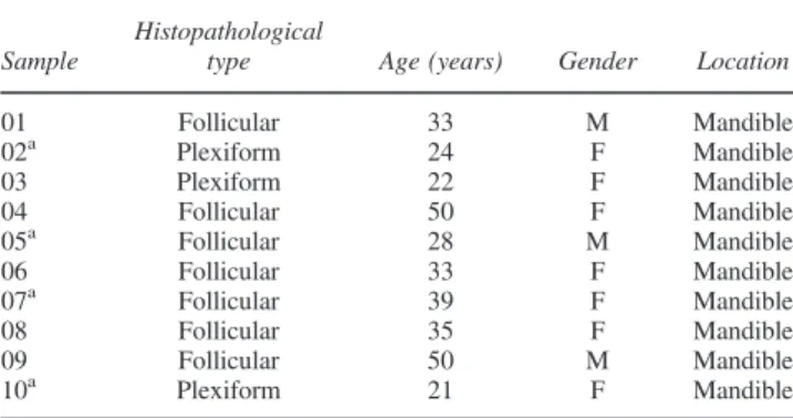
Table 1Clinical data of the multicystic ameloblastoma samples included in the methylation assay
Sample
Histopathological
type Age (years) Gender Location
01 Follicular 33 M Mandible
02a Plexiform 24 F Mandible
03 Plexiform 22 F Mandible
04 Follicular 50 F Mandible
05a Follicular 28 M Mandible
06 Follicular 33 F Mandible
07a Follicular 39 F Mandible
08 Follicular 35 F Mandible
09 Follicular 50 M Mandible
10a Plexiform 21 F Mandible
a
Samples #02, 05, 07 and 10 were included in the qPCR for the evalua-tion of the transcripevalua-tional levels ofTNFRSF25and BCL2L11.All sam-ples, but #04, were BRAFV600E mutation-positive (tested by TaqMan qPCR, data not shown). F=female; M=male.
dna_methylation_data_analysis.php), according to manu-facturer’s instructions. The Excel Template normalises the cycle threshold (Ct) values of both digests with the mock digestion values to calculate and report the percentage of the gDNA that is methylated and unmethylated.
RNA isolation and reverse transcription
Total RNA was isolated from frozen tissue samples using TRIzolâreagent (Ambion, Carlsbad, CA, USA) following the manufacturer’s instructions. The RNA concentration and purity were analysed by spectrophotometry using NanoDrop 2000TM
(Thermo Fisher Scientific, Waltham, MA, USA) and integrity was checked using Agilent 2100 Bioanalyzer instrument, with the Agilent RNA 6000 Nano Kit (Agilent, Waldbronn, Germany). The isolated RNA was treated with DNase I (Invitrogen, Carlsbad, CA, USA) and converted into complementary DNA (cDNA) using SuperScriptâ VILOTM
cDNA System Kit (Invitro-gen, Carlsbad, CA, USA) as outlined by the manufacturer.
Reverse-transcription quantitative polymerase chain reaction (RT-qPCR)
The relative expressions of the genes that showed the highest differential methylation status were investigated by RT-qPCR. Four multicystic ameloblastomas and four den-tal follicles were included in this transcription experiment. Due to the limited amount of tissue available from the tumours and dental follicles, the total RNA could not be extracted from all samples. Power Up SYBRâGreen Mas-ter Mix (Applied Biosystems, Austin, TX, USA) and the following primers were used: TNFRSF25 forward: 5’
GCC ACC CTG ACC TAC ACA TAC C 3’ and reverse: 5’ CCA GCT TCA TCT GCA GTA ACC A 3’ (68-bp amplicon); BCL2L11 forward: 5’ CAC AAA CCC CAA GTC CTC CTT 3’ and reverse: 5’ TGG AAG CCA TTG CAC TGA GA 3’ (60-bp amplicon) and 28S forward 5’
TTG AAA ATC CGG GGG AGA G 3’ and reverse 5’
ACA TTG TTC CAA CAT GCC AG 3’ (100-bp ampli-con). The primer pairs PCR showed ~100% efficiency. The 28S was selected as the reference gene, as it showed stable expression among tissue samples (Diniz et al, 2016). The normal oral mucosa was used as calibrator. All experiments were performed in duplicate and run on a Ste-pOnePlus instrument (Applied Biosystems, Foster City, CA, USA). Analysis of relative gene expression was per-formed with the 2(-Delta Delta CT) method (Pfaff, 2001).
Statistical analysis
The Kolmogorov–Smirnov was performed to evaluate the
normality of the data’s distribution. The Mann–Whitney
tests were used to compare gene expression between the groups, and it was performed using GraphPad Prism soft-ware, version 6.0 (GraphPad Softsoft-ware, CA, USA).P val-ues<0.05 were considered statistically significant.
Results
DNA methylation status
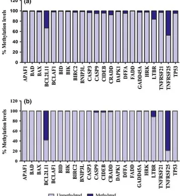
Two genes (TNFRSF25and BCL2L11) presented the most discrepant methylation status. TNFRSF25 methylation frequency in ameloblastomas (47.2%) was lower than in
dental follicles (79.3%). TheBCL2L11gene promoter also showed lower methylation levels in ameloblastomas (33.2%) compared with control (58.3%) (Figure 1).
Gene expression profiling
Higher BCL2L11 expression levels were found in ameloblastomas compared with dental follicles (P < 0.05) (Figure 2). No difference was observed amongTNFRSF25
expression levels in ameloblastoma and dental follicle group (P> 0.05).
Discussion
Apoptosis is known to be involved in tumourigenesis because the insufficiency of cell death may result in
Figure 1Methylation levels percentage in (a) ameloblastoma and (b) dental follicle pools. Note thatBCL2L11 andTNFRSF25showed lower methylation rates in the tumours (a) than in the control group (b) [Colour figure can be viewed at wileyonlinelibrary.com]
Figure 2Gene expression ofBCL2L11andTNFRSF25in dental follicles (DF) and ameloblastomas (AM). Increased expression of BCL2L11was observed in ameloblastomas, while there was no difference in the mRNA transcriptional levels ofTNFRSF5between the two groups
tumour growth (Danial and Korsmeyer, 2004). Epigenetic alterations may lead to suppression of apoptosis-related genes, as well as suppression or activation of a variety of other genes related to critical cell processes. Up to now, DNA methylation is the most investigated epigenetic change in tumours. However, there are few studies addressing DNA methylation changes in odontogenic tumours (Moreira et al, 2009; Gomes et al, 2010a; Guimar~aes et al, 2015) and to date, no study addressed the relevance of DNA methylation profile of apoptosis-related genes in ameloblastomas.
In the present study, we investigated the methylation sta-tus of 22 apoptosis-related genes in multicystic ameloblas-toma and two of them, TNFRSF25and BCL2L11,showed important differences in the methylation levels compared with dental follicles. A hypomethylated profile of both genes was observed in ameloblastoma compared with the dental follicle. Noteworthy, gene expression results for
BCL2L11 were consistent with this finding as we found increased expression levels in ameloblastomas. On the other hand, no difference was observed in TNFRSF25
between both groups. Such discrepancy may be explained by the molecular heterogeneity of the tumoral tissues included in the analysis, which is a well-described feature in malignant and benign neoplasias (Gomeset al, 2016).
TheBCL2L11gene, also known asBIM,codifies a BH3-only pro-apoptotic member of the Bcl-2 family, which con-stitutes one of the most relevant classes of regulators of apoptosis (Czabotar et al, 2014). The Bcl2-L-11 protein can bind to and neutralise pro-survival Bcl-2 proteins or directly activate pro-apoptotic effectors Bax and Bak (Czabotaret al, 2014). Activation of Bax and Bak leads to oligomerization to form pores in the mitochondrial outer membrane (MOM) followed by MOM permeabilization, cytochromecrelease and consequent activation of effector caspases leading to apoptosis (Sionovet al, 2015).
Balance in the intracellular expression levels of pro-apoptotic and anti-pro-apoptotic proteins is crucial for regulat-ing apoptosis. Many studies showed that ameloblastoma has two relatively distinct patterns of Bcl-2 family pro-teins: anti-apoptotic members are predominantly expressed in the outer layer, which is composed of columnar cells with reverse polarity. On the other hand, the pro-apoptotic members are mainly expressed in the inner layer, which is formed by loosely arranged angular cells (Sandra et al, 2001; Luo et al, 2006). However, Kumamoto and Ooya (2008) detected immunohistochemical expression of BH3-only proteins, including Bcl2-L-11, in the outer layers of ameloblastic tumours. These authors suggested that inter-actions with other anti-apoptotic proteins may suppress apoptosis initiated by the BH3-only proteins. This study also observed that benign and malignant ameloblastic tumours, as well as tooth germs, present similar immuno-histochemical expression of BH-only proteins, including Bcl2-L-11, indicating that these proteins may have a role in apoptosis of normal and neoplastic odontogenic epithe-lium. Further, it was observed a distinct expression pattern between histopathological subtypes of ameloblastoma, suggesting that BH3-only proteins, such as Bcl2-L-11, may be involved in the tumour cell differentiation (Kuma-moto and Ooya, 2008).
Alterations of Bcl2-L-11 isoforms are reported in sev-eral diseases, including cancer, but its role in the apoptosis of neoplastic cells can be more complicated. Bcl2-L-11 downregulation is involved in cell transformation and reduced the sensitivity of malignant neoplastic cells to var-ious chemotherapeutic drugs (Sionov et al, 2015). In con-trast, a recent study showed that it is highly expressed in prostate and breast cancer cells, which could be mediated by the transcription factor E2F1. Additionally, Bcl2-L-11 silencing caused cell apoptosis. These findings suggest that Bcl2-L-11 can promote cell survival in addition to its apoptosis-inducing function (Gogadaet al, 2013).
Previous studies agree that pro-apoptotic genes, such as CD95 and PUMA, can also promote the growth of tumours depending on context and specific cell type. This non-apoptotic activity is mediated by different pathways, such as c-Jun N-terminal kinases (JNK), leading to expres-sion of proteins related to tumour initiation (Chen et al, 2010; Tang et al, 2011). Furthermore, the presence of inhibitory proteins can repress the major function of pro-apoptotic members (Tang et al, 2011). The available data do not allow us to conclude that the same mechanism is true for BCL2L11 in ameloblastoma.
On the basis of our results, the transcription of the apoptosis-related gene BCL2L11 is possibly regulated by promoter DNA methylation in ameloblastoma. The biolog-ical significance of thisfinding in ameloblastoma pathobi-ology remains to be clarified.
Acknowledgements
RSG, CCG and MGD are research fellows at the National Coun-cil for Scientific and Technological Development (CNPq), Brazil.
This work was supported by grants from CNPq, Coordination for the Improvement of Higher Education Personnel (CAPES) and Research Support Foundation of the State of Minas Gerais (FAPEMIG)/Brazil.
Author contribution
RSG, CCG, and MGD conceived and designed the study. SFSC, NBP, KMA, WHC and KC performed the experiments. RSG, CCG and SFSC drafted the paper. NBP and SFSC performed statistical analysis. All authors revised the final version of the manuscript. Authors also confirm to have no conflict of interest.
References
Abiko Y, Nagayasu H, Takeshima Met al(2007). Ameloblastic carcinoma ex ameloblastoma: report of a case-possible involvement of CpG island hypermethylation of the p16 gene in malignant transformation.Oral Surg Oral Med Oral Pathol Oral Radiol Endod103: 72–76.
Amm HM, MacDougall M (2016). Molecular signalling in benign odontogenic neoplasia pathogenesis. Curr Oral Health Rep3: 82–92.
Brown NA, Rolland D, McHugh JB (2014). Activating FGFR2-RAS-BRAF mutations in ameloblastoma.Clin Cancer Res20: 5517–5526.
Chen L, Park SM, Tumanov AV et al (2010). CD95 promotes tumour growth.Nature465: 492–496.
Czabotar PE, Lessene G, Strasser A, Adams JM (2014). Control of apoptosis by the BCL-2 protein family: implications for physiology and therapy.Nat Rev Mol Cell Biol15: 49–63.
Danial N, Korsmeyer S (2004). Cell death: critical control points.
Cell116: 205–219.
Delpu Y, Cordelier P, Cho WC, Torrisani J (2013). DNA methylation and cancer diagnosis. Int J Mol Sci 14: 15029–
15058.
Demokan S, Dalay N (2011). Role of DNA methylation in head and neck cancer.Clin Epigenetics2: 123–150.
Dhanuthai K, Chantarangsu S, Rojanawatsirivej S et al (2012). Ameloblastoma: a multicentric study. Oral Surg Oral Med Oral Pathol Oral Radiol113: 782–788.
Diniz MG, Gomes CC, Guimar~aes BVet al(2015). Assessment of BRAFV600E and SMOF412E mutations in epithelial odon-togenic tumours.Tumour Biol36: 5649–5653.
Diniz MG, Duarte AP, Villacis RAet al(2016). Rare copy number alterations and copy-neutral loss of heterozygosity revealed in ameloblastomas by high-density whole-genome microarray analy-sis.J Oral Pathol Med. doi:10.1111/jop.12505.
Dong Y, Zhao H, Li H, Li X, Yang S (2014). DNA methylation as an early diagnostic marker of cancer (review). Biomed Rep 2: 326–330.
Elmore S (2007). Apoptosis: a review of programmed cell death.
Toxicol Pathol35: 495–516.
Esteller M (2007). Epigenetic gene silencing in cancer: the DNA hypermethylome.Hum Mol Gen16: 50–59.
Filizzola AI, Bartholomeu-dos-Santos TC, Pires FC (2014). Ameloblastomas: clinicopathological features from 70 cases diagnosed in a single oral pathology service in an 8-year per-iod.Med Oral Patol Oral Cir Bucal19: e556–e561.
Fregnani ER, da Cruz Perez DE, de Almeida OPet al(2010). Clin-icopathological study and treatment outcomes of 121 cases of ameloblastomas.Int J Oral Maxillofac Surg39: 145–149.
Gardner DG, Heikinheimo K, Shear M, Phillipsen HP, Coleman H (2005). Ameloblastomas. In: Barnes L, Eveson JW, Reichart P, Sidransky D, editors.World Health Organization Classifi ca-tion of Tumours. Pathological and Genetics of Head and Neck Tumours. ARC Press: Lyon, pp. 296–300.
Gogada R, Yadav N, Liu J et al (2013). Bim, a proapoptotic protein, up-regulated via transcription factor E2F1-dependent mechanism, functions as a prosurvival molecule in cancer.
J Biol Chem288: 368–381.
Gomes CC, Brito JA, Andrade CI, Gomez RS (2010a). DNA methyltransferase expression in odontogenic cysts and tumours.Oncol Lett1: 143–146.
Gomes CC, Duarte AP, Diniz MG, Gomez RS (2010b). Review article: current concepts of ameloblastoma pathogenesis.
J Oral Pathol Med39: 585–591.
Gomes CC, Diniz MG, Gomez RS (2014). Progress towards per-sonalized medicine for ameloblastoma.J Pathol232: 488–491.
Gomes CC, Galv~ao CF, do Carmo AC, Pereira NB, Gomez RS (2016). Intratumor molecular heterogeneity in pleomorphic adenoma of the salivary glands. Oral Surg Oral Med Oral Pathol Oral Radiol121: 158–163.
Gopisetty G, Ramachandran K, Singal R (2006). DNA methyla-tion and apoptosis.Mol Immunol43: 1729–1740.
Guimar~aes D, Antunes D, Duarte C, Ferro L, Nunes F (2015). DNA methyltransferase immunohistochemical expression in odontogenic tumours.J Oral Pathol Med44: 59–66.
Heikinheimo K, Kurppa KJ, Laiho Aet al (2015). Early dental epithelial transcription factors distinguish ameloblastoma from keratocystic odontogenic tumor.J Dent Res94: 101–111.
Hertog D, Bloemena E, Aartman IHA, van-der-Waal I (2012). Histopathology of ameloblastoma of the jaws; some critical observations based on a 40 years single institution experience.
Med Oral Patol Oral Cir Bucal17: e76–e82.
Kerr JF, Wyllie AH, Currie AR (1972). Apoptosis: a basic bio-logical phenomenon with wide-ranging implications in tissue kinetics.Br J Cancer 26: 239–257.
Khojasteh A, Khodayari A, Rahimi Fet al(2013). Hypermethy-lation of p16 tumor-suppressor gene in ameloblastic carci-noma, ameloblastoma, and dental follicles. J Oral Maxillofac Surg71: 62–65.
Kumamoto H, Ooya K (2005a). Expression of tumor necrosis factor alpha, TNF-related apoptosis-inducing ligand, and their associated molecules in ameloblastomas. J Oral Pathol Med 34: 287–294.
Kumamoto H, Ooya K (2005b). Detection of mitochondria medi-ated apoptosis signalling molecules in ameloblastomas. J Oral Pathol Med34: 565–572.
Kumamoto H, Ooya K (2008). Immunohistochemical detection of BH3-only proteins in ameloblastic tumors. Oral Dis 14: 550–555.
Kumamoto H, Kimi K, Ooya K (2001). Immunohistochemical analysis of apoptosis-related factors (Fas, Fas igand, caspase-3 and single-stranded DNA) in ameloblastomas.J Oral Pathol Med30: 596–602.
Kurppa KJ, Caton J, Morgan PRet al(2014). High frequency of BRAF V600E mutations in ameloblastoma. J Pathol 232: 492–498.
Luo HY, Yu SF, Li TJ (2006). Differential expression of apopto-sis-related proteins in various cellular components of ameloblastomas.Int J Oral Maxillofac Surg35: 750–755.
Mendenhall WM, Werning JW, Fernandes R, Malyapa RS, Men-denhall NP (2007). Ameloblastoma. Am J Clin Oncol 30: 645–648.
Moreira PR, Guimar~aes MM, Guimar~aes AL et al (2009). Methylation of P16, P21, P27, RB1 and P53 genes in odonto-genic keratocysts.J Oral Pathol Med38: 99–103.
Pfaff MW (2001). A new mathematical model for relative quan-tification in real-time RT-PCR.Nucleic Acids Res29: e45. Rizzardi C, Leocata P, Ventura Let al(2009). Apoptosis-related
factors (TRAIL, DR4, DR5, DcR1, DcR2, apoptotic cells) and proliferative activity in ameloblastomas. Anticancer Res 29: 1137–1142.
Sandra F, Nakamura M, Mitsuyasu T, Shiratsuchi Y, Ohishi M (2001). Two relatively distinct patterns of ameloblastoma: an anti-apoptotic proliferating site in the outer layer (periphery) and a pro-apoptotic differentiating site in the inner layer (cen-ter).Histopathology39: 93–98.
Sionov R, Vlahopoulos S, Granot Z (2015). Regulation of Bim in heath and disease.Oncotarget6: 6–16.
Spruijt CG, Vermeulen M (2014). DNA methylation: old dog, new tricks?Nat Struct Mol Biol21: 949–954.
Sweeney RT, McClary AC, Myers BR et al (2014). Identifi ca-tion of recurrent SMO and BRAF mutaca-tions in ameloblas-tomas.Nat Genet46: 722–725.
Tang D, Lotze M, Kang R, Zeh H (2011). Apoptosis promotes early tumorigenesis.Oncogene30: 1851–1854.
Ushijima T (2005). Detection and interpretation of altered methy-lation patterns in cancer cells.Nat Rev Cancer5: 223–231.