525 525 525 525 525 Mem Inst Oswaldo Cruz, Rio de Janeiro, Vol. 99(5): 525-530, August 2004
Antileishmanial IgG and IgE Antibodies Recognize Predominantly
Carbohydrate Epitopes of Glycosylated Antigens in Visceral
Leishmaniasis
AM Atta/+, R Colossi/+ +, M LB Sousa-Atta, SM B Jeronimo* , M D SB Nascimento* * , GF Bezerra* * , G O rge* * * , EM Carvalho* * *
Departamento de Análises Clínicas e Toxicológicas, Laboratório de Pesquisa em Imunologia, Faculdade de Farmácia, Universidade Federal da Bahia, Rua Barão de Geremoabo s/no, Campus de Ondina, 40171-970 Salvador, BA Brasil *Departamento de
Bioquímica, Universidade Federal do Rio Grande do Norte, Natal, RN, Brasil **Departamento de Patologia, Universidade Federal do Maranhão, São Luiz, MA, Brasil ***Serviço de Imunologia do Hospital Universitário Professor Edgard Santos, Universidade
Federal da Bahia, Salvador, BA, Brasil
The specificity of human antileishmanial IgG and IgE antibodies to glycosylated antigens of Leishmania chagasi was evaluated. An ELISA was performed with soluble leishmanial antigen (SLA) and a panel of 95 sera including samples from patients with subclinical infection (SC) and visceral leishmaniasis (VL), subjects cured of visceral leishmaniasis (CVL), and from healthy individuals from endemic areas (HIEA). Antileishmanial IgG were verified for 18 (40%) of 45 SC subjects (mean absorbance of 0.49 ± 0.17). All nine sera from VL patients had such antibody (0.99 ± 0.21), while 11 (65%) of 17 CVL individuals were seropositive (0.46 ± 0.05). Only three (12%) of 24 HIEA controls reacted in IgG-ELISA. Antileishmanial IgE was detected in 26 (58%) of 45 SC patients (0.35 ± 0.14), and in all VL patients (0.65 ± 0.29). These antibodies were also detected in 13(76%) of 17 CVL subjects (0.42 ± 0.14) while all HIEA controls were seronegative. There was no correlation between antileishmanial IgG and IgE antibody absorbances. Mild periodate oxidation at acid pH of SLA carbohydrates drastically diminished its antigenicity in both IgG and IgE-ELISA, affecting mainly the antigens of 125, 102, 94, and 63 kDa as demonstrated by western immunoblotting.
Key words: visceral leishmaniasis - anti-carbohydrate antibodies - IgG - IgE
Leishmania chagasi is an obligatory intracellular
para-site that causes human visceral infection in South and Central Americas. The human natural habitat of this pro-tozoan is the mononuclear phagocyte from bone marrow, spleen, lymph nodes, gut, and liver. Exuberant splenom-egaly, hepatomsplenom-egaly, blood pancytopenia, polyclonal ac-tivation, and hypergammaglobulinemia characterize vis-ceral leishmaniasis (VL). A high mortality is observed in VL patients when antimonial chemotherapy is omitted or ineffective (Badaró & Duarte 1997). In addition to the clas-sical clinical form of the disease, studies in areas of L. chagasi transmission have identified individuals that were
exposed to Leishmania and presented anti-Leishmania
IgG antibodies or a positive intradermal Montenegro re-action (Badaró & Reed 2001). Since these individuals had no clinical manifestation typical of VL they were consid-ered having a subclinical L. chagasi infection (SC).
An intense polyclonal antibody synthesis against
Leishmania polysaccharides, glycolipids, glycoproteins,
and also proteins is easily detected in VL (Badaró & Reed 2001). Such antibodies are well demonstrated in patient sera by immunoassays like direct agglutination test (DAT),
+Corresponding author. Fax: +55-71-332.1580. E-mail:
ajatta@ig.com.br
++CAPES fellowship
Received 12 January 2004 Accepted 19 July 2004
indirect fluorescent antibody test (IFAT), enzyme-linked immunosorbent assay (ELISA) or western immunoblot-ting, having different isotypes and affinities, which may be associated with clinic and immune features of the Leish-mania infection (Ghosh et al. 1995, Shiddo et al. 1996,
Anam et al. 1999).
Previously, others and we reported the occurrence of antileishmanial IgE antibodies in sera from VL patients, which were associated with active disease (Atta et al. 1998, Anam et al. 1999). However, a study has demonstrated that specific IgE antibodies to cross-reactive carbohy-drate determinants (CCDs) shared by glycosylated anti-gens from different sources affect the in vitro diagnosis of allergic diseases (Mari et al. 1999). Also, the specific reactivities of IgE antibodies with glycoprotein antigens are improved after mild periodate oxidation at acid pH of their glycosylated epitopes, which destroys carbohydrate residues and does not affect the antigenicity of peptide epitopes (Afferni et al. 1999, Mari et al. 1999). Although several reports already demonstrated the antigenicity of
Leishmania glycolipids and glycoproteins during the IgG
immune response observed in leishmaniasis (Kutner et al. 1991, Palatnik de Sousa et al. 1993, Shreffler et al. 1993), the reaction of glycosylated epitopes with antileishmanial IgE antibodies in this disease is unknown. The aim of this work was to investigate the occurrence of IgG and IgE antibodies to glycosylated epitopes of L. chagasi
526 526 526 526
526 Antileishmanial IgG and IgE Antibodies • AM Atta et al.
MATERIALS AND METHODS
Sera - The serologic survey of antileishmanial IgG and
IgE antibodies was carried out with a panel of 95 human sera from Brazilian endemic areas of L. chagasi (Rio Grande
do Norte and Maranhão), including serum samples from 24 healthy individuals (HIEA), from 45 subjects having SC infection, 9 sera from patients suffering of VL and sera from 17 persons cured of VL by antimonial therapy (CVL). Subclinical L. chagasi infection was defined by clinical
exams, the presence of serum IgG antibodies to Leishma-nia and/or a positive Montenegro intradermal test for
leish-maniasis in the absence of clinical diagnosis or previous history of VL.
L. chagasi antigen - In order to obtain leishmanial
soluble antigen (SLA), 109 stationary promastigotes from a cloned human strain of L. chagasi (MHOM-BR 86-BA),
grown in LIT medium supplemented with calf fetal serum and antibiotics, were washed three times in PBS (10 mM phosphate buffer containing 150 mM NaCl, pH 7.4) and disrupted with 6 mM CHAPS (3-[(3-Cholamidopropyl) dimethylammonio]-1-propane-sulfonate) in 50 mM Tris-HCl buffer containing 150 mM NaCl, pH 7.5, and protease inhibitors (PMSF, leupeptin, antipain, ortho-phenantroline, and TPCK). After centrifugation at 13,000 g for 15 min at 4°C, the supernatant was evaluated for protein content and stored at – 20°C until used as SLA.
IgG and IgE immunoassays - Antileishmanial IgG and
IgE antibodies were determined through immunoassays performed into round-bottom wells of polystyrene microtiter plates (Nunc, Maxsorp, Denmark) coated with 500 ng of L. chagasi protein. After antigen sensitization,
the free reactive sites of the wells were blocked with 1% BSA. Immune reactions of antileishmanial IgG antibodies were carried out with 100 µl of sera diluted at 1/500 in PBS-BSA for 1 h at room temperature (RT). After, the wells were thrice washed with PBS containing 0.05% Tween 20 (PBS-T) and incubated in the same conditions with 100 µl of goat anti-human IgG peroxidase conjugate (Sigma Chemi-cal Co., St. Louis, MO) diluted 1/5000 in PBS-BSA. The wells were again washed three times with PBS-T, and the reactions were revealed with 100 µl of citrate-phosphate buffer containing hydrogen peroxide plus TMB (3,3', 5,5'-tetramethylbenzidine), during 30 min in the dark (RT). Fol-lowing the addition of 50 µl of 2N sulfuric acid, the absor-bances were measured at 450-600 nm in a Diamedix BP-12 ELISA reader.
Antileishmanial IgE antibodies were investigated in IgG depleted sera by treatment with purified sheep IgG anti-human IgG (RF-Absorbent, Dade Behring, Ger-many) as previously described (Sousa-Atta et al. 1999). To perform these assays, the wells coated with L. chagasi
antigens were first incubated with 100 µl of IgG depleted sera diluted 1/6 in PBS-BSA for 18 h at 4°C. After the wells were washed with PBS-T, and incubated with 100 µl of goat anti-human IgE peroxidase conjugate (Sigma Chemi-cal Co.) diluted 1/2500 in PBS-BSA for 2 h at 37o C. The reactions were revealed with hydrogen peroxide plus TMB as described in IgG immunoassay. The cut-off of both immunoassays were determined statistically (Frey et al. 1998) using 10 sera from healthy individuals (HI) from a
free area of leishmaniasis (Salvador, Northeast Brazil). All of them were seronegative for rheumatic diseases (rheu-matoid arthritis, SLE) and infections (American trypano-somiasis, syphilis, and viral hepatitis).
The participation of glycosylated epitopes of L. chagasi antigens in the immune reactions with
antileish-manial IgG and IgE antibodies was evaluated after mild periodate oxidation at acid pH of SLA (Woodward et al. 1985).
In this test, leishmanial antigens were first adsorbed into the wells of ELISA plates and their carbohydrates oxidized to aldehyde with 100 µl of 10 mM sodium metaperiodate in 50 mM acetate buffer, pH 4.5, during 1 h at room temperature in the dark. After incubation, the wells were washed one time with sodium acetate buffer, being the new formed aldehyde groups reduced to alcohol through the incubation with 100 µl of 50mM sodium boro-hydride in PBS (Woodward et al. 1985). The wells were washed three times with PBS and immediately used in immunoassays. In order to evaluate the effect of acid treat-ment on the protein epitopes of leishmanial antigens, con-trol wells coated with SLA were incubated with the pH 4.5 acetate buffer only, for 1 h. The proportion of carbohy-drate contribution to the antibody immune reaction in ELISA was calculated comparing the absorbances ob-tained in the controls (100%) and tests. These analysis of carbohydrate antigenicity were carried out using 12 and 16 subclinical sera that were reactive in the immunoas-says to detect antileishmanial IgG and IgE antibodies re-spectively, while all 9 VL serum samples were used in both tests.
Western immunoblotting - In order to identify
par-tially the leishmanial glycoproteins reacting with anti-car-bohydrate antibodies, SLA was fractionated by 10% SDS-PAGE after treatment with 2-mercaptoethanol and electrotransferred to a PVDF membrane. After transfer, strips of the membrane containing the leishmanial anti-gens was first incubated with 0.5% Tween 20 in PBS to block residual free reactive sites (1 h at RT), thrice washed with PBS-T and then treated for mild periodate oxidation at acid pH as described for ELISA. The effect of acid treat-ment on the antigenicity of SLA was also investigated, treating strips of PVDF membrane containing fraction-ated SLA for 1 h at RT with the pH 4.5 acetate buffer only (Woodward et al. 1985). The membrane strips, control and tests, were washed with PBS-T, and incubated with VL sera diluted at 1/100 in PBS-T for 3h at 37oC. After, they were washed with PBS-T, and then incubated with goat anti-human IgG peroxidase conjugate (Sigma Chemical Co.) diluted at 1/2000 in PBS-T for 1 h at room temperature. The electroblots were revealed using as substrate for the HRP reaction diaminobenzidine (DAB) in PBS containing 0.03% hydrogen peroxide.
Statistical analysis - The absorbances obtained in
527 527 527 527 527 Mem Inst Oswaldo Cruz, Rio de Janeiro, Vol. 99(5), August 2004
RESULTS
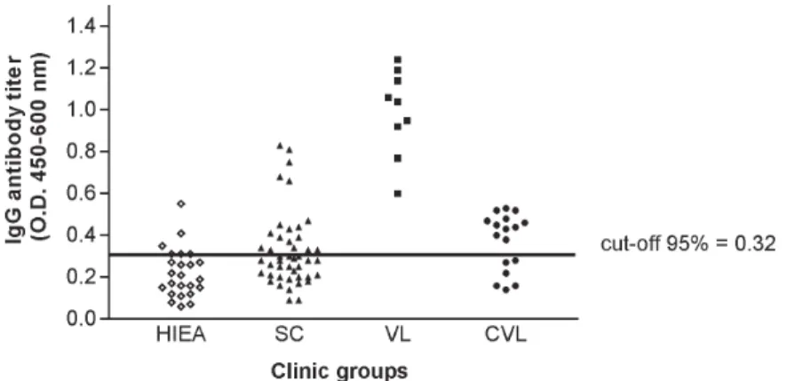
Antileishmanial IgG antibodies were detected in 18 out of 45 (40%) sera from SC subjects, presenting this clinical group a mean absorbance of 0.49 ± 0.17 (CI 95% = 0.40 to 0.57). While all nine (100%) VL patients presented IgG antibodies to Leishmania (OD = 0.99 ± 0.21, CI 95% =
0.83 to 1.15), 11 out of 17 (65%) CVL subjects were serop-ositive in this test (OD = 0.46 ± 0.05, CI 95% = 0.43 to 0.49). Only three out of 24 (12%) healthy individuals from en-demic areas were seropositive for antileishmanial IgG an-tibodies, showing ELISA absorbances ranging from 0.38 to 0.53 (Fig. 1).
IgE antibodies to L. chagasi were detected in 26 out
of 45 (58%) SC individuals, with a mean absorbance of 0.35 ± 0.14 (CI 95% = 0.30 to 0.41). In this group, 15 pati-ents also had antileishmanial IgG antibodies, but 11 (24%) and three (7%) of them presented only IgE or IgG immune response against the parasite, respectively. As verified for antileishmanial IgG antibodies, all nine (100%) VL pa-tients were also seropositive for IgE antibodies to Leish-mania (OD = 0.65 ± 0.29, CI 95% = 0.42 to 0.87) while 13
out of 17 (76%) individuals cured of VL presented IgE antibodies against this parasite (OD = 0.42 ± 0.14, CI 95% = 0.34 to 0.51). IgE antibodies to Leishmania were not
observed in the healthy inhabitants from endemic area (Fig. 2).
The mean absorbances of antileishmanial IgG and IgE antibodies differed in SC, VL, and CVL groups (Kruskal -Wallis test, P < 0.0001 and P = 0.014, respectively), being both more elevated in the group of visceral leishmaniasis. There was no correlation in the Spearman rank-order test between the absorbances of the tests to detect antileish-manial IgG and IgE antibodies in SC group (r = 0.256, P > 0.05) or VL one (r = – 0.033, P > 0.05).
While the antileishmanial IgG test using sera from VL and HIEA groups presented 100% sensitivity and 89% specificity, IgE immunoassay was 100% specific and sen-sitive with the same sera.
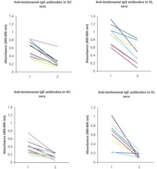
Treatment of L. chagasi antigen by sodium
meta-periodate caused a significant change in the absorbances of the immunoassays for antileishmanial IgG and IgE anti-bodies in SC and VL sera (Fig. 3). A mean absorbance of 0.24 ± 0.13 was verified in the ELISA for antileishmanial IgG antibodies in the SC group after antigen periodate oxidation while of 0.42 ± 0.27 was observed for VL sera. The controls of these reactions using antigen without periodate treatment presented means of antibody absor-bances of 0.53 ± 0.20 and 0.94 ± 0.24 (Mann-Whitney test, P = 0.0002 and P < 0.0001, respectively).
The absorbances of the ELISA for antileishmanial IgE antibodies also diminished after periodate oxidation of SLA, presenting the SC group a mean absorbance of 0.14
Fig. 1: titers of antileishmanial IgG antibodies in indirect ELISA with Leishmania chagasi promastigote antigen and sera from different clinical groups from endemic area of visceral leishmaniasis. HIEA (healthy individuals, n = 24); SC (subclinical, n = 45); VL (visceral leishmaniasis, n = 09); CVL (cured of visceral leishmaniasis, n = 17)
528 528 528 528
528 Antileishmanial IgG and IgE Antibodies • AM Atta et al.
± 0.07 (reaction control = 0.39 ± 0.14; Mann-Whitney test, P < 0.0001), while the VL one showed a mean of 0.14 ± 0.06 against 0.65 ± 0.29 of the reaction control (Mann-Whitney test, P < 0.0001). Such chemical treatment did not change the absorbances of the negative sera used to obtain the cut-off of both immunoassays.
Western immunoblotting analysis (Fig. 4) showed that antileishmanial IgG antibodies reacted strongly with glycosylated epitopes of leishmanial polypeptides of 125, 102, 94, and 63 kDa, which lost their immunoreactivity after periodate oxidation of their carbohydrate residues. A discrete loss of antigenicity was also verified for 32 and 26-28 kDa antigens. The immune reactions of 55, 42-44, and 38 kDa leishmanial polypeptides with these antibod-ies were not affected by carbohydrate oxidation. On the other hand, there was no effect of acid treatment on the antigenicity of protein epitopes of SLA.
DISCUSSION
While a high concentration of serum antileishmanial antibodies is observed in VL patients, individuals with the SC form present low titers of these immunoglobulins (Badaró & Duarte 1997). In this work, we demonstrated that SC individuals might produce simultaneously antileishmanial IgG and IgE antibodies during L chagasi
infection. Furthermore most of the antibodies against L. chagasi recognized predominantly glycosylated epitopes.
Previously others and we observed that both anti-leishmanial IgG and IgE antibodies could be detected si-multaneously in VL patients (Atta et al. 1998, Anam et al. 1999), the titers of these antibodies decreasing after cure. Such decrease of antibody titers was also documented in a following-up study of Indian VL patients (Kumar et al. 2002).
529 529 529 529 529 Mem Inst Oswaldo Cruz, Rio de Janeiro, Vol. 99(5), August 2004
Although antileishmanial IgG antibodies were detected in some healthy individuals from the endemic areas of leishmaniasis, these antibodies may represent immune cross-reaction or document a naturally resolving subclini-cal leishmanial infection.
One important observation here obtained was the in-tense reaction of antileishmanial IgG and IgE antibodies with glycosylated epitopes of Leishmania antigens, that
presented a significant decrease in antigenicity after periodate oxidation of the carbohydrate residues. The binding of human IgE antibodies to carbohydrate epitopes of plant and invertebrate N-glycans is usually associated with the recognition of an alpha (1,3)-fucose linked to the proximal N-acetylglucosamine and also by a beta (1,2)-xylose linked to the core manose, being these antibodies induced after pollen sensitization and insect stings (Fotisch K & Vieths 2001, Petersen & Mundt 2001, van Ree 2002). Leishmania is a highly glycosylated parasite
that has glycoconjugates that react with lectins present-ing different carbohydrate specificities (Andrade & Saraiva 1999). Such exuberant glycosylation could con-tribute for crossed reactions between antileishmanial IgE antibodies and food and indoor allergens that shared the carbohydrate structures of these Leishmania
glycocon-jugates. However, we verified in this study that IgE antileishmanial antibodies reacting strongly and specifi-cally with glycosylated epitopes of L. chagasi
promas-tigote are not detected in sera from healthy individuals from both leishmaniasis endemic and non-endemic areas. We already demonstrated that subjects presenting respi-ratory allergy do not react in antileishmanial IgE
immu-noassays (Atta et al. 1998). Additionally, we also verified that glycosylated epitopes of horseradish peroxidase bear-ing alpha (1,3)-fucose and beta (1,2)-xylose are not recog-nized by antileishmanial IgE antibodies in immunoassays using this enzyme as antigen (data not shown). Such ex-clusive antigenicity of glycosylated epitopes of L. chagasi
glycoconjugates observed in human visceral leishmania-sis are not found in others parasite diseases such as hu-man schistosomiasis, where glycosylated epitopes of gly-coproteins of Schistosoma egg provoke significant crossed
immune reactions in the serologic tests, which only are eliminated after antigen oxidation with sodium meta-periodate (Noya et al. 2000).
Western immunoblotting of periodate oxidized SLA with antileishmanial IgG antibodies, showed that carbo-hydrate epitopes of glycoproteins with molecular weights of 63, 94, 102, and 125 kDa are strongly recognized in these immune reactions. On the other hand, the mild periodate oxidation of SLA at acid pH did not have any deleterious effect on protein epitopes of leishmanial anti-gens, which preserved their antigenicity after 1h incuba-tion in pH 4.5 acetate buffer. Leishmanial antigens of 50, 42-44, and 38 kDa did not react with anti-carbohydrate antibodies, and probably contribute for the immune reac-tivity observed in the immunoassays for IgG and IgE antileishmanial antibodies after periodate oxidation of SLA.
Some of these glycoproteins reacting with antileish-manial antibodies to glycosylated epitopes may be im-portant leishmanial bioactive molecules, such as the sur-face metallopeptidase Gp63, which is involved in both parasite metabolism and infectivity (Bouvier et al. 1987). Furthermore, Leishmania glycolipids (Avila et al. 1991)
could induce both carbohydrate IgE and IgG anti-bodies, as demonstrated in schistosomiasis (van der Kleij et al. 1999). At present, we do not know the biological role played by these anti-carbohydrate IgG and IgE antibod-ies in the host-Leishmania relationship nor the factors
influencing their production. However, the involvement of them in the interaction of Leishmania with cell
recep-tors and possible participation in mechanisms of down regulation of leishmanial glycoprotein enzymes such as Gp63 should be investigated.
REFERENCES
Afferni C, Iacovacci P, Barletta B, Di Felice G, Tinghino R Mari A, Pini C 1999. Role of carbohydrate moieties in IgE bind-ing to allergenic components of Cupressus arizonica pollen
extract. Clin Exp Allergy 29: 1087-1094.
Anam K, Afrin F, Banerjee D, Pramanik N, Guha SK, Goswami RP, Saha SK, Ali N 1999. Differential decline in Leishma-nia membrane antigen-specific immunoglobulin G (IgG),
IgM, IgE and IgG subclass antibodies in Indian kala-zar patients after chemotherapy. Infect Immun 67: 6663-6669.
Andrade AF, Saraiva EM 1999. Lectin-binding properties of different Leishmania species. Parasitol Res 85: 576-581.
Atta AM, D’Oliveira Jr A, Correa J, Atta MLB, Almeida RP, Carvalho EM 1998. Antileishmanial IgE antibodies: a marker of active disease in visceral leishmaniasis. Am J Trop Med Hyg 59: 426-430.
Avila JL, Rojas M, Acosta A 1991. Glycoinositol phospholip-ids from American Leishmania and Trypanosoma spp: par-Fig. 4: effect of mild periodate oxidation of Leishmania chagasi
530 530 530 530
530 Antileishmanial IgG and IgE Antibodies • AM Atta et al.
tial characterization of the glycan cores and the human hu-moral immune response to them. J Clin Microbiol 29:
2305-2312.
Badaró R, Duarte MISL 1997. Leishmaniose visceral (calazar). In R Veronesi, R Focaccia (eds), Tratado de Infectologia,
Atheneu, São Paulo, p. 1234-1259.
Badaró R, Reed SG 2001. Leishmanioses. In SL Ávila, AW Ferreira (eds), Diagnóstico Laboratorial das Principais Doenças Infecciosas e Auto-Imunes, Guanabara Koogan,
Rio de Janeiro, p. 255-262.
Bouvier J, Etges R, Bordier C 1987. Identification of the promastigote surface protease in seven species of Leish-mania. Mol Bioch Parasitol 24: 73-79.
Fotisch K, Vieths S 2001. N- and O-linked oligosaccharides of allergenic glycoproteins. Glycoconj J 18: 373-390.
Frey A, Di Canzio J, Zurakowski D 1998. A statistically de-fined endpoint titer determination method for immunoas-says. J Immunol Methods 221:35-41.
Ghosh AK, Dasgupta S, Ghose AC 1995. Immunoglobulin G subclass-specific antileishmanial antibody response in In-dian kala-azar and post-kala-azar dermal leishmaniasis. Clin Diagn Lab Immunol 3: 291-296.
Kumar P, Pai K, Tripathi K, Pandey HP, Sundar S 2002. Immunoblot analysis of the humoral immune response to
Leishmania donovani polypeptides in cases of human
vis-ceral leishmaniasis: its usefulness in prognosis. Clin Diagn Lab Immunol 9: 1119-1123.
Kutner S, Pellerini P, Breniere SF, Desjeux P, Dedet JP 1991. Antigenic specificity of the 72-kilodalton major surface gly-coprotein of Leishmania braziliensis braziliensis. J Clin Microbiol 29: 595-599.
Mari A, Iacovacci P, Afferni C, Barletta B, Di Felice G, Tinghino R, Pini R 1999. Specific IgE to cross-reactive carbohydrate
determinants strongly affect the in vitro diagnosis of aller-gic diseases. J Allergy Clin Immunol 103: 1005-1111. Noya BA, Colmenares C, Lanz H, Caracciolo MA, Losada S,
Noya O 2000. Schistosoma mansoni: immunodiagnosis is
improved by sodium metaperiodate, which reduces cross-reactivity due glycosylated epitopes of soluble egg antigen.
Exp Parasitol 95: 106-112.
Palatnik-de-Sousa CB, Dultra HS, Borejevic R 1993. Leishma-nia donovani surface glycoconjugate GP36 is the major immunogen component of the fucose-manose ligand (FML).
Acta Trop 53: 59-72.
Petersen A, Mundt C 2001. Investigations on the carbohydrate moieties of glycoprotein allergens. J Chromatogr B 756:
141-150.
Shreffler W, Burns JM, Badaró R 1993. Antibody responses of visceral leishmaniasis patients to gp63, a major surface gly-coprotein of Leishmania species. J Infect Dis 167: 426-430.
Shiddo AS, Huldt G, Nilsson LA, Ouchterlony O, Thorstensson R 1996. Visceral leishmaniasis in Somalia. Significance of IgG subclasses and IgE response. Immunol Lett50: 87-93. Sousa-Atta MLB, Araújo MI, D’Oliveira Jr A, Ribeiro de Jesus A, Almeida RP, Atta AM, Carvalho EM 1999. Detection of specific IgE antibodies in parasite diseases. Braz J Med Biol Res 32: 1101-1105.
van der Kleij D, Tielens AGM, Yazdanbakhsh M 1999. Recog-nition of schistosome glycolipids by immunoglobulin E: possible role in immunity. Infec Immun 67: 5946-5950.
van Ree R 2002. Carbohydrate epitopes and their relevance for the diagnosis and treatment of allergic diseases. Int Arch Allergy Immunol 129:189-197.