Corresponding author: MSc. Anderson Pacheco dos Santos. e-mail: anderson.santos@butantan.gov.br
Received 10 November 2017 Accepted 4 April 2018
Short Communication
Effect of chaotropes in Chagas disease and leishmaniasis
cross-reacting serology assays for epidemiological surveys
Anderson Pacheco dos Santos
[1], Maria Esther de Carvalho
[2],
Luciana Regina Meirelles
[3]and Heitor Franco de Andrade Junior
[3][1]. Seção de Hepatite, Laboratório de Virologia, Instituto Butantan, São Paulo, SP, Brasil.
[2]. Laboratório de Imunoepidemiologia, Superintendência de Controle de Endemias, Secretaria de Estado da Saúde de São Paulo, São Paulo, SP, Brasil. [3]. Laboratório de Protozoologia, Instituto de Medicina Tropical de São Paulo, Universidade de São Paulo, São Paulo, SP, Brasil.
Abstract
Introduction: Serological cross-reactivity between leishmaniasis and Chagas disease, especially at low titers, leads to difficulties of the seroepidemiological interpretation. Methods: We have studied the ability of urea as a chaotrope to select high-avidity antibodies in IgG ELISA, thus reducing low-avidity IgG cross-reactivity in serologically positive samples in both assays. Results: Using 0.5M urea for diluting the sample efficiently defined leishmaniasis or double infections in high-avidity IgG ELISA and eliminated false-positive results. Conclusions: The use of a chaotropic diluting agent is useful for improving the specificity of Chagas disease and leishmaniasis immunoassays.
Keywords: Serology. Chagas disease. Leishmaniasis. ELISA. IFAT. IgG antibody avidity.
Chagas disease and American leishmaniasis, two chronic endemic diseases in the Americas, are caused by protozoa of the order Kinetoplastida and the family Trypanosomatidae, belonging to the genera Leishmania and Trypanosoma, respectively1. The differential diagnosis of Chagas disease and leishmaniasis are usually easy on clinical grounds in symptomatic patients. However, in asymptomatic patients, serology is a conflicting tool frequent in case of both infections; dual serological positivity could occur due to cross-reactive antibodies, owing to the genetic proximity of the two pathogens, and also due to double infections, owing to the high prevalence of both infections in the same area. Avidity assays are used to detect high-affinity antibodies; they use chaotropic agents, which eliminate low-avidity antibodies2. The chaotrope could be used as a dissociating agent to elute bound immunoglobulin G (IgG) or to dilute the IgG, thus blocking low-affinity binding, by affecting the hydrogen bridges and other binding forces between the antigen and antibody3. These chaotropes can also eliminate non-specific antibodies, by their inclusion in the sample diluting solvent for high-avidity enzyme linked immunosorbent assay (ELISA)3.
Serum samples from patients with Chagas disease (n = 10) were provided by Dante Pazzanese Institute of Cardiology, São Paulo State Secretary of Health; leishmaniasis patients serum
samples (n = 10) were provided by the Seroepidemiology Laboratory of the Tropical Medicine Institute of the State of São Paulo [Instituto de Medicina Tropical de São Paulo (IMTSP)]. All samples were previously serologically assayed in their original laboratories and stored at -20°C until use. Field samples were also provided by Superintendency for the Control of Endemics [Superintendência de Controle de Endemias (SUCEN)] from the Program for the Control of Chagas Disease, originating from residents in four geographic regions of the State of São Paulo, Brazil, resulting in eleven available samples that were positive for both infections. Leishmania amazonensis promastigotes cultures were kept in the Laboratory of Protozoology in IMT, in Corning® culture bottles with Roswell Park Memorial Institute (RPMI 1640) medium (Cultilab®), as recommended4, containing 20% of sterile inactivated fetal calf serum, 1µg/L calf hemin and 0,01mg/mL gentamycin, in Biological Oxygen Demand (BOD) incubators, under controlled temperature conditions (24oC). The cultures were daily monitored by phase-contrast microscopy (40X objective). After an incubation period of five days, the promastigote suspension obtained was centrifuged under refrigeration (4oC) at 3,000rpm for 20 minutes. Purified
Leishmania amazonensis promastigotes were added to 20 volumes of distilled water and disrupted by sonication (Sonic Dismembrator, Quigley-Rochester Inc., USA) in an ice bath using ten 30-second periods at 40Hz each; they were checked for lysis using phase contrast microscopy.
content of the supernatant was determined, after which it was stored at -70°C until use. Polystyrene 96-well microtiter plates certified for high protein binding (Costar®) were used. The plates were sensitized with soluble L. amazonensis antigen diluted in carbonate-bicarbonate buffer (0.1M, pH 9.5; 100µL per well), at the concentrations of 1µg and 10µg. After incubation for 20 hours at 6°C, the plates were washed thrice, for five minutes each, with phosphate-buffered saline [(PBS); 0.02M, pH 7.2], containing 0.05% of polyoxyethylene sorbitan monolaurate (Tween 20 - Sigma®) (PBS-T) and blocked with a 0.3% solution of skimmed milk (Molico®) in PBS-T for one hour at 37°C. After blocking and three washes with PBS-T (100µL/well), the control serum samples, positive and negative, at dilutions of 1:100, 1:200, 1:400, 1:800, 1:1,600, 1:3,200, and 1:6,400 were added to the wells in duplicate, and the plate was incubated at 37°C for one hour. After three further washes, a conjugated anti-human IgG peroxidase, (Sigma®) at a dilution of 1:20,000 (100µL per well), was added to each well and the plate was incubated again for one hour at 37°C.
After washing, the revelation of the reaction was performed by addition of 0.05M sodium citrate solution (pH 5.8), containing 0.4mg/mL of ortho-phenylenediamine (OPD), and 0.03% hydrogen peroxide (H2O2). The reaction was stopped after 30 minutes by adding 4N HCl. The absorbance [optical density (OD)] readings were performed at 492 nm using a colorimeter equipped with an automatic evaluation system for microplates (Labsystems Multiskan MS®).
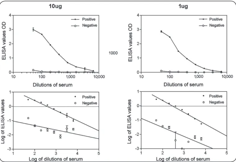
For chaotrope ELISA, the standardization of the optimal concentration of dissociating agents (chaotropes) was made by using the 20 serum samples previously assayed for both infections. The chaotrope used in the standardization of the ELISA was a urea solution further diluted to the following concentrations: 0.125M, 0.25M, 0.5M, 1M, and 2M. The samples were used at the following dilutions: 1:100, 1:400, 1:1,600, and 1:6,400. This diluent was used for sample dilution, and the ELISA was performed as described above. The titers for an optical density value of 1 in ELISA could be estimated in each sample by log-log transformation. In order to establish if a low antigen concentration would be sufficient to eliminate cross-reaction, we tested serially diluted standard positive and negative samples using 1µg/mL and 10µg/mL concentrations of the soluble antigenic extract in solid phase in ELISA. Both assays showed high levels of antibody detection and were equally accurate in discriminating positive and negative controls until the most diluted sample, presenting a typical log response of titers. After analyzing the curve by nonlinear regression, it was seen that a lower antigen concentration in the solid phase yielded better negative results, without affecting the positive sera results, with parallel estimated regression, while higher concentration tends to cross curves between the negative and positive serum samples. Thus, we have chosen the lower concentration (1µg antigen/mL) for use in the ELISA. We tested several chaotrope concentrations in sample dilutions of both infections to verify the effects on IgG ELISA cross-reactive antibodies. The reactions were performed with the chaotrope (urea) concentration varying from 0.125M to up to 2M. We used two standard dilutions of sera, 1/100 and 1/400, and estimated
the serum titers for 1.0 ELISA OD by plotting dilutions in log-log plots. We used the 20 serum samples previously screened externally by using commercial Indirect Immunofluorescence Antibody Test (IFAT): ten seropositive for Chagas disease and ten seropositive for leishmaniasis. Interestingly, at the higher dilution, the Leishmania patients’ sera reacted more intensely than the Chagas patients’ sera if the co-infected conditions were removed; this allows a better distinction of some sera. Despite these effects, based only on the ELISA absorbances, cross reactive antibodies were not completely eliminated.
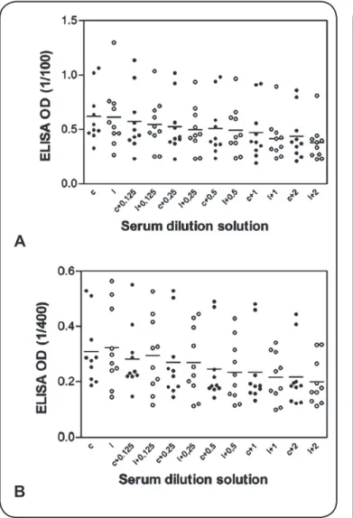
For further analysis, we performed the determination of reactive titers for each chaotrope concentration; these data are shown in Figure 1A and Figure 1B. Titers are more proportional for antibody concentration in ELISA due to the exquisite sensitivity of enzymatic reactions. It is clearly seen in Figure 1A that two Chagas patients’ serum samples and one leishmaniasis serum sample presented no significant decay of titer despite the increasing chaotrope concentration, showing high avidity. We also estimated the ratio of the decay of those reactions according to the mean of the absorbance of reactive samples, which was expressed as OD in specific dilution or titers (Figure 1B). This shows that at the 1/100 dilution, there is an inflexion point in this ratio in case of 0.5M urea in the diluent. The same inflexion was observed for 0.25M urea when the 1/400 dilution was analyzed, while titer the shows an intense decay. Due to feasibility, we define that this assay would be performed best using 0.5M urea in a 1/100 dilution of the serum sample, as titer determination is complex and costly, and the 1/100 dilution usually has low OD results. For studying the use of this methodology in serum samples, we tested cross-reacting positive samples from the state of São Paulo, Brazil; these results are shown in Figure 2A and Figure 2B. The OD values associated with one sample remained invariant, but a reduction was observed in case of the further ten samples (Figure 3).
The dilutions of samples in PBS and 0.5M urea yielded the best results in terms of discrimination of cross-reactions (Figure 3B). Three positive samples presented no decrease of high-avidity ELISA reactivity, suggesting that those samples represented double infections (Figure 3C).
Six other samples presenting low-grade reactivity in conventional ELISA showed negative high-avidity ELISA reactivity; these represented only the cross-reacting Chagas disease sera.
Besides these defining results, this assay also presented two samples negative for both assays; this represents a screening error or true false-positive reaction for both assays . This result suggests that high-avidity ELISA could be used with field samples for improving the resolution of ELISA assays in epidemiological studies (Figure 3C).
FIGURE 1: Evaluation of the effect of antigen concentration in the solid phase for the detection of anti-Leishmania IgG antibodies. The upper graphs indicate
the optical density obtained in duplicate wells, with the SEM. The lower graphs indicate the log-log conversion and linear regression assays of the same data. Note the almost parallel slope of curves in case of the lower antigen concentration. ELISA: enzyme linked immunosorbent assay; OD: optical density;
IgG: immunoglobulin G; SEM: standard error of the mean.
For epidemiological studies, the diagnosis must be conducted in serum samples. The overlapping geographical distribution of Chagas disease and leishmaniasis leads to incorrect estimations due to the diverse occurrence of either Trypanosoma cruzi or
Leishmania cases in a given region; such incorrect estimations
could be minimized by using an immunoassay specific enough to produce a significant reduction of such cross-reactions5,6.
We standardized a high-avidity ELISA that yielded good results despite standard sera presenting true cross-reactions. This determination allows to choose a chaotrope concentration that is feasible for use with field samples. The strategies available for overcoming these difficulties for the diagnosis of leishmaniasis in epidemiological studies were reported as the use of a lower cut-off than that recommended for Chagas disease7, the use of different antigens, including recombinant antigens8,in serological reactions9,10, analysis of sera by western blot with different Leishmania antigens utilized for the serological diagnosis of American cutaneous leishmaniasis11, or subsequent retest by means of the polymerase chain reaction (PCR)12 and flow cytometry13. All these procedures have sampling or feasibility problems due to high cost or
FIGURE 2: Effect of Urea (chaotrope) in anti-Leishmania IgG ELISA initial
serum dilution solution. Codes: c = Chagas patients samples (closed dots); l = Leishmaniasis patients samples (open dots); the bars represent the mean values. A: Serum samples diluted 1/100. Serum samples diluted 1/400. ELISA: enzyme linked immunosorbent assay; OD: optical density;
IgG: immunoglobulin G.
A
B
FIGURE 3: Determination of anti-Leishmania IgG titers in the serum samples
from Chagas or Leishmaniasis patients, after serially diluting the sera using different chaotropes in the diluent solution. c = Chagas patients samples (closed dots); l = Leishmaniasis patients samples (open dots); the bars represent the mean values. A: Effect of chaotrope on the titer of standard sera. Titers are expressed as the serum dilution that gives a 1.0 OD ELISA reaction.
B: Reactivity proportion in OD and titers from sera of both diseases, in different chaotrope concentrations. C: Field cross-reacting samples for leishmaniasis and Chagas disease tested by high-avidity leishmaniasis ELISA. The open dots indicate high-avidity ELISA, while the closed dots indicate conventional anti-Leishmania ELISA. The shaded area represents
the negative area for high-avidity ELISA using mean + 2 SD of the negative samples. ELISA: enzyme linked immunosorbent assay; OD: optical density;
IgG: immunoglobulin G; SD: Standard Deviation.
In this study, we propose the use of a sample dilution solution containing 0.5M urea as a chaotrope, which promotes the efficient performance of ELISA by reducing cross-reactions, especially with regards to retesting near cut-off samples. The use of chaotropes as sample diluents has proved to be useful and adequate for investigating the incidence and prevalence of leishmaniasis in areas with prevalence of American trypanosomiasis.
Conflict of interest
The authors declare that there is no conflict of interest.
Financial support
This work was used partially for the master’s dissertation in Parasitology [Departamentto de Parasitologia, Instituto de Biologia, Universidade de Campinas (UNICAMP)] of AP Santos. HF Andrade Jr was a fellow of Conselho Nacional de Desenvolvimento Científico e Tecnológico (CNPq) and
A
B
Fundação Faculdade de Medicina (FFM). This work was partially supported by Laboratórios de Investigação Médica 49 (LIM 49), Hospital das Clínicas da Faculdade de Medicina da Universidade de São Paulo (HCFMUSP) and Laboratório de Imunoepidemiologia,Superintendência de Controle de Endemias (SUCEN).
REFERENCES
1. de Morais CG, Castro Lima AK, Terra R, dos Santos RF, Da-Silva SA, Dutra PM. The Dialogue of the Host-Parasite Relationship:
Leishmania spp. and Trypanosomacruzi infection. BioMed Res Int
2015;2015:324915.
2. Berth M, Grangeot-Keros L, Heskia F, Dugua JM, Vauloup-Fellous C. Analytical issues possibly affecting the performance of commercial human cytomegalovirus IgG avidity assays. Eur J Clin
Microbiol Infect Dis. 2014;33(9):1579-84.
3. Hedman K, Lappalainen M, Makela O. Recent primary toxoplasma
infection indicated by a low avidity of specific IgG. J Infect Dis. 1989;159(4):736-40.
4. Trager W, Jensen JB. Human malaria parasites in continuous culture. Science. 1976;193(4254):673-5.
5. Vexenat AC, Santana JM, Teixeira ARL. Cross-reactivity of antibodies in human infections by the kinetoplastid protozoa
Trypanosoma cruzi, Leishmania chagasi and Leishmania (Viannia) braziliensis. Rev Inst Med Trop S Paulo. 1996;38(3):177-85. 6. Camargo ME, Rebonato C. Cross-reactivity in fluorescence tests for
Trypanosoma and Leishmania antibodies. Amer J Trop Med Hyg.
1969;18(4):500-5.
7. Rodríguez-Bonfante C, Amaro A, García M, Wohlert LEM, Guillen P, García RA, et al. Epidemiología de la enfermedad de Chagas en el município Andrés Eloy Blanco, Lara, Venezuela: infestación triatomínica y seroprevalencia en humanos. Cad Saúde Pública.
2007;23(5):1133-40.
8. Passos VMA, Volpini AC, Braga EM, Lacerda PAF, Ouaissi A, Lima-Martins MVC, et al. Diferencial serodiagnosis of human
infections caused by Trypanosoma cruzi and Leishmania spp. Using ELISA with a recombinant antigen (rTc 24). Mem Inst Oswaldo Cruz. 1997;92(6):791-3.
9. Vale AM, Fujiwara RT, da Silva Neto AF, Miret JA, Alvarez DCC, da
Silva JCF, et al. Identification of Highly Specific and Cross-Reactive
Antigens of Leishmania Species by Antibodies from Leishmania (Leishmania) chagasi Naturally Infected Dogs. Zoonoses Public
Health. 2009;56(1):41-8.
10. Guimarães MCS, Celeste BJ, Castilho EA, Mineo JR, Diniz JMP.
Imunoenzymatic assay (ELISA) in mucocutaneous leishmaniasis, Kala-azar, and Chagas disease: an epimastigote Trypanosoma cruzi
antigen able to distinguish between anti-Trypanosoma and
anti-Leishmania antibodies. Am J Trop Med Hyg. 1981;30(5):942-7.
11. Gonçalves CCM, Reiche EMV, Abreu Filho BA, Verzignassi-Silveira TG, Felizardo TC, Maia KR, et al. Evaluation of antigens from various Leishmania species in a Western Blot for diagnosis of american tegumentary leishmaniasis. Am J Trop Med Hyg.
2002;66(1):91-102.
12. Degrave W, Fernandes O, Thiemann O, Wincker P, Britto C, Cardoso A, et al. Detection of Trypanosoma cruzi and Leishmania
using the Polimerase Chain Reaction. Mem Inst Oswaldo Cruz.
1994;89(3):367-8.
13. Teixeira-Carvalho A, Campos FMF, Geiger SM, Rocha RDR, de Araújo FF, Vitelli-Avelar DM, et al. FC-TRIPLEX Chagas/
Leish IgG1: a multiplexed flow cytometry method for differential
serological diagnosis of Chagas disease and leishmaniasis. PLoS
One. 2005;10(4):e0122938.
14. Franck FM, Fernández MM, Taranto NJ, Cajal SP, Margni RA, Castro E, et al. Characterization of human infections by Leishmania spp in the Northwest of Argentina: immune response, double infection with Trypanosoma cruzi and species of Leishmania
involved. Parasitol. 2003;126(Pt 1):31-9.
15. Tiburcio MG, Anversa L, Kanunfre KA, Ferreira AW, Rodrigues Júnior V, Silva LA. Anti-Leishmania infantum IgG antibody avidity
in visceral leishmaniasis. Clin Vaccine Immunol. 2013;20(11):