545 545 545 545 545 Mem Inst Oswaldo Cruz, Rio de Janeiro, Vol. 95(4): 545-551, Jul./Aug. 2000
Analysis of Genetic Diversity of
Trypanosoma cruzi
: an
Application of Riboprinting and Gradient Gel
Electrophoresis Methods
JR Stothard
+, IA Frame, HJ Carrasco, MA Miles
Pathogen Molecular Biology and Biochemistry Unit, Department of Infectious and Tropical Diseases, London School of Hygiene and Tropical Medicine, Keppel Street, London WC1E 7HT, UK
Analysis of restriction fragment length polymorphism (RFLP) profiles derived from digestion of polymerase chain reaction (PCR) products of the ribosomal 18S from Trypanosoma cruzi yields a typi-cal ‘riboprint’ profile that can vary intraspecifitypi-cally. A selection of 21 stocks of T. cruzi and three outgroup taxa: T. rangeli, T. conorhini and Leishmania braziliensis were analysed by riboprinting to assess divergence within and between taxa. T. rangeli, T. conorhini and L. braziliensis could be easily differentiated from each other and from T. cruzi. Phenetic analysis of PCR-RFLP profiles indicated that, with one or two exceptions, stocks of T. cruzi could be broadly partitioned into two groups that formally corresponded to T. cruzi I and T. cruzi II respectively. To test if ribosomal 18S sequences were homogeneous within each taxon, gradient gel electrophoresis methods were employed utilising either chemical or temperature gradients. Upon interpretation of the melting profiles of riboprints and a sec-tion of the 18S independently amplified by PCR, there would appear to be at least two divergent 18S types present within T. cruzi. Heterogeneity within copies of the ribosomal 18S within a single genome has therefore been demonstrated and interestingly, this dimorphic arrangement was also present in the outgroup taxa. Presumably the ancestral duplicative event that led to the divergent 18S types preceded that of speciation within this group. These divergent 18S paralogues may have, or had, different func-tional pressures or rates of molecular evolution. Whether or not these divergent types are equally tran-scriptionally active throughout the life cycle, remain to be assessed.
Key words: riboprinting 18S paralogy denaturing gradient gel electrophoresis -temperature gradient gel electrophoresis
At the molecular DNA level, the ability to characterise stocks, strains and clones of kinetoplastid protozoa is fundamental to under-standing the epidemiology of their respective dis-eases (Tibayrenc 1998a) and that of Trypanosoma cruzi and Chagas disease presents no exception (Macedo & Pena 1998). For most simplistically, molecular markers discriminate and define clones or strains of the pathogen, providing taxonomic characters, upon which further DNA assays can be developed e.g. sensitive tools for diagnostics. By the shared presence of taxonomic characters, molecular data have the potential to infer
evolu-Funded by The Wellcome Trust.
+Corresponding author. Fax: +442079425518. E-mail:
Present address: Biomedical Parasitology Division, De-partment of Zoology, The Natural History Museum, Cromwell Road, London SW7 5BD, UK
Received 13 April 2000 Accepted 15 May 2000
tionary relationships or, at the very least, groups of organisms on the basis of similarity i.e. their phenetic patterns (Li 1997). Over the last 10 years, DNA typing of kinetoplastid parasites has under-gone impressive developments, primarily accruable to development and implementation of polymerase chain reaction (PCR) based assays. So much so that a new synthetic field of research has been defined and generically known as ‘integrated genetic epi-demiology of infectious diseases’ (Tibayrenc 1998b).
As researchers attempt to find natural ‘demes’ or divisions, the taxonomy within and between the isolates that are collectively known as T. cruzi is attracting considerable attention. Substantial het-erogeneity is known to exist but there has been a general reluctance to name formal taxa (Momen 1999). The partial realisation of the divisions, how-ever, has led to the nomenclature of T. cruzi I, T. cruzi II and T. cruzi to try to acknowledge formally this intraspecific variation (Anon 1999). T. cruzi I is equivalent to principal zymodeme 1 (Z1) and T.
546 546 546 546
546 Genetic Diversity of T. cruzi JR Stothard et al.
exhibit hybrid-like characters, or await further characterisation, or are from principal zymodeme 3 (Z3) (Miles et al. 1981) are collectively referred to as T. cruzi [without the group designation suffix (Anon 1999)]. Z2 isolates were originally de-scribed from central and eastern Brazil, in domes-tic transmission cycles, whereas Z1 isolates were predominantly sylvatic when found sympatric with Z2 but in the north of the Amazonian basin were in both domestic and sylvatic cycles. Z3 isolates were rarely isolated from humans and are almost exclusively associated with burrowing animals such as the armadillo. In conjunction with the prob-lematic strain taxonomy, the reproductive biology of T. cruzi is also complex. Clonal propagation of
T. cruzi is thought to predominate (Tibayrenc 1998a) though there is evidence of sexuality in sylvatic transmission cycles (Bogliolo et al. 1996, Carrasco et al. 1996) and evidence of genetic ex-change has been found in the laboratory (Stothard et al. 1999).
This paper assesses the impact that molecular characterisation of the 18S gene has had upon strain typing of T. cruzi. The paper draws upon recent work and focuses upon methods of gradient gel electrophoresis, which are also known as mutation analysis.
RIBOPRINTING
Analysis of sequence variation within the ribo-somal 18S has proven useful for phylogenetic pur-poses within the Trypanosomatidae (Noyes 1998, Briones et al. 1999, Stevens & Gibson 1999, Wright et al. 1999), though the use of this region for phy-logenetic inference has not been without debate (Stothard in press). In T. cruzi, there are approxi-mately 110 copies of the 18S per nucleus, the organisation of which is slightly unusual in com-parison to other protozoa (Pulido et al. 1996). On a less finer scale than DNA sequencing, variation within the 18S can be detected by restriction en-zymes since many enen-zymes, especially those with four base pair recognition sites e.g. AluI, have sev-eral cutting sites within the 18S. By using a bat-tery of such frequently cutting enzymes, the 18S can be easily ‘typed’ and quickly scanned for vari-able nucleotides without the need for DNA se-quencing. Moreover, through the use of polymerase chain reaction (PCR), the amplification products can be directly digested and separated by conven-tional agarose or polyacrylamide electrophoresis to produce a ‘fingerprint-like’ profile that can be easily, cheaply achieved within a single working day. Indeed as riboprinting is less sensitive than DNA sequencing, this might even be an advanta-geous way to analyse variation within the 18S. For example, as the 18S has been shown to be
paralogous (Stothard et al. 1997); as long as the enzyme cutting sites do not fall within these paralogous regions, the associated problems of paralogy/orthology disappear (Stothard in press). In contrast, if the restriction sites were in the paralogous regions, the riboprint profile could be interpreted as an incomplete digestion (Fig. 1).
The process of generating PCR-RFLP (restric-tion fragment length polymorphism) profiles from the 18S is also known as riboprinting (Clark 1992). Riboprinting allows detection of cryptic genetic variation within species, organism misiden-tifications and culture mix-ups, independent veri-fication of DNA sequences, and the rapid genera-tion of data useful in phylogenetic analyses (Clark et al. 1995). Pioneering studies using this method-ology revealed variation between species of
Entomoeba (Clark & Diamond 1991) and was later applied upon sylvatic strains of T. cruzi from North America, coining the nomenclature of ‘ribodemes’ to group isolates of T. cruzi with the same riboprint (Clark & Pung 1994). Stothard et al. (1998a) ex-panded the number of isolates examined by riboprinting to 21, with better coverage of Z3. Through the inclusion of outgroup taxa T. rangeli, T. conorhini and Leishmania braziliensis an assess-ment was made of the evolutionary divergence within T. cruzi and to explore the ability of riboprinting to be used as a diagnostic assay. For example, T. rangeli is transmitted by the same triatomine vectors as T. cruzi and mixed infections may occur in both vertebrate and invertebrate hosts, however, only T. cruzi infections are pathogenic in man (Hoare 1972). The 18S was amplified, us-ing the primers SSU1-5’ GATCTGGTTGATT CTGAA and SSU2-5’GATCCAGCTGCAGGTT CA, to produce a fragment of approximately 2000 bp. A selection of 10 restriction enzymes was used to sequentially digest the 18S and RFLP patterns were revealed by PAGE and silver staining (Stothard et al. 1998a). Both interspecific and in-traspecific variation were revealed. For example, digestion with CfoI differentiated T. cruzi from the other species: T. rangeli, T. conorhini and L. braziliensis (Stothard et al. 1998a). To date, CfoI riboprint profiles within T. cruzi were invariant and would suggest that this profile is ‘stable’ for all T. cruzi isolates, though examination of further iso-lates is warranted. In contrast, digestion with
547 547547 547 547 Mem Inst Oswaldo Cruz, Rio de Janeiro, Vol. 95(4), Jul./Aug. 2000
Fig. 1: schematic representation of the problem of gene paralogy within a hypothetical gene family using restriction fragment length polymorphism (RFLP) studies. Two taxa, taxon 1 and taxon 2, are presented. The tandemly repeated genes within the gene family of taxon 1 are homogeneous (uniformly grey) such that the RFLP patterns for MspI and CfoI are the same for the repetitive copies. In taxon 2, however, the gene array is not homogeneous. The white region (of approximately 200 bp) is divergent between the repetitive copies. Whilst the CfoI profile is constant between the copies, as there are no restriction sites within this divergent region, the MspI profile is variable. The white region has lost a MspI cutting site and in addition, is considerable different to the comparable region of the DNA sequence of grey. The MspI RFLP profile of taxon 2 might be interpreted as an incomplete digestion but in reality is direct evidence for gene paralogy. RFLP data generated from cutting sites outside the white region would be free from evolutionary paralogy but those within are not. Minor bands in riboprints might be good evidence for such paralogous variation [e.g. see Fig. 1 A of Clark & Pung (1994)].
After pooling the riboprint profiles generated from the 21 stocks of T. cruzi, an estimate of pairwise phenetic distance between stocks of T. cruzi was calculated (Stothard et al. 1998a). This data matrix was analysed by principal coordinate analysis and relationships between isolates were plotted for the first three factors that summarised 85% of the total variance. Interestingly a bi-polar
548 548 548 548
548 Genetic Diversity of T. cruzi JR Stothard et al.
this bi-polar grouping remained ambiguous and perhaps if there was better taxonomic sampling of such isolates they may even form a new, coherent group. Similarly the relationship of CL Brener is somewhat speculative as it remained singularly placed (Stothard et al. 1998a).
GRADIENT GEL ELECTROPHORESIS
A variety of advanced electrophoretic methods can be used to screen DNA sequences that are ob-tained by conventional PCR, for sequence changes by direct physical separation (Cotton 1997). These methods for mutation detection are also collectively known as mutational analysis and include, amongst others, single strand conformational polymorphism (SSCP) analysis and gradient gel electrophoresis (GGE). Collectively these methods share the abil-ity to separate DNA fragments of identical size but of differing sequence that would otherwise co-mi-grate during conventional electrophoretic separa-tion. These methods, whilst widely applied in bio-medical research, are beginning to be adopted within parasite strain typing (Stothard et al. 1997, Gasser & Zhu 1999).
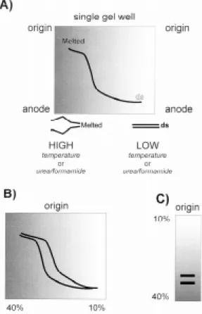
Typically, a double stranded DNA fragment has a characteristic melting profile upon the disasso-ciation of this homoduplex into its respective single stranded chains (Cotton 1997). This melting pro-file is reversible, giving rise to a sigmoid transi-tion curve (Fig. 2A). Double stranded DNA mo-bility is greater than that of single stranded or par-tially melted DNA. This profile is also sequence dependent, such that differing DNA sequences have different melting profiles despite being iden-tical in length (Fig.2B). Gradient gels can utilize either a chemical (DGGE) or temperature (TGGE) method. Gels can also be perpendicular (the gradi-ent is at right angles to the direction of electro-phoresis; Fig. 2A, B) or parallel (the gradient is in the same direction of electrophoresis; Fig. 2C). The precise gradient is usually determined empirically but computer programs, such as MELT 87 (Lerman & Silverstein 1987), can predict the behaviour of a specified sequence and an optimal gradient (Cot-ton 1997). Gradient gel electrophoresis can detect single point mutations between test and reference DNA. It is also a good method to separate com-plex mixtures of DNA that would otherwise co-migrate during conventional electrophoresis. As such, gradient gel methods are ideally suited to study the variation in gene families such as the ri-bosomal RNA complex (Schlötterer 1995).
DENATURING GRADIENT GEL ELECTROPHORESIS (DGGE)
DGGE has the ability to separate DNA frag-ments of identical size but differing sequence iden-tity through the utilization of a chemical gradient,
Fig. 2: basic theory of gradient gel electrophoresis. A: a chemi-cal or temperature gradient is formed perpendicular to the di-rection of electrophoresis. A DNA fragment is loaded across the single well and migrates towards the anode. Where the gradient is insufficient to melt the fragment, the mobility is the same as that of double stranded (ds) DNA. As the gradient increases the fragment progressively melts and is electro-phoretically retarded. The melting curve (sigmoidal in this in-stance) is dependent upon the sequence composition; B: two DNA fragments of identical size, but differing sequence, will have different melting profiles. This electrophoretic mobility shift allows separation of the two types; C: to facilitate further comparison (i.e. lane to lane), parallel gels (gradient in the same direction of electrophoresis) are used. The two fragments illustrated would co-migrate under standard PAGE or agarose electrophoresis, however, gradient gels permit direct separa-tion.
549 549549 549 549 Mem Inst Oswaldo Cruz, Rio de Janeiro, Vol. 95(4), Jul./Aug. 2000
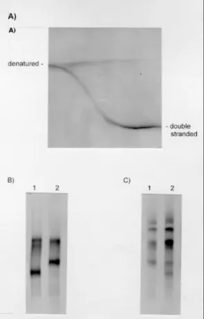
Upon non-denaturing PAGE, a single 450bp am-plification product was seen and no size variation was apparent between taxa (Stothard et al. 1997). Through the use of perpendicular gels, a chemical gradient from 10% to 25% was shown to denature the fragment. The sigmoidal transition profile is illustrative of a single melting domain within the amplified fragment; in this particular instance the PCR product was derived from a bacterial plasmid carrying the 18S from T. cruzi (Fig. 3A).
PCR products were then directly obtained from PCR amplification from T. cruzi genomic DNA preparations. Parallel gels that covered the same chemical gradient range (Fig. 3B) then separated these products. Variation was immediately detect-able between the homoduplex molecules of Z1/Z3 and Z2. As two bands were apparent in each lane, there would appear to be at least two divergent 18S sequence types present within each stock exam-ined. SSCP analysis could also detect variation between Z1/Z3 and Z2. By using heteroduplex analysis (Stothard et al. 1997), a finer level of reso-lution was achieved by finding variation between Z1 and Z3. In addition, as extra-bands were visu-alized upon heteroduplex analysis of Z1 and Z2, the presence of at least two 18S divergent types within each stock was further confirmed. More im-portantly each of these two types differed between stocks (Fig. 3B).
TEMPERATURE GRADIENT GEL ELECTROPHORE-SIS (TGGE)
To further screen the whole 18S gene for addi-tional regions of paralogy, riboprint profiles were subjected to TGGE analysis. Riboprint profiles, obtained from seven enzymes: HaeIII, AluI,
Sau3A, ScrFI, MspI, DdeI and CfoI, were sepa-rated by TGGE covering a thermal gradient from 45oC to 65oC. Riboprint profiles from CfoI and
DdeI exhibited split melting curve profiles char-acteristic of regions of paralogy. Unfortunately, as the fragment shown to be paralogous for DdeI di-gestion was not conserved across T. cruzi as there was RFLP variation, this profile was not examined further (Stothard et al. 1998b). The riboprint pro-file for CfoI was invariant within T. cruzi and was therefore further characterized. In total, five iso-lates of T. cruzi covering the principal zymodemes and T. conorhini, T. rangeli and L. braziliensis were shown to exhibit a profile indicative of paralogy [see fragment B in Fig. 1, Fig. 2 (Stothard et al. 1998b)].
To ascertain the region of the 18S that was shown to be paralogous, TGGE profiles were electroblotted onto nylon membranes and followed by hybridization with the 18S V1, V2 regions char-acterized by DGGE analysis (see above). The
frag-ment, labeled B, was shown to hybridize therefore the paralogous regions of the 18S were presum-ably confined only to the V1, V2 regions. Interest-ingly, this gene dimorphism is present in T. rangeli, T. conorhini and Leishmania spp. For
Leishmania, however, there would appear addi-tional regions of paralogy within the 18S [see frag-ment labeled * Fig. 2 (Stothard et al. 1998b)]. This fragment clearly contains three divergent types. What is more, in consideration with linkage to the dimorphic V1, V2 regions, there could be up to six
550 550 550 550
550 Genetic Diversity of T. cruzi JR Stothard et al.
divergent types present within the genome of L. braziliensis. If we assume that Leishmania was ancestral to other trypanosomes, it would appear that at least two divergent 18S types have been lost within Trypanosoma. Conversely, if we infer that if Trypanosoma was ancestral, Leishmania would require two new duplicative events to give rise to these paralogous copies.
CONCLUSIONS
The ability to differentiate T. cruzi with the ri-bosomal 18S has shed new light on the interpreta-tion of the evoluinterpreta-tionary relainterpreta-tionships within the trypanosomes. The assumption that this gene fam-ily is homogeneous within a genome is clearly mis-leading and the dynamics of this gene family are more complicated than prevously thought. Firstly the problem of sequence paralogy is a fundamen-tal issue within this group. The shared duplicative state of the V1, V2 regions would appear to be ancestral (plesiomorphic) to the group as a whole. Interpretation of the analysis of Briones et al. (1999) is particularly interesting in this light; the separate analyses of the variable regions of the 18S derived conflicting tree topologies, indicating sub-stantive rate heterogeneity across the 18S. This might not be unexpected given that certain regions of it seem to be afflicted by evolutionary paralogy. Secondly, the polarity of interpretation of the 18S in terms of duplications or losses of the divergent types needs further clarification. Most pessimisti-cally, how confident can we be of the current 18S data seeing as T. cruzi could have lost two thirds of the molecular evidence?
The relative insensitivity of riboprinting there-fore has advantages in light of this sequence paralogy. The restriction sites studied so far ap-pear to fall outside these paralogous regions and without recourse to the gradient gel methods their very existence might have remained undetected. In explanation, the contemporary way to derive 18S sequence data is to pool several PCR products and cycle sequence the template using overlapping sets of primers. The occurrence of split chromatogram peaks may well have gone unnoticed seeing as cer-tain templates can be preferentially amplified/se-quenced in comparison to the mixture as a whole. Riboprinting confirms the existence of two bio-logical lineages within T. cruzi, corresponding to
T. cruzi I and T. cruzi II, but indicates that if there were greater taxonomic sampling especially of Z3, new groups might come to light. Perhaps if riboprinting, using only HaeIII and CfoI, were to be used in conjunction with the DNA typing as-says of the 24S D7 domain by Souto et al. (1999) this combined methodology could lead to a very powerful diagnostic tool for nearly all kinetoplastid
infections. It would be interesting to explore the potential of these assays in a more clinical setting.
ACKNOWLEDGEMENTS
To Jamie Stevens for his invitation to the ‘Work-shop on Molecular Evolution of Trypanosomes’.
REFERENCES
Anon 1999. Recommendations from a Satellite Meet-ing. Mem Inst Oswaldo Cruz 94: 429-432. Bogliolo AR, Lauria-Pires L, Gibson WC 1996.
Poly-morphisms in Trypanosoma cruzi: evidence of ge-netic recombination. Acta Trop 61: 31-40. Briones MRS, Souto RP, Stolf BS, Zingales B 1999.
The evolution of two Trypanosoma cruzi subgroups inferred from rRNA genes can be correlated with the interchange of American mammalian faunas in the Cenozoic and has implications to pathogenicity and host specificity. Mol Biochem Parasitol 104: 219-232.
Carrasco HJ, Frame IA, Valente SA, Miles MA 1996. Genetic exchange as a possible source of genomic diversity in sylvatic populations of Trypanosoma cruzi. Am J Trop Med Hyg 54: 418-424.
Clark CG 1992. Riboprinting: a molecular approach to the taxonomy of protozoa. In JJ Lee, AT Soldo (eds),
Protocols in Protozoology, Allen Press, Lawrence, Kansas, p. D-4.1-D-4.4.
Clark CG, Diamond LS 1991. The Laredo strain and other Entamoeba histolytica-like amoebae are En-tamoeba moshkovskii. Mol Biochem Parasitol 46: 11-18.
Clark CG, Pung OJ 1994. Host specificity of ribosomal DNA variation in sylvatic Trypanosoma cruzi from North America. Mol Biochem Parasitol 66: 175-179.
Clark CG, Martin DS, Diamond LS 1995. Phylogenetic relationships among anuran trpanosomes as revealed by riboprinting. J Euk Microbiol 42: 92-96. Cotton RGH 1997. Mutation Detection, Oxford
Univer-sity Press, Oxford, 198 pp.
Gasser RB, Zhu XQ 1999. Sequence-based analysis of enzymatically amplified DNA fragments by muta-tion detecmuta-tion techniques. Parasitol Today 15: 462-465.
Hoare CA 1972. The Trypanosomes of Mammals – A Zoological Monograph, Blackwell Scientific Publi-cations, Oxford, 749 pp.
Lerman LS, Silverstein T 1987. Computational simula-tion of DNA melting and its applicasimula-tion to denatur-ing gradient gel electrophoresis. Methods Enzymol 155: 482-501.
Li W-H 1997. Molecular Evolution, Sinauer Associates, Inc., Massachusetts, 487 pp.
Macedo AM, Pena SDJ 1998. Genetic variability of Try-panosoma cruzi: implications for the pathogenesis of Chagas disease. Parasitol Today 14: 119-124. Miles MA, Povoa MM, Souza AA, Lainson R, Shaw JJ,
Ketteridge DA 1981. Chagas disease in the Amazon Basin: II. The distribution of Trypanosoma cruzi
551 551551 551 551 Mem Inst Oswaldo Cruz, Rio de Janeiro, Vol. 95(4), Jul./Aug. 2000
Miles MA, Toyé PJ, Oswald SC, Godfrey DG 1977. The identification by isoenzyme patterns of two dis-tinct strain-groups of Trypanosoma cruzi circulat-ing independently in a rural area of Brazil. Trans R Soc Trop Med Hyg 71: 217-225.
Momen H 1999. Taxonomy of Trypanosoma cruzi: a commentary on characterization and nomenclature.
Mem Inst Oswaldo Cruz 94: 181-184.
Noyes HA 1998. Can trypanosome trees be trusted?
Parasitol Today 14: 49-50.
Pulido M, Martinez-Calvillo S, Hernandez R 1996. Try-panosoma cruzi rRNA genes: a repeated element from the non-transcribed spacer is locus specific.
Acta Trop 62: 163-170.
Schlötterer C 1995. Temperature-gradient gel electro-phoresis as a screening tool for polymorphisms in multigene families. Electrophoresis 16: 722-728. Sheffield VC, Beck JS, Nichols B, Cousineau A, Lidral
AC, Stone EM 1992. Detection of multiallele poly-morphisms within gene sequences by GC-clamped denaturing gradient gel electrophoresis. Am J Hum Gen 50: 567-575.
Souto RP, Vargas N, Zingales B 1999. Trypanosoma rangeli: discrimination from Trypanosoma cruzi
based on a variable domain from the large subunit ribosomal RNA gene. Exp Parasitol 91: 306-314. Stevens JR, Gibson WC 1999. The molecular evolution
of trypanosomes. Parasitol Today 15: 432-437. Stothard JR 2000. Trypanosome trees and homologies.
Parasitol Today16: 173.
Stothard JR, Frame IA, Miles MA 1997. Use of poly-merase chain reaction-based single strand confor-mational polymorphism and denaturing gradient gel electrophoresis methods for detection of sequence variation of ribosomal DNA of Trypanosoma cruzi. Int J Parasitol 27: 339-343.
Stothard JR, Frame IA, Miles MA 1999. Genetic diver-sity and genetic exchange in Trypanosoma cruzi: dual drug-resistant “progeny” from episomal transformants. Mem Inst Oswaldo Cruz 94: 189-193. Stothard JR, Frame IA, Carrasco HJ, Miles MA 1998a. On the molecular taxonomy of Trypanosoma cruzi
using riboprinting. Parasitology 117: 243-247. Stothard JR, Frame IA, Carrasco HJ, Miles MA 1998b.
Temperature gradient gel electrophoresis (TGGE) analysis of riboprints from Trypanosoma cruzi. Parasitology 117: 249-253.
Tibayrenc M 1998a. Genetic epidemiology of parasitic protozoa and other infectious agents: the need for an integrated approach. Int J Parasitol 28: 85-104. Tibayenc M 1998b. Integrated genetic epidemiology of infectious diseases: the Chagas model. Mem Inst Oswaldo Cruz 93: 577-580.
Wright ADG, Li S, Feng S, Martin DS, Lynn DH 1999. Phylogenetic position of the kinetoplastids,
Cryptobia bullocki, Cryptobia catostomi, and
Cryptobia salmositica and monophyly of the genus