Nutr Hosp. 2011;26(3):528-536 ISSN 0212-1611 • CODEN NUHOEQ S.V.R. 318
Original
Factors associated with oxidative stress in women with breast cancer
F. G. K. Vieira1, P. F. Di Pietro1, B. C. B. Boaventura1, C. Ambrosi1, G. Rockenbach1,M.ª A. Fausto2, C. G. Crippa3and E. L. Da Silva1
1Post-Graduate Program in Nutrition. Santa Catarina Federal Universiy. Florianópolis. SC. Brazil. 2Food Department.
Federal University of Ouro Preto. Ouro Preto. Minas Gerais. Brazil. 3Department of Tocoginecology. Federal University of
Santa Catarina. Florianópolis. Santa Catarina. Brazil.
FACTORES ASOCIADOS CON ESTRÉS OXIDATIVO EN MUJERES CON CÁNCER
DE MAMA
Resumen
Objetivo:Evaluar la asociación entre las variables fisio-lógicas, físicas, de estilo de vida y nutricionales y de los biomarcadores de estrés oxidativo en mujeres con cáncer de mama.
Métodos:Este estudio transversal se realizó en 55 mujeres diagnosticadas de cáncer de mama. Se analizó el grado de estrés oxidativo midiendo los hidroperóxidos lipídicos (HL), las sustancias reactivas del ácido tiobarbi-túrico (TBARS), las proteínas carbonilo, el glutatión reducido (GSH) de sangre completa y la capacidad antio-xidante sérica (CA). Los datos de la dieta se obtuvieron mediante cuestionario de frecuencia de alimentos. Se usó la regresión linear para determinar la asociación entre las variables estudiadas y los biomarcadores de estrés oxida-tivo. Los datos de las proteínas carbonilo no se incluyeron en los análisis de regresión linear puesto que no mostra-ron una distribución normal, incluso después de la trans-formación logarítmica y de otro tipo.
Resultados:Después de ajustar para el aporte de ener-gía, el consumo de pollo y de productos lácteos con alto contenido en grasas se asoció con un aumento en los nive-les de HL, mientras que el consumo de vitamina E se aso-ció con una disminuaso-ción de los niveles de HL (R2 = 23,8%). El consumo de aceite se asoció con un aumento de los niveles de TBARS (R2 = 6,82%). El estado de los ganglios linfáticos axilares se asoció con un descenso de los niveles de GSH (R2 = 9,31%). La mayor edad se asoció directa-mente con los niveles de CA, mientras que la grasa de ori-gen animal y el consumo de dulces se asoció con niveles bajos de CA (R2 = 41,42%).
Conclusión:El consumo de pollo, vitamina E, lácteos (especialmente de aquellos con alto contenido en grasa), aceites y grasas de origen animal, así como dulces, junto con el estado de los ganglios axilares y la edad podrían ser determinantes importantes en el estrés oxidativo de muje-res con cáncer de mama.
(Nutr Hosp. 2011;26:528-536)
DOI:10.3305/nh.2011.26.3.4812
Palabras clave: Cáncer de mama. Estrés oxidativo. Ingesta diaria. Factores predictivos.
Abstract
Objective:To assess the association between physiolog-ical, physphysiolog-ical, lifestyle and nutritional variables and oxidative stress biomarkers in women with breast cancer. Methods:This cross-sectional study was conducted on 55 women newly diagnosed with breast cancer. The extent of oxidative stress was analyzed by the measure-ment of plasma lipid hydroperoxides (LH), thiobarbi-turic acid reactive substances (TBARS), protein car-bonyl, whole blood reduced glutathione (GSH) and serum antioxidant capacity (AC). Diet data were obtained from food frequency questionnaire. Linear regression was used to determine the association between the variables studied and oxidative stress biomarkers. The protein car-bonyl data was not included in the linear regression analyses since the data did not show a normal distribu-tion, even after logarithmic and other transformations.
Results:After adjusting for energy intake, the intake of chicken and high-fat dairy products was associated with increased levels of LH, while vitamin E intake was associ-ated with decreased LH levels (R2 = 23.8%). Intake of oils was associated with increased levels of TBARS (R2 = 6.82%). Positive axillary lymph node status was associ-ated with decreased levels of GSH (R2 = 9.31%). Increas-ing age was directly associated with levels of AC, while animal fat, dairy product, and sweet food intakes were associated with low levels of AC (R2 = 41.42%).
Conclusion:Intake of chicken, vitamin E, dairy prod-ucts (particularly high-fat dairy prodprod-ucts), oils, animal fat, and sweet foods, along with axillary lymph node sta-tus and age, may be important determinants of oxidative stress in women with breast cancer.
(Nutr Hosp. 2011;26:528-536)
DOI:10.3305/nh.2011.26.3.4812
Key words: Breast cancer. Oxidative stress. Dietary intake. Predictive factors.
Correspondence: Patricia Faria Di Pietro. Programa de Pós-Graduacão em Nutrição. Centro de Ciências da Saúde.
Universidade Federal de Santa Catarina. Campus Universitário. Cep 88040-900 Trindade. Florianópolis/SC. Brazil. E-mail: fariadipietro@gmail.com
Introduction
Breast cancer, the most common cancer among women worldwide, accounts for the highest morbidity and mortality.1 Annually, around 1 million new
patients are diagnosed with breast cancer and 400,000 women die from the disease.2The etiology of breast
cancer is multifactorial and several risk factors associ-ated with breast cancer may exert their effects via gen-eration of an oxidative stress status.
Oxidative stress is caused by an unfavorable bal-ance between reactive oxygen species (ROS) and antioxidant defenses. ROS are generated during nor-mal cellular metabolism, as a result of the influence of various environmental factors, as well as during pathological processes.3Oxidative stress is
responsi-ble for DNA, lipid and protein damage and it plays an important role in the development and progression of many human diseases, including breast cancer.4
Fur-thermore, there is a great interest in the measurement of oxidative stress biomarkers in breast cancer patients. Several markers of oxidative stress are cur-rently available, such as lipid hydroperoxides (LH) and thiobarbituric acid reactive substances (TBARS), which have been used extensively as markers of lipid peroxidation, as well as protein car-bonyl, that is the most frequently used biomarker of protein oxidation in epidemiological and clinical studies.5-8
To control the overproduction of ROS, the cells are protected against oxidative stress by antioxidant detoxifying mechanisms, which include nonenzy-matic antioxidants such as reduced glutathione (GSH), vitamins A, C and E and various antioxidant enzymes.9Erythrocyte glutathione has been
com-monly employed as a biomarker of oxidative stress because GSH is a reducer compound widely distrib-uted in cells. In addition, the levels of erythrocyte GSH may reflect the glutathione activity in other tis-sues.10Due to the complexity involved in measuring
all known antioxidants separately and the interac-tions among different antioxidant compounds, sev-eral methods have been developed to assess the antioxidant capacity (AC) of serum or plasma.11,12
These methods provide an overview of the biological interactions between individual antioxidants and how efficiently these translate into host cell protec-tion during periods of oxidative stress.13
Identification of potentially modifiable factors that affect oxidative stress in breast cancer patients is an increasingly important task. Dietary intake repre-sents one of a set of factors that have received con-siderable attention because of its ability to either reduce or promote oxidative stress.14-16Therefore, in
the present study, we investigated the association between blood biomarkers of oxidative stress and physiological, physical, lifestyle and nutritional variables in newly diagnosed women with breast cancer.
Subjects and methods
Subjects
The population of this cross-sectional study was selected from 226 women admitted between October 1, 2006 and July 31, 2007, attending by breast surgery in the Carmela Dutra Maternity Hospital, Florianópolis, Santa Catarina, Brazil. One-hundred fifty nine women were ineligible for inclusion in the study: 65 (28%) were excluded because they had already started some type of neoadjuvant cancer treatment; 90 (39%) had benign tumors without suspicion of malignancy and; 4 (2%) had previous history of cancer. Thus, there were 67 eligible women, of which one (2%) refused to par-ticipate and 11 (5%) had benign tumors diagnosis after the interview. Finally, 55 women newly diagnosed with a first primary in situor invasive breast cancer were interviewed. All subjects were classified anato-mopathologically according to the Tumor-Node-Metastasis system.17There were no age or race
restric-tions. None of the patients were pregnant or lactating. The study was approved by the ethical committee of the Federal University of Santa Catarina and of Carmela Dutra Maternity Hospital.
Blood samples
Blood samples were obtained by venous arm puncture using a vacuum system (Vacutainer) in a tube containing EDTA and a tube with serum-separation gel after overnight fasting between 7 and 8 am. Plasma and serum were immediately obtained by centrifugation (1000 x g, 10 min, 4°C). To quantify the reduced glutathione (GSH), an aliquot of EDTA-blood was hemolyzed and immediately preserved in trichloroacetic acid medium. Plasma samples were used for measurement of TBARS, LH and protein carbonyls and the serum was used to measure the AC. TBARS and LH levels were analyzed immediately after sample collection. The remaining plasma, serum and acid extracts were stored at -70oC for no longer than 1 month for other
biochemi-cal determinations. All measurements were performed in duplicate.
Reagents
hexahydrate and potassium phosphate monobasic were purchased from Vetec Química Fina (Rio de Janeiro, RJ-Brazil). All other chemicals and reagents used in the study were of analytical grade and obtained from standard commercial suppliers.
Biochemical analysis
Plasma lipid hydroperoxides (LH) were determined using ferrous oxidation-xylenol orange (FOX), as described by Jiang et al.18The method is based on the
fast oxidation of Fe2+to Fe3+in acid medium mediated
by lipid peroxides. In the presence of xylenol orange, Fe3+forms a complex (Fe3+-xylenol orange), which is
measured spectrophotometrically at 560 nm. The FOX reagent (1.4 mL), containing 250 mM H2SO4, 4.4 mM
BHT, 1 mM xylenol orange, and 2.5 mM iron and ammonium sulfate in methanol, was added to aliquots of plasma. Subsequently, the mixture was kept at room temperature for 30 min, the tubes were centrifuged (1,000 x g, 5 min) and the absorbance was read. A stan-dard hydrogen peroxide curve was used to quantify the LH.
Lipid peroxidation of plasma was estimated through the detection of derivative products from oxidation, stances that react with thiobarbituric acid–reactive sub-stances (TBARS), mainly malondialdehyde, according to a procedure previously described by Esterbauer and Cheeseman.19Aliquots of plasma or deionized water
(blank) were mixed with 0.5 mL 20% TCA containing 0.5 N HCl and 50 µL of 10 mM BHT. Thiobarbituric acid at 1% was added and the mixture was incubated at 100o C for 1 h. After cooling in an ice-bath, 2.5 mL of
butyl alcohol was added, vortex-mixed and centrifuged at 1000 x g for 5 min. The absorbance of the super-natant was measured at 532 nm against the blank. Recently prepared 1,1,3,3-tetramethoxypropan (TMP) was used as the standard.
The carbonyl content in plasma protein was deter-mined using the reagent 2,4 dinitrophenylhydrazine (DNPH) as described by Levine et al.8One hundred
microliters of plasma were mixed with 600 µL of 10 mM DNPH in 2 N HCl in 1.5 mL test tubes. Six hun-dred microliters of 2 N HCl were added to 100 µL of plasma as the blank. The tubes were vortex-mixed every 10 min and after 1 h of incubation at room tem-perature in the dark, 600 µL of 20% TCA was added. After a further 10 min of incubation all samples were centrifuged at 11,000 x g for 5 min. The supernatant was then discarded and the precipitate was washed 3 times with 800 µL of a mixture of ethanol:ethyl acetate, 1:1 (v/v) and centrifuged after 10 min. Finally, the pro-tein precipitate was dissolved in 900 µL of 6 M guani-dine hydrochloride in 20 mM KH2PO4.The samples were then incubated again for 1 h at 37o C. The
absorbance of the test sample was measured at 360 nm against a guanidine solution. Total protein concentra-tion was determined through measuring the absorbance
of the blank at 280 nm, using bovine serum albumin as the standard. The concentration of carbonyls (nmol/mg protein) was calculated using a molar extinction coeffi-cient (ε) of 22.000.
The concentration of thiol compounds of low molec-ular weight in whole blood, such as GSH, was evalu-ated according to the method of Beutler et al.10Firstly,
an aliquot of the total EDTA-blood was hemolyzed with cold water and the proteins were precipitated by the addition of 30% TCA. Aliquots of 50 µL of the hemolyzed sample and 50 µL of 10 mM DTNB were mixed in tubes containing 0.8 mL of 200 mM phos-phate buffer, pH 8.0. After 3 min, the absorbance of the thiolate anion was measured at 412 nm. Commercial GSH was used as the standard.
The antioxidant capacity (AC) of the serum samples was determined using the ferric-reducing antioxidant power (FRAP) assay as described by Benzie and Strain.11In this procedure, the antioxidants present in
the serum are evaluated as reducers of Fe3+to Fe2+,
which is chelated by TPTZ to form a Fe2+-TPTZ
com-plex with maximum absorbance at 593 nm. Ten micro-liters of serum were mixed with 1 mL of reagent con-taining 1.7 mM FeCl3and 0.8 mM TPTZ, prepared in
300 mM sodium acetate, pH 3.6. The samples were incubated for 15 min at 37°C and the absorbance was measured at 593 nm. The results were calculated using Trolox as standard and were expressed as Trolox equivalents.
Data collection
The main questionnaire was administered by a trained interviewer through in-person interviews con-ducted at the Nutrition Service Unit of the Carmela Dutra Maternity Hospital and lasted ~ 1 h. Information was collected on known and suspected risk factors for breast cancer, including cigarette smoking, alcohol intake, menstrual and reproductive histories, hormone use, physical activity, prior medical history and other study variables. During each interview, height and weight were measured (Filizola, São Paulo, Brazil) using standard procedures20to obtain body mass index
(BMI). The participants were then classified according to categories defined by the World Health Organiza-tion.21
Usual dietary intake was obtained using a food fre-quency questionnaire, previously validated in Brazil,22
containing 94 food items classified into ten groups: cereals, fruits, vegetables, beans, meat and eggs, dairy products, oils and fat, sweet foods, alcoholic beverages and non-alcoholic beverages. For each food item the participants determined the size of the portion con-sumed with the help of an album containing color pho-tographs of products23or household measures of
grams and milliliters of fruits, doughnuts, lard, cream and yerba mate infusions were obtained according to the assay described by Griswold.24The grams of
por-tions for polenta,a dish made from boiled cornmeal, were obtained from Ben25and for the other foods from
Pinheiro et al.26
Dietary intake was analyzed in terms of grams and milliliters of specific food groups determined by tradi-tional food groups27and the a-priorihypotheses. For
each subject, individual dietary intake was converted to a monthly frequency weighted variable (in grams or milliliters) according to the reported portion size. As described by Block et al.,15these individual values were
summed to create the specific food groups and divided by 30.5 to determine an average daily level of con-sumption (grams or milliliters per day). Food intake was also analyzed for energy and nutrient content using the nutrient database of the United States Department of Agriculture (USDA) (issue 20, full version, 2008).28
Food nutritional values which were not available in this nutrient database were obtained from Brazilian food tables.29,30
Statistical analysis
Statistical analyses were performed using Stata 10.0 software, and in all cases the level of significance was established as 5%. The variables of this study were classified into: (a) variables related to cancer (anato-mopathological stage, axillary lymph node status and tumor size); (b) variables related to individuals (age,
menopausal status, use of dietary supplements, use of medicaments, concomitant diseases, use of oral contra-ceptives, physical activity and cigarette smoking); (c) anthropometric variables; and (d) dietary intake vari-ables (food and beverage groups, energy and nutrient content).
The dietary intake data were analyzed as continuous or categorized variables (1 = intake greater than 50th
percentile).
The Shapiro-Wilk test was used to evaluate whether or not the distribution of the variables was normal. The mean values of two groups were compared using the Student’s t-test and the means of more than two groups were assessed using Analysis of Variance followed by the Bonferroni multiple-comparison test. When the data distribution was non-parametric, the Mann-Whit-ney Utest or Kruskall-Wallis test (for medians) was used. The chi-square test and the Fisher’s exact test were used for comparing categorical variables.
Linear regression was used to estimate the associa-tion between the cancer-related, individual, anthropo-metric and dietary intake variables, and the plasma LH and TBARS levels, whole blood GSH levels and serum AC levels. Linear regression was not carried out on the plasma protein carbonyl levels since the data distribu-tion was not normal, even after logarithmic and other transformations. The variables with p < 0.25 in the uni-variate linear regression analysis were employed in the multivariate regression model. Variables that showed collinearity or low frequency were excluded from the multivariate model, whilst variables with more than two categories were transformed into indicator Table I
Characteristics of newly diagnosed breast cancer women (n = 55), Florianópilis, SC-Brazil
Mean ± SD1or percentage Median Range
Age (years) 51.2 ± 10.5 50.0 33-78
Ductal carcinoma (%) 94.6
Positive axillary lymph node status (%) 41.8 Anatomopathological stage (%)
0 5.5
I 32.7
II 40.0
III 21.8
Body mass index (kg/m2) 28.1 ± 4.9 27.3 17.8-40.4
Smoking (%)
Nonsmoker 76.4
Current smoking 23.6
Postmenopausal status (%) 54.6
Plasma LH2(µmol/L) 0.89 ± 0.37 0.84 0.12-1.85
Plasma TBARS2(µmol/L) 4.93 ± 0.87 4.79 3.09-6.84
Plasma protein carbonyl (nmol/mg) 0.68 ± 0.25 0.63 0.27-1.48
Whole blood GSH2(µmol/L) 77.2 ± 17.5 75.9 28.5-116.3
Serum AC2(µmol/L) 664.4 ± 153.3 686.2 409.7-979.5
1SD, standard deviation.
(dummy) variables. The final models for the linear regression analysis were constructed using stepwise regression analysis to select the minimum set of predic-tors that significantly (p< 0.05) maximized the model R2.
In order to validate the final predictive regression model, we estimated the optimism in the R2statistic
using an exhanced internal validation technique. The optimism is the mean difference between the R2of the
original regression model and the R2yielded when β
-coefficients derived from bootstrap samples (1000 rep-etitions) are applied to the original dataset31.
Results
Table I gives the characteristics and the levels of oxidative stress biomarkers of the study participants.
When the mean levels of plasma LH and TBARS, and the median of plasma protein carbonyl were ana-lyzed according to cancer-related, individual, and anthropometric variables, no significant differences were detected (data not shown). Blood GSH level was not associated with the individual and anthropometric variables but was associated with the cancer-related variables. For example, women with positive axillary lymph node status had significantly (p = 0.0001) lower GSH levels (69.99 ± 14.76 mol/L) than women with negative axillary lymph node status (82.33 ± 17.72 mol/L). Serum AC was not associated with cancer-related or anthropometric variables. However, post-menopausal women or women over 50 years of age had significantly higher AC levels than premenopausal women (postmenopausal: 707.7 ± 158.3 vs
pre-menopausal: 612.3 ± 131.9 mol/L; p = 0.02) or women with age lower than 50 years (> 50 y: 719.9 ± 150.8 vs ≤
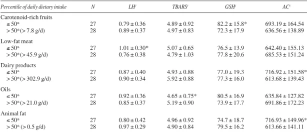
50 y: 618.1 ± 141.7 mol/L; p = 0.01) (data not shown). Table II shows the dietary intake variables associ-ated with biomarkers of oxidative stress. Enhanced intake of low-fat meat, which includes roasted or boiled red meat, pork, chicken or fish, was signifi-cantly associated with lower levels of plasma LH. The enhanced intake of oils was positively associated with high levels of plasma TBARS. The levels of blood GSH were significantly lower in women with increased intake of carotenoid-rich fruits. The levels of serum AC were significantly lower in women with high intake of dairy products and animal fat. Variables related to dietary intake were not significantly associ-ated with plasma protein carbonyls (data not shown).
In the univariate linear regression analysis, plasma LH levels were significantly related to daily intake of chicken (β = 0.16; p = 0.03), low-fat dairy products (β
= -0.0005; p = 0.04), oils (β = -0.01; p = 0.01) and low-fat meat (β = -0.25; p = 0.01) (data not shown). Table III reports the final model of the linear regres-sion for plasma LH levels after adjusting for energy intake. This final model explained 23.80% (R2
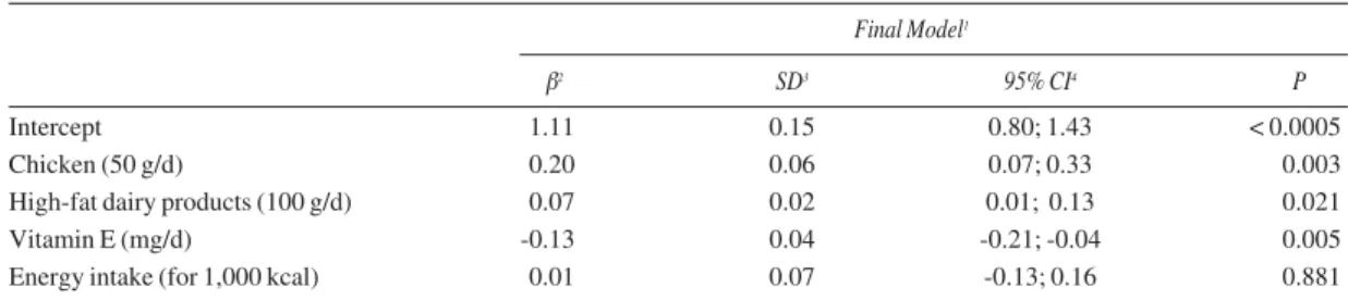
adjusted) of the plasma LH variability. There was no significant interaction between the variables of the model. The final model indicated that the daily intake of chicken and high-fat dairy products were associ-ated with an increase in the level of LH; while the daily intake of vitamin E was associated with an decrease in LH levels (table III). The internal valida-tion of the regression model for plasma LH levels yielded a very low estimate of the over optimism for R2of 0.56745 (< 1%).
Table II
Dietary intake variables associated with levels of markers of oxidative stress in newly diagnosed breast cancer women (n = 55), Florianópolis, SC-Brazil
Percentile of daily dietary intake N LH1 TBARS1 GSH1 AC1
Carotenoid-rich fruits
≤ 50th 27 0.79 ± 0.36 4.89 ± 0.92 82.2 ± 15.8* 693.19 ± 164.54
> 50th (> 7.8 g/d) 28 0.89 ± 0.37 4.97 ± 0.83 72.3 ± 17.9 636.56 ± 138.89
Low-fat meat
≤ 50th 27 1.01 ± 0.30* 5.07 ± 0.65 76.5 ± 13.9 642.40 ± 155.13
> 50th (> 45.9 g/d) 28 0.76 ± 0.38 4.79 ± 1.03 77.8 ± 20.6 685.53 ± 151.24
Dairy products
≤ 50th 27 0.87 ± 0.40 4.93 ± 0.88 77.0 ± 19.3 716.92 ± 151.58*
> 50th (> 302.9 g/d) 28 0.90 ± 0.34 5.92 ± 0.88 77.3 ± 16.0 613.68 ± 139.43
Oils
≤ 50th 27 0.92 ± 0.36 4.65 ± 0.75* 80.5 ± 16.9 635.84 ± 127.82
> 50th (> 21.0 g/d) 28 0.85 ± 0.37 5.19 ± 0.90 73.9 ± 17.7 691.86 ± 172.23
Animal fat
≤ 50th 27 0.80 ± 0.42 4.96 ± 0.92 74.7 ± 18.7 716.93 ± 149.96*
> 50th (> 0.5 g/d) 28 0.97 ± 0.29 4.90 ± 0.84 79.5 ± 16.2 613.66 ± 141.11
1µmol/L; LH, lipid hydroperoxides; TBARS, thiobarbituric acid reactive substances; GSH, reduced glutathione; AC, antioxidant capacity.
Among all variables studied, only intake of oils (β = 0.54; p = 0.02) was positively associated with plasma TBARS levels in the univariate linear regression analy-sis (data not shown). After adjusting for energy intake, intake of oils explained 6.82% (R2adjusted) of the
vari-ability in the TBARS levels. Furthermore, increased consumption of oils (> 21g/d) was associated with an increase in the plasma TBARS of 0.52 µmol/L (p = 0.028) (data not shown).
In the univariate linear regression analysis, blood GSH levels were significantly associated with the posi-tive axillary lymph node status (β = -12.33; p = 0.009), daily intake of carotenoid-rich fruits (β = -0.18; p = 0.03) and consumption of more than 7.8 g per day of carotenoid-rich fruits (β = -9.9; p = 0.04) (data not shown). After adjusting for energy intake, the linear regression model explained 9.31% (R2adjusted) of the
blood GSH variability showing that only a positive axillary lymph node status was associated with a decrease in the GSH levels of 12.41 µmol/L (p = 0.009) (data not shown).
Variables significantly associated with serum AC levels in the univariate linear regression analysis were age (β = 6.11; p = 0.001), menopausal status (β = 95.41; p = 0.02), daily intake of dairy products (β = -26.62; p = 0.005), high-fat dairy products (β = -0.275; p = 0.02), animal fat (β = -58.93; p = 0.01), sweet foods (β = -75.06;
p = 0.001), energy intake (β = -0.064; p = 0.01), lipids (β = -1.79; p = 0.02), carbohydrates (β = -0.392; p = 0.01), saturated fat (β = -6.66; p = 0.001) and monoun-saturated fat (β = -6.38; p = 0.01) (data not shown). After adjusting for energy intake, the final model show that increasing age was associated with an enhanced serum AC, while that the daily intake of animal fat, dairy products and sweet foods were associated with a decrease in the serum AC. The final model explained 41.42% (R2adjusted) of the variability in serum AC
levels (table IV). There was no significant interaction between the variables of the model. The internal vali-dation of the regression model for serum AC levels yielded a very estimate of the over optimism for R2of
5.96%.
Discussion
One mechanism by which some physiological, phys-ical, lifestyle and nutritional factors may help to pre-vent cancer is through the modulation of oxidative stress status. The most common approach to the mea-surement of free radical and oxidative stress has been by determining products of free radical reactions with biological macromolecules.6It has been shown that
oxidative stress can contribute to cancer development.4
Table III
Final model of linear regression for plasma lipid hydroperoxide levels (LH) (µmol/L) in breast cancer women (n = 55), Florianópolis, SC-Brazil (model R2= 0.2380)
Final Model1
β2 SD3 95% CI4 P
Intercept 1.11 0.15 0.80; 1.43 < 0.0005
Chicken (50 g/d) 0.20 0.06 0.07; 0.33 0.003
High-fat dairy products (100 g/d) 0.07 0.02 0.01; 0.13 0.021
Vitamin E (mg/d) -0.13 0.04 -0.21; -0.04 0.005
Energy intake (for 1,000 kcal) 0.01 0.07 -0.13; 0.16 0.881
1Adjusted for dietary energy intake; 2β = Coefficient estimates; 3SD = Standard Deviation; 4CI = Confidence Interval.
Table IV
Final model of linear regression for serum antioxidant capacity (AC) levels (µmol/L) in newly diagnosed breast cancer women (n = 55), Florianópolis, SC-Brazil (model R2= 0.4142)
Final Model*
β1 SD2 95% CI3 P
Intercept 493.77 109.51 273.68; 713.85 < 0.0005
Age (years) 5.48 1.65 2.15; 8.81 0.002
Animal fat (5 g/d) -41.19 18.43 -78.23; -4.41 0.030
Dairy products (100 g/d) -26.31 7.98 -45.18; -15.07 0.002
Sweet foods (50 g/d) -56.51 22.90 -102.55; -10.25 0.017
Energy intake (for 1,000 kcal) 22.57 26.16 -30.00; 75.15 0.392
In addition, oxidative stress biomarkers have been shown to be elevated in the blood and in malignant breast biopsies of breast cancer patients.5,9,16Results
from the current study expand upon previously pub-lished data by providing an insight into the associations between several variables cancer-related, individual, anthropometric and dietary intake variables, and oxida-tive stress biomarkers assessed by lipid peroxidation (plasma levels of LH and TBARS), protein oxidation (plasma levels of protein carbonyl) and by the mea-surement of antioxidants (blood GSH and serum AC), in women newly diagnosed with breast cancer.
Overall, a small number of significant associations between cancer-related variables and oxidative stress biomarkers were found in our study population. The extent of oxidative stress was not affected by the level of malignancy of the breast cancer (advanced anato-mopathological status and tumor size) in the women studied, according to previous studies.5,9,32Of the
vari-ables related to cancer, only positive axillary lymph node status was inversely associated with blood levels of GSH. In fact, a decreased content of erythrocyte GSH has been reported in several diseases including breast cancer.33,34The lower levels of GSH seen in
breast cancer patients with positive axillary lymph node status support the hypothesis that glutathione sta-tus is inversely related to malignant transformation.33, 34
Despite some conflicting results, it has been observed that in healthy populations an older age, post-menopausal status, non use of dietary supplements, absence of regular physical activity and cigarette smoking may contribute to the enhancement of oxida-tive stress.15,16,35 Contrary to our expectations, we
observed increased serum AC levels in older and post-menopausal women with breast cancer. The positive association of age and postmenopausal status with serum AC levels might be intriguing and unexpected. However, our results are in agreement with those of other studies. For example, a cross-sectional study on healthy individuals also showed high serum AC levels in older subjects13and a positive association between
age and erythrocyte GSH has been reported.6In
addi-tion, increased activity of blood glutathione peroxidase and superoxide dismutase, two antioxidant enzymes, was found in postmenopausal compared to pre-menopausal breast cancer women.5
Here, we found no evidence of a significant associa-tion between smoking, intensity of physical activity or BMI and oxidative stress. Although cigarette smoking is a well known environmental oxidant, previous stud-ies relating cigarette smoking to markers of oxidative stress have provided conflicting results.6,15,16An excess
or lack of physical exercise can produce an imbalance between ROS and antioxidants, leading to oxidative stress.36In addition, obesity may also be associated
with oxidative stress.6,37However, in agreement with
other studies,6,16in this study we found no association
between intensity of physical activity or BMI and oxidative stress status.
It has been largely believed that fruits and vegetables (as antioxidant rich foods) and food products with high content of polyunsaturated fatty acid (PUFAs) (as pro-oxidants) are the most diet-related constituents closely linked to oxidative stress. However, considering that several studies have failed to observe a positive associ-ation between blood levels of antioxidants and protec-tion of cells against oxidative damage,38or between
diet enriched with PUFAs and an increase in lipid per-oxidation,39further studies are warranted to better
elu-cidate the role of dietary intake in the modulation of oxidative stress. In the current study, the consumption of high oils diet (rich in PUFAs) by breast cancer women showed positive association with TBARS in the multivariate linear regression analysis. These results are consistent with an earlier study on women previously treated for breast cancer, which a polyunsat-urated fat intake was associated with elevated lipid per-oxidation.37In addition, the results for the multivariate
analysis also indicated that chicken and high-fat dairy products intake was positively associated with lipid peroxidation measured by plasma LH levels, while vit-amin E intake was negatively associated with lipid per-oxidation. For healthy individuals, dietary fat and satu-rated fat intake, which are found in foods such as chicken and high-fat dairy products, have been associ-ated with increasing oxidative stress markers.40The
protective role of dietary vitamin E against lipid perox-idation has also been previously described in breast cancer patients.37and, due to its free radical scavenger
activity and lipophilic profile, vitamin E may con-tribute to a decreased susceptibly of PUFAs in cell membranes to oxidation.3
The present study showed some surprising results such as the negative relationship between daily intake of carotenoid-rich fruits and blood GSH levels in the univariate linear regression analysis. However, this association was no longer significant after adjusting for energy intake in the final model, indicating therefore that not all associations found in this study are neces-sarily causal. Regardless, further studies would be nec-essary to confirm or refute this negative association. There was no significant interaction between the vari-ables of the model.
In the multivariate analysis, the intake of animal fat, dairy products and sweet foods by the breast cancer women was associated with decreased serum AC lev-els, as has also been described for healthy subjects.41
On the other hand, consumption of fruits and vegeta-bles were not associated with serum AC of breast can-cer patients. Although fruit and vegetable intake can decrease oxidative stress, including the enhancement of AC,16,37contrary results have been reported for
healthy people.42
suggest-ing that our model is valid31. Second, our results are
derived from a convenience sample, which may inhibit the validity of conclusive statements regarding a cause-effect relationship between biomarkers of oxidative stress status and the variables for women newly diag-nosed breast cancer examined. However, our study has the important advantage that all samples were collected before surgery and onset of chemotherapy or radiation. It has been shown that recent treatment promotes signifi-cant alterations in the level of oxidative stress markers in cancer patients.43Another advantage of our study is the
use of multiple markers of oxidative stress status. Fur-thermore, the results indicated that some of the proposed biomarkers of oxidative stress status are associated with important variables that represent significant risk factors or prognosis factors for breast cancer. However, the observed associations are not consistent for all of the dif-ferent biomarkers, indicating that they cannot be used interchangeably.
Conclusion
In summary, based on the results of this study, we suggest that intake of chicken, vitamin E, dairy prod-ucts (mainly high-fat dairy prodprod-ucts), oils, animal fat and sweet foods, along with the axillary lymph node status and age, may be important determinants of oxidative stress in breast cancer women.
Acknowledgements
We acknowledge the Postgraduate Program in Nutrition of the Federal University of Santa Catarina, Coordenação de Aperfeiçoamento de Pessoal de Nível Superior (CAPES) and Conselho Nacional de Desen-volvimento Científico e Tecnológico (CNPq) for financial help.
References
1. Parkin DM, Bray F, Ferlay J, Pisani P. Global Cancer Statistics, 2002. CA Cancer J Clin2005; 55: 74-108.
2. World Health Organization. The World Health Report. Geneva: WHO; 1997.
3. Sies H. Biochemistry of oxidative stress. Angew Chem Int Ed Engl1986; 25: 1058-1071.
4. Gago-Domínguez M, Jiang X, Castelao JE. Lipid peroxidation, oxidative stress genes and dietary factors in breast cancer pro-tection: a hypothesis. Breast Cancer Res2007; 9: 201-211. 5. Ray G, Batra S, Shukla NK, Deo S, Raina V, Ashok S et al.
Lipid peroxidation, free radical production and antioxidant sta-tus in breast cancer. Breast Cancer Res Treat2000; 59: 163-70. 6. Trevisan M, Browne R, Ram M, Muti P, Freudenheim J, Carosella AM et al. Correlates of markers of oxidative status in the general population. Am J Epidemiol2001; 154: 348-56. 7. Shacter E. Quantification and significance of protein oxidation
in biological samples. Drug Metab Rev2000; 32: 307-6. 8. Levine RL, Garland D, Oliver CN, Amici A, Climent I, Lenz
AG, et al. Determination of carbonyl content in oxidatively modified proteins. Methods Enzymol1990; 186: 464-478.
9. Polat MF, Taysi S, Gul M, Cikman O, Yilmaz I, Bakan E, et al. Oxidant/antioxidant status in blood of patients with malignant breast tumor and benign breast disease.Cell Biochem Funct
2002; 20: 327-331.
10. Beutler E, Duron O, Kelly BM. Improved method for the determi-nation of blood glutathione. J Lab Clin Med1963; 61: 882-90. 11. Benzie IFF, Strain JJ. The ferric reducing ability of plasma
(frap) as a measure of antioxidant power: the frap assay. Anal Biochem1996; 239: 70-76.
12. Cao G, Prior RL. Comparison of different analytical methods for assessing total antioxidant capacity of human serum. Clin Chem1998; 44: 1309-15.
13. Maxwell SR, Dietrich T, Chapple ILC. Prediction of serum total antioxidant activity from the concentration of individual serum antioxidants. Clin Chim Acta2006; 372: 188-194. 14. Lasheras C, Gonzalez S, Huerta JM, Lombardia C, Ibanez R,
Patterson AM et al. Food habits are associated with lipid perox-idation in an elderly population. J Am Diet Assoc2003; 103: 1480-87.
15. Block G, Dietrich M, Norkus EP, Morrow JD, Hudes M, Caan B et al. Factors associated with oxidative stress in human popu-lations. Am J Epidemiol2002; 156: 274-85.
16. Rossner PJr, Gammon MD, Terry MB, Agrawal M, Zhang FF, Teitelbaum SL et al. Relationship between urinary 15-F2t-isoprostane and 8-oxodeoxyguanosine levels and breast cancer risk. Cancer Epidemiol Biomarkers Prev2006; 15: 639-44. 17. Brasil. Ministério da Saúde. Secretaria de Atenção à Saúde.
Ins-tituto Nacional de Câncer. TNM: classificação de tumores malignos. 6. ed. Rio de Janeiro: INCA; 2004.
18. Jiang ZY, Hunt JJ, Wolff SP. Ferrous ion oxidation in the pres-ence of xylenol orange for detection of lipid hydroperoxide in low density lipoprotein. Anal Biochem1992; 202: 384-89. 19. Esterbauer H, Cheeseman K. Determination of aldehydic lipid
peroxidation products: malonaldehyde and 4-hydroxynonenal.
Methods Enzymol1990; 186: 407-21.
20. Jelliffe JD, Jelliffe EFP. Community nutritional assessment with especial reference to less technically developed countries. 2thed. London: Oxford University press; 1989.
21. World Health Organization. Obesity: preventing and managing the global epidemic. Report of a WHO Consultation on obesity. Geneva: WHO; 2000.
22. Sichieri R, Everhart MD. Validity of a brazilian frequency questionnaire against dietary recalls and estimated energy intake. Nutr Res1998; 19: 1649-59.
23. Zabotto CB. Registro fotográfico para inquéritos dietéticos. Campinas: Unicamp; 1996.
24. Griswold RM. Estudo Experimental dos Alimentos. São Paulo: Edgard Blücher; 1972.
25. Ben ML. Quanto pesa?: tabela de pesos e medidas de alimentos. Porto Alegre: Ediplat; 2007.
26. Pinheiro ABV, Lacerda EMA, Benzecry EH, Gomes MCS, Costa VM. Tabela para avaliação de consumo alimentar em medidas caseiras. 2thed. São Paulo: Atheneu; 2004.
27. Brasil. Ministério da Saúde. Secretaria de Atenção à Saúde. Departamento de Atenção Básica. Coordenação-Geral da Polí-tica de Alimentação e Nutrição. Guia Alimentar para a popula-ção brasileira: promovendo a alimentapopula-ção saudável. Brasília: Ministério da Saúde; 2006.
28. United States Department of Agriculture (USDA). Agricultural Research Service. USDA National Nutrient Database for Stan-dard Reference, release 20. [food composition table]; 2007 [Available in http://www.nal.usda.gov/fnic/foodcomp [2008 Jan 28]].
29. NEPA/UNICAMP. Núcleo de Estudos e Pesquisas em Alimen-tação/ Universidade Estadual de Campinas. Tabela Brasileira de Composição de Alimentos – TACO, versão 2. Campinas: NEPA-UNICAMP; 2006.
30. Philippi ST. Tabela de composição de alimentos: suporte para decisão nutricional. 2thed. São Paulo: Coronário; 2002.
31. Harrell Jr FE. Regression modeling strategies. New York: Springer; 2001.
can-cer patients: relationship to tumor progression. J Nutr1996; 126: 1201S-07S.
33. Yeh CC, Hou MF, Tsai SM, Lin SK, Hsiao JK, Huang JC, et al. Superoxide anion radical, lipid peroxides and antioxidant status in the blood of patients with breast cancer. Clin Chim Acta
2005; 361: 104-11.
34. Kumaraguruparan R, Subapriya R, Kabalimoorthy J, Nagini S. Antioxidant profile in the circulation of patients with fibroade-noma and adenocarcifibroade-noma of the breast. Clin Biochem2002; 35: 275-79.
35. Lesgards JF, Durand P, Lassarre M, Stocker P, Lesgards G, Lanteaume A et al. Assessment of lifestyle effects on the over-all antioxidant capacity of healthy subjects. Environ Health Perspect2002; 110: 479-86.
36. Bloomer RJ. Effect of exercise on oxidative stress biomarkers.
Adv Clin Chem2008; 46: 1-50.
37. Thomson CA, Giuliano AR, Shaw JW, Rock CL, Ritenbaugh CK, Hakim IA et al. Diet and biomarkers of oxidative damage in women previously treated for breast cancer. Nutr and Cancer
2005; 51: 146-54.
38. Bianchini F, Elmstahl S, Martinez-Garciá C, van Kappel AL, Douki T, Cadet J et al. Oxidative DNA damage in human lym-phocytes: correlations with plasma levels of -tocopherol and carotenoids. Carcinogenesis2000; 21: 321-24.
39. Higdon JV, Liu J, Du SH, Morrow JD, Ames BN, Wander, RC. Supplementation of postmenopausal women with fish oil rich in eicosapentaenoic acid and docosahexaenoic acid is not associated with greater in vivo lipid peroxidation compared with oils rich in oleate and linoleate as assessed by plasma malondialdehyde and F2-isoprostanes. Am J Clin Nutr2000;
72: 714-22.
40. Van Zeeland AA, De Groot AJL, Hall J, Donato F. 8-Hydroxy-deoxyguanosine in DNA from leukocytes of healthy adults: relationship with cigarette smoking, environmental tobacco smoke, alcohol and coffee consumption. Mutat Res1999; 439: 249-57.
41. Miller ER, Erlinger TP, Sacks FM, Svetkey LP, Charleston J, Lin PH et al. A dietary pattern that lowers oxidative stress increases antibodies to oxidized LDL: Results from a random-ized controlled feeding study. Atherosclerosis2005; 183: 175-82.
42. Haldar S, Rowland IR, Barnett YA, Bradbury I, Robson PJ, Powell J et al. Influence of habitual diet on antioxidant status: a study in a population of vegetarians and omnivores. Eur J Clin Nutr2007; 61: 1011-22.