online | memorias.ioc.fiocruz.br Human host counteracts a wide array of parasitic
dis-eases; however, the crucial aspect is the failure of the host immune system to clear these parasites despite antigen recognition. The underlying presumption is that immuno-modulation by parasites induces regulatory T cells, which, in turn, suppress anti-parasite effector cells, leading to a diffused immune response. Regulatory T cells (Tregs) are defined as a “self-check” mechanism to inhibit autoim-mune reactions, which develop naturally in both humans and mice (Sakaguchi 2000, 2004, 2005). Several types of regulatory cells exist; some are induced in response to infectious challenge and the others are considered natural regulators (Belkaid & Rouse 2005). Investigation of human polymorphisms in the promoter region of immune genes will possibly reflect the level of susceptibility to parasitic infections and of Treg expression (Belkaid & Rouse 2005). Gene expression is instigated by an ordered transcriptional process in the promoter region of the gene and is regulated by various types of cis-acting DNA sequence elements lo-cated at the promoter region. Consequently, polymorphisms in the promoter regions (regulatory polymorphisms) can direct substantial changes in gene expression. One such gene of interest that plays a key role in altering suscepti-bility to a parasitic infection is the signal transducer and activator of transcription 6 (STAT6) gene locus.
The STAT6 gene belongs to the STAT family of tran-scription factors and is believed to be responsible for mediat-ing various cytokine-induced responses. In general, STATs play a role in signal transduction and in the activation of
transcription. In the cytoplasm, these STAT proteins exist in an inactive form and are phosphorylated by the Janus ki-nase family of tyrosine kiki-nases following cytokine-receptor interactions (Kaplan et al. 1996). On phosphorylation, these proteins form dimers then translocate to the nucleus and bind to defined DNA sequences to regulate gene transcrip-tion (Schindler & Darnell 1995). STAT6 proteins are par-ticularly involved in the interleukin-4 signalling pathway, where they are activated by stimulation of IL4 and IL13 (Kaplan et al. 1996). Recent studies have demonstrated that STAT6 gene promoter polymorphisms are associated with high infection levels in urinary schistosomiasis. In particu-lar, one promoter polymorphism (rs324013) in STAT6 was found to be associated with the control of infection levels (He et al. 2008). Similarly, a STAT6 variant (rs324015) was associated with resistance to Ascaris in children from an endemic area in China (Peisong et al. 2004). A very recent study in the context of active inflammation in asthma pa-tients (Pillemer et al. 2009) concluded that blocking the dominant stimulatory effects of IL4 and IL13 via STAT6 on disease-associated Th2 cells is expected to result in a beneficial effect in asthma patients.
In the current study, we aim to identify regulatory single nucleotide polymorphisms (SNPs) on the STAT6 gene locus from 40 unrelated Gabonese individuals who had earlier infection episodes with various parasites. These identified regulatory SNPs that are pre-described in the database were further validated for their allelic gene expression, which may possibly correlate with vari-ous physiological responses.
SUBJECTS, MATERIALS AND METHODS
Genomic DNA isolation - A total of 40 DNA samples obtained from unrelated Gabonese individuals were used to identify SNPs in the promoter region of STAT6 genes by direct sequencing. Blood samples were collect-ed from adult patients with uncomplicatcollect-ed malaria at the
Financial support: EU-TRANCHI (INCO-CT-2006-032436) VTP and SB equally contributed to the work.
+ Corresponding author: [email protected] Received 25 June 2010
Accepted 12 November 2010
Molecular characterization of regulatory polymorphisms
in the promoter region of the
STAT6
gene in a Gabonese population
Thirumalaisamy Palanichamy Velavan1/+, Silke Bechlars1, Jürgen Tomiuk2, Peter G Kremsner1,3, Jürgen FJ Kun1
1Institute for Tropical Medicine 2Institute for Human Genetics, University of Tübingen, Tübingen, Germany 3Medical Research Unit, Albert Schweitzer Hospital, Lambaréné, Gabon
Parasites remain competent invaders of host immunity. Their invasion strategies have proven to impact immunore-levant genes leading to diversity among gene families. We focussed on signal transducer and activator of transcription (STAT6) factor that plays a fundamental role in signal transduction and activation of transcription. Recent studies have highlighted the role of STAT6 variants in control of infection levels. We identified and investigated regulatory single nucleotide polymorphisms (SNPs) in the promoter regions of the STAT6 gene in a group of Gabonese individu-als exposed to a variety of parasitic infections. Three promoter variants were identified in 40 individual subjects. We further validated these promoter variants for their allelic gene expression using transient transfection assays. One promoter variant, rs3024944 (G/C), revealed an altered expression of the marker gene. The identification of function-altering SNPs in the promoter may facilitate studying parasite susceptibility in association studies.
Medical Research Unit of the Albert Schweitzer Hospital (Lambaréné, Gabon) between August-November 2004. The study was approved by the local ethical committee of the International Foundation of the Albert Schweitzer Hospital. DNA was isolated using the QIAamp DNA-Blood Mini Kit (Qiagen, Hilden, Germany).
Sequencing and SNP identification - For sequenc-ing analysis, gene and genomic sequences (NM003153) were obtained from the SNPper database (http://snpper. chip.org/) and polymerase chain reaction (PCR) prim-ers were designed using the PRIMER3 software (Rozen & Skaletsky 2000) (www-genome.wi.mit.edu/cgi-bin/ primer/primer3_www.cgi). Primers were designed to am-plify the promoter regions of the STAT6 gene. Fragments of the promoter of the STAT6 gene were amplified by PCR using the primer - STAT6-forward (5’-TTGTGGTCAG-GGAAGTTGTG-3’), STAT6-intern01 (5’-GGACCTAG-GAGTTGGCTGGCATCGAG-3’), intern02 (5’-CCCCA-GTCCTGATCCCCCACGTG-3’) and with STAT6-reverse (5’-CATGCACTCATAGAGGCCC-3’) (MWG Operon, Ebersberg, Germany). The promoter region of the STAT6 gene was amplified using STAT6-forward and STAT6-re-verse primer pairs in a singleplex reaction for each sam-ple. The amplified promoter region is 764 bp upstream from the transcriptional starting point. PCR amplifica-tions were carried out in 20 µL reaction volumes with 5 ng of genomic DNA, 1 x PCR buffer (20 mM Tris-HCl pH 8.4, 50 mM KCl, 1.5 mM of MgCl2; Qiagen), 0.125 mM of dNTPs, 0.5 mM of each primer and 1 U Taq DNA polymerase (Qiagen, Hilden, Germany) on a PTC-200 Thermal Cycler (MJ Research, USA). Thermal cycling parameters were as follows: initial denaturation at 94ºC for 2 min, followed by 40 cycles of 30 s at 94ºC denatur-ation, 30 s at 64ºC annealing temperature, 1 min 30 s at 72ºC extension, followed by a final extension of 2 min at 72ºC. PCR products were cleaned using Exo-SAP-IT (USB, Affymetrix, USA) and 1 µL of the purified prod-uct were directly used as templates for sequencing, using the BigDye Terminator v.2.0 Cycle Sequencing Kit (Ap-plied Biosystems, USA) on an ABI 3100 DNA Sequencer, according to the manufacturer’s instructions. The prim-ers employed for sequencing PCR were STAT6-forward, STAT6-intern02 and STAT6-reverse. DNA polymor-phisms were identified when assembled with reference sequence of the STAT6 gene (NM003153) on chromo-some 12 (position chr12:55,775,462-55,791,428) obtained from SNPper database (http://snpper.chip.org/) using the BioEdit (www.mbio.ncsu.edu/BioEdit/bioedit.html) and Seqscape v.2.5 (Applied Biosystems, USA) program.
Cloning and construct preparation - To confirm that the SNPs were without PCR errors introduced by the Taq polymerase, the respective genomic DNA identified to have SNPs was amplified with primers containing a 15 bp homology to the linearized vector and were cloned into a pGL3 basic vector. In brief, PCR amplifications were carried out in 50 µL reaction volumes with the same pro-gram conditions as previously mentioned. The amplified PCR products were analyzed by electrophoresis in 1.5% agarose gels, with a 100 bp DNA ladder molecular size marker (Invitrogen, Karlsruhe, Germany). PCR products
were eluted from the gel and purified using a Nucleospin Kit (Macherey-Nagel, Düren, Germany). Cloning was done into the pGL3 basic vector using the In-Fusion Ad-vantage PCR Cloning Kit (Clontech, Mountain View, CA). Plasmids were isolated using QIAprep® Spin Miniprep Kit
(Qiagen, Hilden, Germany). To ensure accuracy of the se-quenced promoter regions, several independent plasmids containing inserts were sequenced in both directions using appropriate primer pairs. The plasmid that exhibited true polymorphism was transformed into one shot Escherichia coli (Invitrogen, Karlsruhe, Germany). Two independent colonies were picked from these transformations and DNA was prepared using an EndoFree Plasmid Maxi Kit (Qia-gen, Hilden, Germany).
Transient transfection assays - We tested the activities of the observed polymorphic promoters using Jurkat T cell lines. In principle, four independent transfection experi-ments for each construct in duplicates were performed with Jurkat T cells. Jurkat T cells (0.8 x 10⁶ cells/µL) were grown
in a Roswell Park Memorial Institute (RPMI) 1640-supple-mented medium (Sigma-Aldrich, Hamburg, Germany) con-taining 10% fetal bovine serum, 2 mM L-glutamine and 1% streptomycin-penicillin substrate (Invitrogen, Karlsruhe, Germany). Jurkat cells (0.8 x 10⁶ cells/µL) were transfected
with TransIT reagent (Mirus Bio, Madison, USA) as rec-ommended by the manufacturer. In brief, 120 µL of Tran-sIT reagent was added to 3 mL of RPMI 1640 serum free medium (Sigma-Aldrich, Hamburg Germany) and was in-cubated for 20 min at room temperature (RT). Each of the 24-well plates was then seeded with 0.5 mL (0.8 x 10⁶ cells/
µL) of Jurkat T cells along with 500 ng of plasmid DNA constructs in addition to 20 ng of Renilla and was allowed to incubate for 20 min. After being incubated, 52 µL of TransIT + RPMI serum-free medium mix were suspended across each well. The whole procedure was performed in 2 x 24-well plate formats. After 24 h, one plate was induced with 20 ng/mL Phorbol 12-myristate 13-acetate (PMA, Sigma-Aldrich, Hamburg, Germany) and with 25 µg/mL Concanavalin A (Sigma-Aldrich, Hamburg, Germany). After 24 h, cells were harvested by centrifugation, washed twice with phosphate-buffered saline and lysed in 100 µL of 1 x passive lysis buffer (Promega, Mannheim, Germany). After incubation for 20 min at RT on a rocking platform, 10 µL of the lysate was used for the measurement of lu-ciferase activity in the SIRIUS Luminometer (Berthold de-tection system, Pforzheim, Germany). The experiments are performed using dual luciferase reporter assay system (Pro-mega, Mannheim, Germany). For each experiment, a con-stitutively expressed Renilla luciferase in low amounts was used as positive control (Jüliger et al. 2003); a promoterless plasmid (pGL3 basic) was integrated as a negative control. Each construct was measured eight times, stimulated and non-stimulated with two different DNA preparations. Rela-tive luciferase activity was calculated as luciferase firefly/ luciferase Renilla multiplied by 1,000.
Statistical analysis - Data have been analyzed by StatView (www.statview.com). Comparison of the lu-ciferase activity of the three different STAT6 promot-er variants was analyzed by a t-test (before and aftpromot-er stimulation). In addition, each construct’s activity was compared to the activity of the wild type (common al-leles) at both the induced and non-induced states.The statistically significant level was set as > 0.05. Haplo-type analysis was performed using the Haploview v.4.2 software (Barrett et al. 2005).
RESULTS
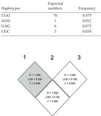
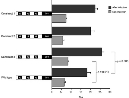
All 40 subjects were sequenced for the promoter re-gion and were analyzed for SNPs. In total, three SNPs were identified in 40 subjects, all of which had already SNP IDs: rs3024941, rs3024943 and rs3024944. Their respective genotype and allelic frequencies are sum-marized in Table I. All three of the observed SNPs were heterozygous. Construct 2 possessed two SNPs, C/G and G/A, which are linked. Four out of eight pos-sible haplotypes were found. The number of haplotypes observed and their corresponding frequencies is rep-resented (Table II). Based on haplotype frequencies, the degree of expected heterozygosity across three loci was 0.227, which corresponds closely to the observed heterozygosity, which is 0.250. The comparison of linkage disequilibrium between three loci in the pro-moter regions of the STAT6 gene locus as inferred from Haploview is represented in Fig. 1.Comparison of the activity of the three different STAT6 promoter vari-ants analyzed by luciferase activity is depicted in the Fig. 2. All four constructs could be induced by phor-bol myristate acetate/concanavalin A significantly (p < 0.0001) when compared before and after induction. The construct 3 variant had a higher luciferase activity in comparison to other two variants and remained sig-nificant in comparison to the wild type both at induced and non-induced state (p = 0.003; p = 0.016). The com-parison of expression levels of the reporter gene be-tween the wild-type alleles and construct 1 and 2 did not show statistically significant differences (p ≥ 0.50). The TF-Search online tool indicated that the SNP in
construct 2 (G/A #rs3024943) is located in a putative transcription factor site myeloid zinc finger-1 (MZF-1). The SNP in construct 1 (C/G #rs3024941) does not al-ter putative transcription factor binding sites when ex-amined, whereas the SNP variant in construct 3 (G/C #rs3024944) is located in a transcription factor binding site for the T cell factor-1 (TCF-1).
TABLE I
Genetic variants identified in the promoter regions of the signal transducer and activator of transcription 6 gene locus
Single nucleotide polymorphisms
(rs#) Polymorphism Flanking sequences Genotype
Analyzed
individuals Allele Frequencya
Allele frequency
(HapMap)b
rs3024941 C > G GCAGG[C/G]AGCAG CC
GC
33 7
C G
0.825 0.175
0.858 0.142
rs3024943 G > A CCTGG[G/A]GAGCC GG
AG
34 6
G A
0.85 0.15
0.820 0.180
rs3024944 G > C GCAGA[G/C]TTTGA GG
CG
37 3
G C
0.925 0.075
0.905 0.095
a: frequency corresponds for both allele and genotypes; b: yoruba population representing sub-Saharan African individual group.
TABLE II
Observedhaplotypes in the promoter regions of the signal
transducer and activator of transcription 6 gene locus
Haplotypes
Expected
numbers Frequency
CGG 70 0.875
GGG 1 0.012
GAG 6 0.075
CGC 3 0.038
DISCUSSION
The critical barrier in control of infections remains to be the failure of the immune system to clear para-sites, despite antigen recognition. Parasites represent a major selective force by influencing the genetic predis-position determined by immune system gene variants (Maizels 2009). Variations in the promoter regions of these immune system genes potentially modify the gene expression levels by changing specificity of transcrip-tion binding sites and/or by altering the kinetics of tran-scription initiation. In our current study, we identified three pre-described SNP variants in the studied Gabo-nese population. When compared to the National Cen-ter for Biotechnology Information HapMap database, all three observed SNP variants were found in the yo-ruba population of Nigeria representing a sub-Saharan African group. The allele frequencies described in the HapMap database corroborate with allele frequencies observed in this study (Table I). A very large cohort will remain obligatory to associate any disease severity with these identified mutations. Interestingly, we found the promoter variant rs3024944 (G/C) exhibited an altered expression of the marker gene when compared to that of the wild type at a basal level and after induction. How-ever, we did not find a difference in expression between the other two constructs compared to the wild type. The SNP in construct 2 (G/A #rs3024943) is located in a tran-scription factor binding site MZF-1. MZF-1 is a Krupple class zinc finger gene encoding a putative transcrip-tional regulator of myeloid differentiation. MZF-1 is ex-pressed in nonhaematopoietic cells as a transcriptional
repressor, whereas in haematopoietic cells it acts as a transcriptional activator (Morris et al. 1995). However, a study concluded that there was no expression of MZF-1 expression levels in Jurkat T cells (Hromas et al. 1991). Therefore the SNPs in this binding site do not lead to altered expression levels. Construct 2 (G/A #rs3024943) was linked to SNP variant (C/G #rs3024941). Although linked, the effect of MZF-1 expression levels in Jurkat T cells might be a lone contribution by construct 2 because construct 1 does not alter any of the transcription fac-tor binding sites when examined. The SNP variant in construct 3 (G/C #rs3024944) is located in a transcrip-tion factor binding site TCF-1. The TCF-1 is a mem-ber of family of genes with homology to high mobility group I (HMG) proteins (Castrop et al. 1995). This fam-ily also includes lymphocyte enhancer-binding factor-l and SRy-related HMG-box (SOX-4). TCF-1, in part with ß-catenin, plays a pivotal role in T cell activation and is specifically expressed in Tlymphocytes (Staal et al. 1999, yu et al. 2010). Therefore individuals with this SNP variant (G/C #rs3024944) may possibly have an el-evated T cell response to infections.
STAT6 gene promoter variants had been previously shown to be associated with the outcome of several hu-man diseases. A major association was inferred between a genetic variant (G/A 4219) of the 3’-UTR regulatory element of the STAT6 gene in an Ascaris infection study (Peisong et al. 2004). The study explicitly concluded that the homozygous state of this genetic variant indicated a strong association with diminished Ascaris burden in children living in an area of endemic infection. Another study concluded, however, that a promoter
phism (rs324013) in STAT6 was found to be associated with the control of infection levels in urinary schistoso-miasis (He et al. 2008). They also detected an additive effect between promoter polymorphisms rs324013 and rs1800925 in the STAT6 gene locus, and both these poly-morphisms affected transcription factor binding when analyzed by an electrophoretic mobility shift assay. A very recent study revealed that constitutive STAT6 sig-nalling in a Th cell impairs the suppressive potential of Tregs. The IL4-STAT6 signalling pathway plays a pre-dominant role in Th2 cells proliferation and of Treg cells (Pillemer et al. 2009). The proliferation of naive T cells to Treg cells is regulated or kept in check by two fac-tors, namely, transforming growth factor-beta (TGFß) and STAT6. TGFß when bound to the specific enhancer region of the FoxP3 promoter induces the conversion of naive T cells to Tregs, whereas STAT6 binding to a spe-cific silencer region of FoxP3 promoter blocks the pro-liferation of naive T cells to Tregs (Takaki et al. 2008). When it comes to infection, the role of STAT6 remains vital in the control of the Treg population. A polymor-phism in the STAT6 promoter that ably alters STAT6 expression can possibly increase proliferation of Tregs, thereby leading to a diffused immune response in the host with an elevated parasitic burden.
The identified regulatory SNPs in this current study will afford useful information for understanding the rel-evance of sequence polymorphisms in populations of different ethnic backgrounds and may serve as a basis to study parasite susceptibility in association studies. These regulatory polymorphisms in the promoter regions of STAT6 may assist in identifying potential targets for the therapeutic control of gene expression. In summary, we identified three SNP variants in the proximal promoter regions of the STAT6 gene by direct sequencing using DNA samples from 40 unrelated Gabonese individuals and investigated the functional activity of these promot-er variants. The detection of such human immune gene polymorphisms will remain crucial in a genetic makeup of the patient population to level of infection and to that of expression of Treg activity.
ACKNOWLEDGEMENTS
To A Weierich, V Galinat and V Grummes, for technical assistance.
REFERENCES
Barrett JC, Fry B, Maller J, Daly MJ 2005. Haploview: analysis and vi-sualization of LD and haplotype maps. Bioinformatics 21: 263-265. Belkaid y, Rouse BT 2005. Natural regulatory T cells in infectious
disease. Nat Immunol 6: 353-360.
Castrop J, van Wichen D, Koomans-Bitter M, van de Wetering M, de Weger R, van Dongen J, Clevers H 1995. The human TCF-1 gene
encodes a nuclear DNA-binding protein uniquely expressed in nor-mal and neoplastic T-lineage lymphocytes. Blood 86: 3050-3059. He H, Isnard A, Kouriba B, Cabantous S, Dessein A, Doumbo O,
Chevillard C 2008. A STAT6 gene polymorphism is associated with high infection levels in urinary schistosomiasis. Genes
Im-mun 9: 195-206.
Hromas R, Collins SJ, Hickstein D, Raskind W, Deaven LL, O’Hara P, Hagen FS, Kaushansky K 1991. A retinoic acid-responsive hu-man zinc finger gene, MZF-1, preferentially expressed in myeloid cells. J Biol Chem 266: 14183-14187.
Jüliger S, Bongartz M, Luty AJ, Kremsner PG, Kun JF 2003. Func-tional analysis of a promoter variant of the gene encoding the in-terferon-gamma receptor chain I. Immunogenetics 54: 675-680. Kaplan MH, Schindler U, Smiley ST, Grusby MJ 1996. STAT6 is
re-quired for mediating responses to IL-4 and for development of Th2 cells. Immunity 4: 313-319.
Maizels RM 2009. Parasite immunomodulation and polymorphisms of the immune system. J Biol 8: 62.
Morris JF, Rauscher FJ 3rd, Davis B, Klemsz M, Xu D, Tenen D, Hromas R 1995. The myeloid zinc finger gene, MZF-1, regulates the CD34 promoter in vitro. Blood 86: 3640-3647.
Peisong G, yamasaki A, Mao XQ, Enomoto T, Feng Z, Gloria-Bottini F, Bottini E, Shirakawa T, Sun D, Hopkin JM 2004. An asthma-associated genetic variant of STAT6 predicts low burden of as-caris worm infestation. Genes Immun 5: 58-62.
Pillemer BB, Qi Z, Melgert B, Oriss TB, Ray P, Ray A 2009. STAT6
activation confers upon T helper cells resistance to suppression by regulatory T cells. J Immunol 183: 155-163.
Rozen S, Skaletsky H 2000. Primer3 on the www for general users and for biologist programmers. Methods Mol Biol 132: 365-386. Sakaguchi S 2000. Regulatory T cells: key controllers of
immuno-logic self-tolerance. Cell 101: 455-458.
Sakaguchi S 2004. Naturally arising CD4+ regulatory T cells for
immunologic self-tolerance and negative control of immune re-sponses. Annu Rev Immunol 22: 531-562.
Sakaguchi S 2005. Naturally arising Foxp3-expressing CD25+ CD4+
regulatory T cells in immunological tolerance to self and non-self. Nat Immunol 6: 345-352.
Schindler C, Darnell JE Jr 1995. Transcriptional responses to polypeptide ligands: the JAK-STAT pathway. Annu Rev Biochem 64: 621-651. Staal FJ, Burgering BM, van de Wetering M, Clevers HC 1999.
Tcf-1-mediated transcription in T lymphocytes: differential role for glycogen synthase kinase-3 in fibroblasts and T cells. Int Immu-nol 11: 317-323.
Takaki H, Ichiyama K, Koga K, Chinen T, Takaesu G, Sugiyama y, Kato S, yoshimura A, Kobayashi T 2008. STAT6 inhibits TGF-beta1-mediated Foxp3 induction through direct binding to the Foxp3 promoter, which is reverted by retinoic acid receptor.
J Biol Chem 283: 14955-14962.