NORVEGICUS) BY 16S RRNA AND LIPL32 GENE SEQUENCING
Kumaresan Vedhagiri 1; Kalimuthusamy Natarajaseenivasan 1*; Shanmugarajan G. Prabhakaran 1; Joseph Selvin 1; Ramasamy Narayanan 1; Yogesh S. Shouche 2; Paluru Vijayachari3; Sivalingam Ratnam4
1
Division of Medical Microbiology, Department of Microbiology, School of Life Sciences, Bharathidasan University,
Tiruchirappalli – 620 024, Tamilnadu, India; 2National Centre for Cell Sciences, Maharastra, Pune – 411 007, India; 3Regional
Medical Research Centre, Indian Council of Medical Research, WHO Collaborative Centre for Diagnosis, Research, Reference
and Training in Leptospirosis, Port Blair – 744 101, Andaman and Nicobar Islands, India; 4Tamilnadu Veterinary and Animal
Sciences University, Chennai – 600 007, India.
Submitted: December 26, 2008; Returned to authors for corrections: July 15, 2009; Approved: August 21, 2009.
ABSTRACT
The main goal of this study was to evaluate the prevalence of leptospirosis among field rodents of
Tiruchirappalli district, Tamil Nadu, India. In total 35 field rats were trapped and tested for seroprevalence by
the microscopic agglutination test (MAT). Isolation of leptospires was performed from blood and kidney
tissues and characterized to serovar level. Genomospecies identification was carried out using 16S rRNA and
lipL32 gene sequencing. The molecular phylogeny was constructed to find out species segregation.
Seroprevalence was about 51.4 %, and the predominant serovars were Autumnalis, Javanica,
Icterohaemorrhagiae and Pomona. Two isolates from the kidneys were identified as serovar Javanica of
Serogroup Javanica, and sequence based molecular phylogeny indicated these two isolates were Leptospira
borgpetersenii.
Key words: Leptospirosis, Leptospira borgpetersenii, lipL32, 16S rRNA.
INTRODUCTION
Rodents are one of the most common carriers for
spreading of Leptospira among all mammals and were first
confirmed by isolation in Indian rats (10). Prevalence of
leptospirosis in tropical and subtropical countries is common
because the countries like India have an ideal niche for the
transmission of leptospires. The agriculture fields, especially
paddy and grain cultivating fields, are the most favorable
places for their survival and procreation. The rodent population
is high in these places because they can get their food
adequately (15). The rat is the primary carrier for spreading of
Leptospira through their excrement especially in the rice fields.
Leptospires are spread by the contaminated urine of the
infected rodents to the cattle through their grazing fields. This
greatly increases the possibilities of spreading leptospires to the
rice field workers (1, 14). In earlier days,
serological identification involved using the microscopic
agglutination test (MAT). This technique was further improved
by using specific monoclonal antibodies for serovar level
identification. Recently, the molecular biology based
identification has been introduced for the genomospecies and
so far seventeen species have been classified (2). Furthermore,
the molecular level identification will be a convenient and
simple procedure to perform when compared with the existing
techniques. The purpose of the present study is to isolate and
characterize the species level of the leptospires of the rodent
(Rattus norvegicus) using 16S rRNA and lipL32 gene
sequencing. In addition, the molecular phylogenetic analysis
were done for the 16S rRNA and lipL32 for the genomospecies
level identification.
MATERIALS AND METHODS
Study area and samples
The study area is in the rural range of the Tiruchirappalli
district located in Tamil Nadu, South India. In the study area,
temperature ranges from 28ºC to 38ºC. and the geographical
position of this area is in North latitude between 10 and 11 - 30'
and East longitude between 77 - 45' and 78 - 50'. The proposed
collection areas were entirely engaged with agricultural
activities especially cultivation of paddy, sugarcane and
groundnut. These are all productive sources for rodent breeding
and many possibilities for the pathogen spreading. The
entrapment of the rodents was attained prior to the harvesting
phase. Thirty five animals were trapped, the maximum amount
of blood collection was attained by direct heart puncture and
serum was separated, kept at -20ºC until use.
Isolation of leptospires
The isolation of Leptospira was made from direct
inoculation of two drops of blood in 5 mL of
Ellinghausen-McCullough-Johnson-Harris semisolid medium (EMJH)
(Difco-USA) with the addition of 0.15 % of agarose (Sigma)
and 10 % Bovine serum albumin (BSA) (5). The trapped field
rats were sacrificed by cervical dislocation and the body
surface was wiped with 70 % ethanol and dissected aseptically.
A sample from both kidneys were acquired using sterile rat
toothed forceps and inoculated into EMJH medium tubes,
incubated at room temperature (30ºC) in the dark and examined
under dark field illumination with intervals of 10 days to check
for the growth of leptospires for at least three months (13, 17).
When growth was perceived, successive transfer was made in
liquid and semisolid media until the growth was sufficiently
abundant.
Microscopic agglutination test (MAT)
MAT was performed on the sera samples collected from
the rats using twelve live leptospiral strains as antigens. The
strains belong to the serogroups Australis (strain
Jez Bratislava), Autumnalis (Akiyami A), Ballum (Mus 127),
Bataviae (Swart), Canicola(Hond Utrecht IV), Icterohaemorrha
giae (RGA), Grippotyphosa (Moskova V), Hebdomadis
(Hebdomadis), Javanica (Poi), Pomona (Pomona), Pyrogenes
(Pyrogenes) and Semaranga (Patoc I). MAT was done at
doubling dilutions starting from 1 in 20. Positive samples were
titrated to their end point. All the strains were maintained in
EMJH medium with periodical subculture. Seven days old
cultures free from contaminations were utilized for the
performance of MAT.
Determination of pathogenic status and virulence
The virulence nature of the isolates was determined in
laboratory animals (5). A group of four 30 days old guinea
pigs (Approximately 150 g) were inoculated intraperitoneally
with 2 x 10⁸ leptospires in a final volume of 1.0 mL. Animals
were confined in separate cages and monitored daily for the
presence of clinical signs and symptoms. The reisolation was
performed from the kidney tissues of the inoculated guinea
pigs for confirmation. All animal procedures carried out in this
study were approved by the Institutional Animal Ethical
Committee (Animal ethics approval No. BDU/ IAEC / 2008/
10).
Confirmation of the pathogenic status was also performed
by the in-vitro system at 13ºC and in the presence of
8-azaguanine (Sigma) with a concentration of 225 µg/mL (8, 9).
The pathogenic strains Jez Bratislava and Wijinberg and the
non-pathogenic strains Patoc I and Veldrat Semaranga were
densities of the cultures (14).
Serological characterization
The identification of the isolate to the serogroup level was
carried out first by MAT using a panel of 39 anti-Leptospira
rabbit anti sera representative of 25 serogroups, following the
standard procedure (3). Subsequently, the isolates were tested
with rabbit anti sera raised against 12 reference strains of 12
local circulating serovars. After the determination of the
serogroup status of the isolates these were further characterized
using mouse monoclonal antibodies (mAbs) of serogroup
Javanica. The isolates were also sent to WHO/FAO/
Collaborating Centre for Reference and Research on
Leptospirosis, Queensland, Australia for further confirmation.
DNA purification
Genomic DNA was extracted from seven days old isolates
grown at 30º C in EMJH medium and harvested by
centrifugation. The pellet was washed twice and dissolved in
500 µL solution I (10 mM Tris, 10 mM of MgCl2, 2 mM of
ethylenediaminetetra acetic acid (EDTA), 400 mM NaCl and
10 mM KCl in 500 ml of Millipore water). 50 µ L of lyzozyme
(5mg/mL dissolved in solution I) was added and followed by
incubation at 37ºC for 15 mins. Then 50 µL of 10 % SDS and 5
µL of Proteinase K (10 mg/mL) was added and incubated at
65ºC for 30 mins. 40 µL of 5 M NaCl and 32 µL of CTAB
were added and again incubated at 65ºC for 30 mins. Then
equal volume of chloroform and isoamylalcohol (24:1) were
added. The content was vortexed and centrifuged at 10,000 g
for 10 mins. The supernatant was mixed with 0.6 % volume of
chilled ethanol and kept in -70ºC for one hour. Centrifuge the
vials at 12,000 g for 30 mins, the pellet was air dried, dissolved
in 50 µL of Tris-EDTA (TE) buffer and stored at -20ºC.
PCR and gene sequencing
The species level identification was carried out by 16S
rRNA and lipL32 gene amplification using the specific primers
as per the earlier reports (6, 20). Primers 16S rRNA
(F)-5’-GGC (F)-5’-GGC GCG TCT TAA ACA TG-3’, 16S rRNA (R)-5’-
GTG CCA GCA GCC GCG GTA A-3’ and lipL32-F-5’- TTA
CCG CTC GAG GTG CTT TCG GTG GTC TGC-3’,
lipL32-R-5’- TGT TAA CCC GGG TTA CTT AGT CGC GTC
AGA-3’ were used. The 16S rRNA and lipL32 fragments were
amplified using a thermal cycler (Eppendorf, Germany).
Briefly each 50 µL PCR reaction mixture contained
approximately 50 ηg of purified DNA, 0.1 M primers, 250
M of each dNTPs (Fermentas), 3 mM MgCl2, 0.5 U of Taq
DNA Polymerase (Fermentas), in 10 mM Tris-HCl (pH 9) and
50 mM KCl. The temperature profile as one cycle at 94ºC for 3
min, 35 cycles at 94ºC for 30 s, 52ºC for 30 s, 72ºC for 1.5 min
and a final extension at 72ºC for 7 min and lipL32 gene were
amplified by initially one cycle of 94ºC for 5 min and 35 cycles
of 94ºC for 90 s, 51ºC for 90 s, 72ºC for 2 min and final
extension at 72ºC for 7 min. Amplified products were
evaluated by agarose gel electrophoresis. Before sequencing,
the 16S rRNA and lipL32 products were purified by
MontageTM PCR purification kit according to manufacturer
instructions (Millipore). The purified products were sequenced
using ABI PRISM model 377 automatic sequencer (Applied
Biosystems, USA) giving 99 percent sequence accuracy. The
sequences were aligned using bioinformatics tools and
deposited in the National Center for Biotechnology
Information (NCBI), Bethesda, USA.
Phylogenetic analysis
The 16S rRNA and lipL32 gene sequences of the rat
isolates and the highly identical sequences for 16S rRNA and
lipL32, used in this study were retrieved from NCBI database
using Basic Local Alignment Search Tool (BLASTn) and
PSI-BLAST tool with default parameters. Sequences with more
significant identity were aligned with ClustalW algorithm
implemented in Molecular Evolutionary Genetics Analysis
(MEGA 4.0) using Smith-Waterman substitution matrix and
trimmed to consensus and Unweighted Pair Group Method
with Arithmetic Mean (UPGMA) trees were constructed with
1000 bootstraps at uniform divergence rates with Jukes and
Cantor evolutionary model and 0.25 gamma distribution factors
BioEdit 7.0.4.1 (7). Posterior probability and conserved regions
among closely related sequences were carried by MEGA 4.0
and BioEdit 7.0.4.1.
RESULTS
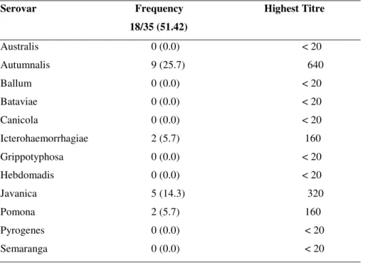
The seroprevalence of the present study revealed that,
serovar Autumnalis was the predominant serogroup followed
by Javanica, Icterohaemorrhagiae and Pomona. The percentage
seroprevalence observed was 25.7 % for Autumnalis, 14.3 %
for Javanica, 5.7 % each for Icterohaemorrhagiae and Pomona
(Table 1). In a total of 35 field rats utilized for this study two
leptospiral isolates were recovered from rat kidneys. The
isolates showed typical morphology and characteristic motility
of the genus Leptospira under dark-field microscopic
examination. These strains grew well in aerobic conditions
both in EMJH semisolid and liquid medium. They failed to
grow in tripticase soy broth in which Leptonema grow well.
The optimum temperature for their growth 28-30ºC and
the optimum pH 7.2-7.4 were recorded. The cells were difficult
to stain by the Gram’s method but stained well by silver
impregnation techniques such as Fontana’s method. The
isolates were coded as R1R and R1L, which showed moderate
growth after 20 days of incubation period. The isolates were
inoculated into guinea pigs for the determination of
pathogenicity and virulence, the inoculated guinea pigs were
died after 12-14 days from the date of inoculation and they
showed the clinical symptoms like dullness, fever and rough
coat before their death. Further, pathogenicity was confirmed
by growth at 13ºC and in the presence of 8-azaguanine. The
saprophytic strains Veldrat, Semaranga and Patoc I reached a
maximum density within 21 days at 13ºC and in the presence
of 8-azaguanine within 15 days of incubation. In contrast, the
growth of the isolates as well as the pathogenic reference
strains Wijnberg and Jez Bratislava were inhibited at 13ºC and
in the presence of 8-azaguanine even after 21 days of
incubation indicating the pathogenic nature of the isolates. The
re-isolation was successful from the kidney tissues of the
guinea pigs in EMJH semisolid medium. These findings
indicate the virulence nature of the isolated strains.
Table 1. Percentage of seroprevalence of leptospires among the field rats (Rattus norvegicus).
Serovar Frequency Highest Titre
18/35 (51.42)
Australis 0 (0.0) < 20
Autumnalis 9 (25.7) 640
Ballum 0 (0.0) < 20
Bataviae 0 (0.0) < 20
Canicola 0 (0.0) < 20
Icterohaemorrhagiae 2 (5.7) 160
Grippotyphosa 0 (0.0) < 20
Hebdomadis 0 (0.0) < 20
Javanica 5 (14.3) 320
Pomona 2 (5.7) 160
Pyrogenes 0 (0.0) < 20
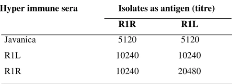
Serological characterizations of the isolates were
performed by agglutination of the isolates with group sera of
25 serovar-specific reference rabbit antisera as a first method
and in the second method the HIS raised for the isolates were
reacted against the serovar Javanica and the corresponding
antigen to determine the heterologous titre (Table 2). The
group sera assay showed a MAT titre of 1 in 5120 and 2560 for
the isolates R1L and R1R respectively for serovar Javanica and
in the second assay it was 5120 for both the isolates. Based on
these findings the two isolates were identified as serovar
Javanica of serogroup Javanica. They were screened against a
panel of mAbs of serovar Javanica for further evaluation and
confirmed as serovar Javanica of serogroup Javanica. A titre of
10240, 2560, 1280, 1280 were observed against the mAbs
98-19, 98-12, 98-8, 20-4 for the isolate R1R and it was observed
as 10240, 5120, 1280 for the isolate R1L respectively. These
findings were also confirmed with WHO/FAO Reference
Laboratory, Brisbane, Australia.
Table 2. MAT results of the hyper immune sera (HIS) of R1R and R1L against the local circulating leptospiral serovar(s)
__________________________________________________
Hyper immune sera Isolates as antigen (titre) R1R R1L Javanica 5120 5120
R1L 10240 10240
R1R 10240 20480
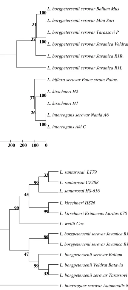
The genetic characterization for the genomospecies level
identification was performed using 16S rRNA sequencing of
two isolates exhibited 732 bp for R1R and 641 bp for R1L
respectively through BLAST alignment (EU159685,
EU159686). The alignment score of these two sequences is
200 and the score value is 1123 bits. The identities between the
sequences were 99 %. The phylogenetic pattern by UPGMA
tree is evidenced that, the isolates R1R and R1L uniquely
clustered with different serovars of L. borgpetersenii with 100
% bootstrap confidence value. The closest neighboring clusters
include strains of L. biflexa, L. kirschneri and L. interrogans
showed 100 % bootstrap confidence values. Based on the
phylogenetic analysis, the genetic nature of the isolates
confirms that they are members of L. borgpetersenii (Fig. 1).
Further the lipL32 gene sequencing of the two isolates
exhibited 715 bp for R1R and 700 bp for R1L respectively
through BLAST alignment (EU526389, EU526390). The Blast
alignment score was found that 200 among the all lipL32
gene sequences, homology was 99 to 100 % and the maximum
identity was 97 to 99 %. The phylogenetic tree evidenced that
these two isolates closely clustered with different serovars like
Ballum, Bataviae, and Tarassovi of L. borgpetersenii. The
closest neighboring groups include serovars of L. kirschneri
and strains L. santarosai clustered 99 % bootstrap confidence
value. Even though the lipL32 is highly analogous protein
present in all pathogenic Leptospira but the phylogenetic
pattern of the present study exhibited the clonality of the
sequences of the isolates with the L. borgpetersenii and due to
that it may also be used to analyze species segregation (Fig. 2).
Based on the lipL32gene sequence of the isolates, it was further
confirmed as belonged to genomospecies L. borgpetersenii.
DISCUSSION
In South India, paddy is a major source of food and most
of the lands are used for its cultivation. It also encourages
rodent propagation in the field. The study area Tiruchirappalli
is fully surrounded by agricultural fields on the river bank of
Cauvery. In the rice cultivation fields the rodent infestation is
very common and they are the major reservoir for the
dissemination of the leptospires in the surrounding
environment (15). Isolation of Leptospira from the rodent
population and species level identification by 16S rRNA and
lipL32 gene sequencing is the first of its kind from
Tiruchirappalli. In earlier studies Lahiri reported
that significant findings in the urban population of the city of
Bombay by isolation of serovar Icterohaemorrhagiae from rats
L. santarosai LT79
L. santarosai CZ288
L. santarosai HS-616
L. kirschneri HS26
L. kirschneri Erinaceus Auritus 670
L. weilii Cox
L. borgpetersenii serovar Javanica R1L
L. borgpetersenii serovar Javanica R1R
L. borgpetersenii serovar Ballum
L. borgpetersenii Veldrat Batavia
L. borgpetersenii serovar Tarassovi
L. interrogans serovar Autumnalis N2
99
88
33 99 47
33
99
45
99
L. borgpetersenii serovar Ballum Mus
L. borgpetersenii serovar Mini Sari
L. borgpetersenii serovar Tarassovi P
L. borgpetersenii serovar Javanica Veldrat
L. borgpetersenii serovar Javanica R1R.
L. borgpetersenii serovar Javanica R1L
L. biflexa serovar Patoc strain Patoc.
L. kirschneri H2
L. kirschneri H1
L. interrogans serovar Nanla A6
L. interrogans Aki C
100 100 100
100 31
37
26 37
0 100 200 300
Figure 1. Phylogenetic pattern for 16S rRNA sequences of leptospiral isolates (R1R and R1L) in
comparison with sequences of representative strains
from L. interrogans, L. kirschneri, L. borgpetersenii
and L. biflexa.
Figure 2. Phylogenetic pattern for lipL32 gene sequences of leptospiral isolates (R1R and R1L)
in comparison with sequences of representative
strains from L. interrogans, L. kirschneri, L.
mice and wistar rats were done from Chennai and the species
level identification of the rat isolates by the gene sequences is a
significant contribution to this present investigation (14). The
Random Amplified Polymorphic DNA (RAPD) fingerprinting
techniques were carried out for the species level identification
of the rat isolates of Andaman Islands as L. interrogans, which
belonged to serovar Icterohaemorrhagiae of serogroup
Icterohaemorrhagiae (16). Even though the RAPD
fingerprinting techniques are simple and easy to perform, the
consistency and reproducibility is lacking. This may be
conquered by the amplification of the specific genes,
sequencing and phylogenetic analysis for the clonality to
identify the specific species of the leptospiral isolates. Two of
the leptospiral isolates R1R and R1L were identified to the
serovar level using group sera, followed by monoclonal
antibody typing and further they were characterized up to
species level using the 16S rRNA and lipL32 gene sequencing.
From slaughtered cattle, two leptospiral strains were isolated
and identified as Canicola and Copenhageni by monoclonal
antibody typing method in Brazil (21). In the present
investigation in addition to monoclonal antibody typing the
sequence based species identification was also performed. The
lipL32 sequences were proved as conserved sequences among
the pathogenic leptospires and they were also utilized for the
species divergence using the phylogenetic approaches (6). The
phylogenetic approach between these isolates showed the
clonality, based on the findings the isolates were identified as
serovar Javanica of L. borgpetersenii. Since the isolation and
serological identification of the leptospires seems to be
cumbersome and the sequence based molecular identification
may make a way for the easy understanding of the species
diversification of the leptospires. As a first step this was
carried out for the isolates from Tiruchirappalli and it has
revealed the existence of L. borgpetersenii species in this
locality. This study also reported that, the species level
identification through molecular tools may explore the
diversification of the leptospiral species in various
geographical areas to find out prevalence and existence of the
leptospires in the clinical samples and environment. While the
Leptospira serovar Javanica has also been isolated from human
cases as well as from the rodent population, this reveals the
transmission of the leptospires from the rodent carriers as per
the present investigation (18). Obviously this approach also
leads for the establishment of the molecular based transmission
dynamics. However the L. borgpetersenii serovar Javanica is a
pathogenic strain, the pathogenicity and the virulence of the
strains could also be demonstrated during the study. A previous
study carried out in Brazil reported that L. noguchii strain Caco
as a sheep isolate and they identified using the 16S rRNA
sequencing and the virulence was demonstrated in
hamsters (4). In another study two strains of serogroup
Grippotyphosa were isolated and identified (12). The rodents
are the major contributory source for the spreading of
leptospires, the isolation of L. borgpetersenii serovar Javanica
strain R1R and R1L from Tiruchirappalli give much affordable
knowledge for the epidemiology of leptospirosis.
ACKNOWLEDGMENTS
This study was supported by grants from the Department
of Science and Technology (DST), Government of India. The
authors thank the Vice-Chancellor, Bharathidasan University
for the facilities and thank Dr. L. D. Smythe, WHO/FAO/
Collaborating centre for reference and Research on
Leptospirosis, Queensland, Australia for the serovar level
confirmation of the leptospiral isolates.
REFERENCES
1. Bahaman, A.R.; Marshall, R.B.; Blackmore, D.K.; Hathway, S.W. (1980). Isolation of Leptospira interrogans serovar Hardjo from sheep in New Zealand. New Zeal. Vet. J. 28.
2. Bharati, A.R.; Nally, J.E.; Ricaldi, J.N. (2003). Leptospirosis: a zoonotic disease of global importance. Lan. Infect. Dis. 3, 757-771.
3. Dikken, H.; Kmety, E. (1978). Serological typing methods of leptospires.
Metho. Microbiol.11, 260–295.
5. Faine, S.; Adler, B.; Bolin, C.; Perolat, P. (1999). Leptospira and leptospirosis. (2nd eds), Melbourne, Victoria, Australia. Medical Science
p. 1- 259.
6. Haake, D.A.; Chao, G.; Zuerner, R.L.; Barnett, J.K.; Barnett, D.; Mazel, M.; Matsunaga, J.; Levett, P.N.; Bolin, C.A. (2000). The leptospiral major outer membrane protein LipL32 is a lipoprotein expressed during mammalian infection. Infect. Immun. 68, 2276–285. 7. Hall, T.A. (1999). BioEdit: BioEdit: a user-friendly biological sequence
alignment editor and analysis programme for Windows 95/98/NT, Nucl. Aci. Symp. Ser. 41, 95-98.
8. Johnson, R.C.; Harris, V.G.; (1967b). Differentiation of pathogenic and saprophytic Leptospira in growth at low temperature. J. Bacteriol. 94, 27-31.
9. Johnson, R.C.; Rogers, P. (1964a). Differentiation of pathogenic and saprophytic Leptospira with 8 – Azaguanine. J. Bacteriol. 88, 1618-623. 10. Knowles, R.; Das Gupta, B.M. (1932). Leptospiral infection in Indian
rats. Ind. J. Med. Res. 54, 611-614.
11. Lahiri, M.N. (1941). Studies on Leptospira Icterohaemorrhagiae in rats in Bombay city. Ind. Med. Gaz. 76, 536-538.
12. Lilenbaum, W.; Morais, Z.M.; Gonçales, A.P.; deSouza, G.O.;
Richtzenhain, L.; Vasconcellos, S.A. (2007). First isolation of
leptospires from dairy goats in Brazil. Braz. J. Microbiol., 38, 507-510.
13. Natarajaseenivasan, K.; Ratnam, S.; Ramadass, P.; Manual, P.S.H. (1996). Persistence of dinger’s rings by Leptospira interrogans
serovar Australis in semi solid EMJH medium. Ind.Vet. J. 73, 571-572.
14. Natarajaseenivasan, K.; Ratnam. S. (1997). A probable virulence factor of Javanica isolates. Ind. J. Ani. Sci. 68, 139-140.
15. Natarajaseenivasan, K.; Boopalan, M.; Selvanayaki, K.; Suresh, S.R.; Ratnam, S. (2002). Leptospirosis among Rice Mill Workers of Salem, South India. Jap.J. Infect.Dis. 55, 170-173.
16. Natarajaseenivasan, K.; Vijayachari, P.; Sharma, S.; Roy, S.; Sugunan. A.P.; Biswas, D.; Sehgal, S.C. (2005). Phylogenetic relatedness among leptospiral strains belonging to same serovar recovered from patients with different clinical syndromes. Infect. Gent. Evol. 5, 185-191. 17. Ratnam, S.; Subramanian, S.; Adinarayanan, N. (1987). Experimental
study with leptospires in Bandicoot Bandicota bengalensis. Ind. J. Experi.Biol. 25, 105-107.
18. Saravanan, R.; Rajendran, P.; Thyagarajan, S.P. (1998). Isolation of
Leptospira Javanica from urine sample of an acute renal failure case in Chennai: India. Ind. J. Med. Microbiol. 16, 61- 63.
19. Tamura, K.; Dudley, J.; Nei, M.; Kumar, S. (2007). MEGA 4: Molecular Evolutionary Genetics Analysis (MEGA) Software Version 4.0. Mole.
Biol. Evol. 24, 1596- 599.
20. Weisberg, W.G.; Barns, S.M.; Pelleiter, D.A.; Lane, D.J. (1991). 16S ribosomal DNA amplification for phylogenetic study. J. Bacteriol. 173, 697-703.