396
Rev Soc Bras Med Trop 50(3):396-398, May-June, 2017 doi: 10.1590/0037-8682-0445-2016
Short Communication
Corresponding author: Dra. Ilana Teruszkin Balassiano.
e-mail: [email protected] Received 19 October 2016 Accepted 24 March 2017
Molecular and serological characterization of
Leptospira
kirschneri
serogroup Pomona isolated from
a human case in a Brazilian rural area
Ilana Teruszkin Balassiano
[1], Juliana Magalhães Vital-Brazil
[1],
Tatiane Mendes Varela Ramos
[1], Loeci Natalina Timm
[2]and Martha Maria Pereira
[1][1]. Laboratório de Zoonoses Bacterianas, Centro de Referência Nacional para Leptospirose, World Health Organization/Pan American Health Organization Centro Colaborador para Leptospirose - Coleção de Leptospira, Instituto Oswaldo Cruz, Fundação Oswaldo Cruz, Rio de Janeiro, RJ, Brasil.
[2]. Fundação Estadual de Produção e Pesquisa em Saúde, Instituto de Pesquisas Biológicas, Laboratório Central, Porto Alegre, RS, Brasil.
Abstract
Introduction: Leptospirosis is an important health concern in Brazil. Currently, information on the epidemiology of the disease
in the rural areas of the country is lacking. Methods: Serological and molecular techniques were used to characterize a clinical
isolate of Leptospira. Results: The strain CLEP00060, isolated from a 59-year-old man in a rural area of Rio Grande do Sul state,
Brazil, was identiied as belonging to L. kirschneri serogroup Pomona serovar Mozdok. Conclusions: This study contributes to
the local epidemiological knowledge of leptospirosis, prevention of the disease by vaccines, and improvements in its diagnosis.
Keywords: Leptospira kirschneri. Leptospirosis. Typing.
Leptospirosis is a worldwide zoonosis, being more common in tropical regions and predominantly found in impoverished populations inhabiting developing countries, where incidence peaks are observed during the raining season1. Although leptospirosis is currently recognized as a disease of epidemic potential with a signiicant impact on public health in many countries, it remains neglected2. This can be explained by its
common incidence in areas where socioeconomic (poverty, lack of water and sanitation, poor housing conditions) and environmental (heavy rains or loods) factors are crucial for the maintenance of leptospires and subsequent occurrence of the disease2,3.
Although considered the deinitive diagnosis for leptospirosis, the isolation of Leptospira is not a timely aid for the clinical
care of human patients, being a method rarely achieved4.
Moreover, the identiication of clinical isolates at the serovar level is a laborious process and is restricted to a few reference laboratories1.
Despite these challenges, identifying the infective serovar is very important for diagnosis and epidemiological studies, because it can provide information related to the host reservoirs
involved in pathogen transmission and, therefore, contribute to the adoption of multidisciplinary control strategies2-4.
Concerning the epidemiology of leptospirosis, there is a lack of information on the countryside areas of Brazil, compared to that on the urban areas where serovars Copenhageni and Icterohaemorrhagiae are predominant2,5. However, it should
be highlighted that there are reports of other serovars found in different species of domestic and wild animals in those areas6,7.
Leptospirosis is an important public health problem in the State of Rio Grande do Sul (Southern Brazil), with an average of 428 cases reported annually. A study conducted using the One Health approach showed that rural populations of the state have an approximately eight times higher risk of contracting leptospirosis than their urban counterparts do, even though the number of reported cases was high in both areas2.
In the present study, we demonstrate the identiication of a clinical isolate obtained from a human anicteric case in a rural area of Southern Brazil.
397
Balassiano IT, et al -Characterization of L. kirschneri
Dice (Opt 1.50%) (Tol 1.5%.2.5%) (H>0.0% S>0.0%) [0.0%.100.0%]
Not I Not I
65 70 75 80 85 90 95 100
L. kirshneri serovar Mozdok strain 5621
CLEP 00060
L. santarosai serovar Tropica strain CZ 229
L. interrogans serovar Pomona strain Pomona
L. interrogans serovar Pomona strain Mezzano
L. interrogans serovar Monjakov strain Monjakov
L. interrogans serovar Pomona strain Comelli
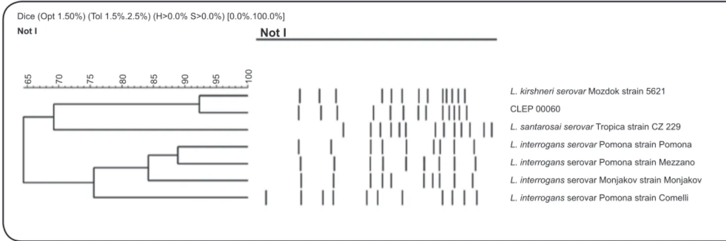
FIGURE 1 - Dendrogram generated by PFGE analysis showing the relationship among the clinical isolate CLEP 00060 and some reference strains from the serogroup Pomona. L.: Leptospira; PFGE: Pulsed Field Gel Electrophoresis; CLEP: Collection of Leptospira.
titers obtained with MAT were as follows: Icterohaemorrhagiae, 400; Australis, 200; and Pomona, 100.
Blood culture was positive for the presence of Leptospira,
visualized using dark field microscopy8. The culture was
purified, maintained by subculturing in Ellinghausen-McCullough-Johnson-Harris (EMJH) medium8, and stored at
the Collection of Leptospira [(CLEP) Oswaldo Cruz Institute/ FIOCRUZ)] under the registration number CLEP 00060.
Microscopic agglutination test was also carried out against reference antisera (obtained from the Royal Tropical Institute, Netherlands) representative of the 15 most prevalent serogroups in Brazil: Icterohaemorrhagiae, Canicola, Grippotyphosa, Pomona, Australis, Bataviae, Ballum, Cynopteri, Javanica, Panama, Pyrogenes, Sejroe, Tarassovi, Autumnalis, and Hebdomadis. The results demonstrated the antigenic relationship between CLEP 00060 and the Pomona serogroup (data not shown).
To identify the strain at the genomospecies level, we used different methodologies previously described as alternative tools to overcome the complex relatedness among serogroups, serovars, and species of Leptospira9-11.
Multilocus sequence typing (MLST)11 was performed and puriied amplicons were sequenced using the Platform of deoxyribonucleic acid (DNA) Sequencing - RPT01A-PDTIS/ FIOCRUZ (http://plataformas.cdts.iocruz.br/). Sequences were aligned with Clustal W and analyzed at the MLST website (http://pubmlst.org/leptospira). The analysis revealed that CLEP 00060 could be clustered with 100% similarity in the ST 117 and most likely belongs to Pomona serogroup (data not shown). Pulsed Field Gel Electrophoresis (PFGE)12 was performed
using the restriction enzyme NotI (Sinapse Biotecnologia,
Brazil). Fingerprints of the clinical isolate and different reference strains from serogroup Pomona were analyzed using GelCompar (Applied Maths) software, and the patterns produced were compared with the Dice coeficient and clustered using the unweighted pair-group method using arithmetic
averages method. The dendrogram revealed a similarity index of approximately 92% between CLEP 00060 and strain 5621 (L. kirschneri serogroup Pomona serovar Mozdok)
(Figure 1), indicating that these two strains belong to closely
related serovars, according to the previously established criteria for PFGE interpretation13.
Collection of Leptospira 00060 and two reference strains (L. kirschneri serogroup Grippotyphosa serovar Grippotyphosa
strain Moskva V and L. interrogans serogroup Pomona serovar
Pomona strain Pomona) were submitted to DNA extraction, followed by laB ampliication by polymerase chain reaction (PCR). The amplicons were digested separately by the restriction enzymes HindIII and HaeIII (Sinapse Biotecnologia, Brazil) and
the ingerprints were analyzed9. DNA was also used to amplify the rrs gene, and the ampliied products were sequenced10,14.
Sequences were compared with reference Leptospira sequences
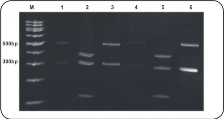
in the genome database of the National Center for Biotechnology Information (NCBI), using the basic local alignment search tool (BLAST) (http://blast.ncbi.nlm.nih.gov/Blast.cgi). The fingerprints generated by flaB-restriction fragment length
polymorphism (RFLP) clearly showed the genetic relationship between the clinical isolate and L. kirschneri (Figure 2).
Phylogenetic studies based on 16S rRNA gene sequences conirmed the result (data not shown).
Taken together, we assume that the isolate CLEP 00060 belongs to L. kirschneri serogroup Pomona and is closely
related to serovar Mozdok, although more reined molecular methods, such as secY sequencing, and a comparison with a
larger number of reference strains at PFGE could improve the identiication. Even though patient sera presented low titers against serogroup Pomona in MAT, the identiication of the strain can still be inferred because MAT presents low accuracy (about 33%) considering the correct prediction of the infective serogroup/serovar15.
Based on the available information, serovar Mozdok is rarely isolated from humans in Brazil7. This is only the second
398
Rev Soc Bras Med Trop 50(3):396-398, May-June, 2017
M 1 2 3 4 5 6
500bp
300bp
FIGURE 2 - Polyacrylamide gel electrophoresis (5%) of the PCR products resulting from the digestion of laB by the restriction endonucleases
HindIII and HaeIII. M: 100bp DNA ladder (Sinapse Biotecnologia, Brazil); 1:L. kirschneri reference strain Moskva V; 2: L. interrogans reference strain Pomona; 3: strain CLEP 00060 (1-3 digested with HaeIII); 4:L. kirschneri reference strain Moskva V; 5:L. interrogans reference strain Pomona; 6: strain
CLEP 00060 (4-6 digested with HindIII). PCR: polymerase chain reaction;
DNA: deoxyribonucleic acid; L.: Leptospira;CLEP: Collection of Leptospira.
Mozdok from a human case in a rural area of Brazil. The irst report7 describes the characterization of two isolates, from
human and canine cases, in Pelotas, also in Rio Grande do Sul and about 240km from Santa Vitória do Palmar. Besides, the isolation of strains belonging to serogroup Pomona is more frequent in animals than in humans. Our inding reinforces the need for further studies with larger numbers of strains to deine the epidemiological situation of leptospirosis in the rural areas of Brazil. It should be noted that awareness about the presence of uncommon serogroups and serovars in our territory would allow, according to the One Health approach, the prevention and control of leptospirosis by employing speciic measures in accordance with our socio-economic scenario, bringing a long-term beneit to the Brazilian population.
Acknowledgements
The authors acknowledge Elisabete Coli for laboratory assistance, André Felipe Mercês Santos for PFGE dendrogram production, and Dr. Eliane de Oliveira Ferreira for revising the text. We are also grateful for access to the Leptospira MLST database, which is located at Imperial College London and is funded by the Wellcome Trust, and the Platform of DNA Sequencing - RPT01A-PDTIS/FIOCRUZ for sequencing procedures.
Financial support
Secretaria de Vigilância em Saúde, Ministério da Saúde.
Conlicts of interest
The authors declare that have no conlicts of interest.
REFERENCES
1. Adler B, de la Peña Moctezuma A. Leptospira and leptospirosis. Vet
Microbiol. 2010; 40 (3-4): 287-96.
2. Schneider MC, Najera P, Pereira MM, Machado G, Dos Anjos CB, Rodrigues RO, et al. Leptospirosis in Rio Grande do Sul, Brazil: an ecosystem approach in the animal-human interface. PLoS Negl Trop Dis. 2015;9(11):e0004095.
3. Ko AI, Goarant C, Picardeau M. Leptospira: the dawn of the molecular genetics era for an emerging zoonotic pathogen. Nat Rev Microbiol. 2009;7(10):736-47.
4. Chiani Y, Jacob P, Varni V, Landolt N, Schmeling MF, Pujato N, et al. Isolation and clinical sample typing of human leptospirosis cases
in Argentina. Infect Genet Evol. 2016; 37: 245-51.
5. Miraglia F, Matsuo M, Morais ZM, Dellagostin OA, Seixas FK, Freitas JC, et al. Molecular characterization, serotyping, and antibioticsusceptibility proile of Leptospira interrogansserovar Copenhageni isolates from Brazil. Diagn Microbiol Infect Dis.
2013;77(3):195-9.
6. Cosate MR, Barouni AS, Moreira EC, Veloso IF, Gomes MT, Salas CE. Molecularcharacterization by LSSP-PCR and DNAsequencing of a pathogenic isolate of Leptospira interrogans from Brazil.
Zoonoses Public Hlth. 2012;59(6):379-88.
7. da Cunha CE, Felix SR, Neto AC, Campello-Felix A, Kremer FS, Monte LG, et al. Infection with Leptospira kirschneri serovar Mozdok: irst report from the southern hemisphere. Am J Trop Med Hyg. 2016;94 (3):519-21.
8. World Health Organization (WHO). Human leptospirosis: guidance for diagnosis, surveillance and control. Geneva: WHO; 2003. 109p.
9. Kawabata H, Dancel LA, Villanueva SY, Yanagihara Y, Koizumi
N, Watanabe H. laB-polymerase chain reaction (laB-PCR) and
its restriction fragment length polymorphism (RFLP) analysis are
an eficient tool for detection and identiication of Leptospira spp. Microbiol Immunol. 2001;45(6):491-6.
10. Fenner JS, Anjum MF, Randall LP, Pritchard GC, Wu G, Errington J, et al. Analysis of 16S rDNA sequences from pathogenic Leptospira serovars and use of single nucleotide polymorphisms for rapid speciation by D-HPLC. Res Vet Sci. 2010;89(1):48-57.
11. Boonsilp S, Thaipadungpanit J, Amornchai P, Wuthiekanun V,
Bailey MS, Holden MT, et al. A single multilocus sequence typing (MLST) scheme for seven pathogenic Leptospira species. PLoS Negl Trop Dis. 2013;7(1):e1954.
12. Ribeiro RL, Machry L, Brazil JM, Ramos TM, Avelar KE, Pereira MM. Technical improvement to prevent DNA degradation of Leptospira spp. in pulsed ield gel electrophoresis. Lett Appl Microbiol. 2009;49(2):289-91.
13. Tenover FC, Arbeit RD, Goering RV, Mickelsen PA, Murray BE, Persing DH, Swaminathan B. Interpreting chromosomal DNA restriction patterns produced by pulsed-ield gel electrophoresis: criteria for bacterial strain typing. J Clin Microbiol. 1995;33(9):2233-9. 14. Weisburg WG, Barns SM, Pelletier DA, Lane DJ. 16S ribosomal DNA