Relationship between retinal sensitivity and disease
activity in patients with psoriasis vulgaris
Helin Deniz Demir,IGo¨knur Kalkan,IISemiha Kurt,IIIAlper Gu¨nes¸,IEngin Sezer,IVU¨ nal ErkorkmazV
IGaziosmanpas¸a University, Faculty of Medicine, Department of Ophthalmology, Tokat, Turkey.IIYildirim Beyazit University, Faculty of Medicine, Department of Dermatology, Ankara, Turkey.IIIGaziosmanpas¸a University, Faculty of Medicine, Department of Neurology, Tokat, Turkey.IVAcibadem University, Faculty of Medicine, Department of Demartology, Istanbul, Turkey.VSakarya University, Faculty of Medicine, Department of Biostatistics, Sakarya, Turkey.
OBJECTIVES:Psoriasis is a hyperproliferative chronic inflammatory skin disease of unknown etiology and ocular structures and visual pathways can also be affected during the course of this disease. Subclinical optic neuritis has previously been observed in psoriatic patients in visual evoked potential studies. This trial was designed to evaluate retinal sensitivity in patients with psoriasis vulgaris.
METHODS:A total of 40 eyes of 40 patients with chronic plaque-type psoriasis and 40 eyes of 40 age- and sex-matched control subjects were included in this study. The diagnosis of psoriasis was confirmed by skin biopsy. The severity was determined using the Psoriasis Area and Severity Index and the duration of the disease was recorded. After a full ophthalmological examination, including tests for color vision and pupil reactions, the visual field of each subject was assessed using both standard achromatic perimetry and short wavelength automated perimetry.
RESULTS:The mean Psoriasis Area and Severity Index was 22.05¡6.409. There were no significant differences in the visual field parameters of subjects versus controls using either method. There were correlations
between disease severity and the mean deviations in standard achromatic perimetry and short wavelength automated perimetry and between disease severity and the corrected pattern standard deviation and pattern standard deviation of short wavelength automated perimetry (r = -0.363, r = -0.399, r = 0.515 and r = 0.369, respectively).
CONCLUSIONS: Retinal sensitivity appears to be affected by the severity of psoriasis vulgaris.
KEYWORDS: Psoriasis; Psoriasis Area and Severity Index; Standard Achromatic Perimetry; Short Wavelength Automated Perimetry; Visual Field.
Demir HD, Kalkan G, Kurt S, Gu¨nes¸ A, Sezer E, Erkorkmaz U¨. Relationship between retinal sensitivity and disease activity in patients with psoriasis vulgaris. Clinics. 2015;70(1):14-17.
Received for publication onSeptember 26, 2014;First review completed onNovember 7, 2014;Accepted for publication onNovember 7, 2014
E-mail: helindeniz@hotmail.com
& INTRODUCTION
Psoriasis is a hyperproliferative chronic inflammatory skin disease of unknown etiology. It is characterized by red and scaly plaques caused by epidermal hyperproliferation. It has a complex pathogenesis including immune system alterations and systemic and/or local factors. Psychological stress, focal infections and some drugs as well as the activation of leucocytes, which control cellular immunity and T cell-dependent inflammatory processes, cause the growth of epidermal and vascular cells in psoriatic skin lesions (1-6).
Ocular structures and visual pathways can also be affected in patients suffering from psoriasis (4,7). Subclinical optic neuritis has previously been shown in psoriatic patients in visual evoked potential studies. Elongation of P100 latency and a reduction in the response amplitude, which designate myelin and axonal involvement of the optic nerve, have also been observed (8,9). Electroretinography responses revealed impairment in overall retinal electrophysiologic functions (10). Visual field assessment is a functional measurement method that documents central and peripheral sensitivity of the retina in both pathological and normal situations (11). Standard achromatic perimetry (SAP) and short wavelength auto-mated perimetry (SWAP) use white and blue light stimuli, respectively, of different sizes and intensities to evaluate the retinal sensitivity and SWAP was found to be more sensitive for detecting early visual field changes. In light of these findings, the objective of this study was to examine the visual field in psoriatic patients without any pathology of the retina and/or optic nerve with the aim of evaluating retinal sensitivity.
Copyrightß2015CLINICS– This is an Open Access article distributed under
the terms of the Creative Commons Attribution Non-Commercial License (http:// creativecommons.org/licenses/by-nc/3.0/) which permits unrestricted non-commercial use, distribution, and reproduction in any medium, provided the original work is properly cited.
No potential conflict of interest was reported.
DOI:10.6061/clinics/2015(01)03
CLINICAL SCIENCE
& MATERIALS AND METHODS
A total of 40 eyes of 40 patients with newly diagnosed plaque-type psoriasis and 40 eyes of 40 age- and sex-matched control subjects were included in the study. The diagnosis of psoriasis was based on both histopathological and clinical findings. The severity was determined using the Psoriasis Area and Severity Index (PASI) and the duration of the disease recorded. Possible reasons for exclusion included the following: a history of neurological diseases, space-occupying lesions, optic neuropathy, high refractive errors, glaucoma, retinal pathology, color vision defects and cataracts, the use of systemic and/or topical retinoic acid or immunosuppressive medication, and undergoing ultravio-let therapy. Each patient underwent a full ophthalmological examination, including measurement of pupil reactions and application of the Ishihara test for color vision.
All visual field tests were performed using the central 30-2 threshold strategy with a Humphrey Field Analyzer (750i; Humphrey Instruments, San Leandro, California, USA) with SAP (using a Goldmann size III stimulus) and SWAP (using a Goldmann size V stimulus). Each test was performed by the same technician and the patients were instructed to rest before the examination of the other eye. For each subject, only one eye with reliable visual field parameters (fixation loss of less than 20%, false positive rate of less than 33%, false negative rate of less than 33%) was included in the study. Global indices (mean deviation (MD), pattern standard deviation (PSD), short-term fluctuation (SF) and corrected pattern standard deviation (CPSD)) were evalu-ated. This study was conducted according to the guidelines of the Declaration of Helsinki and patients provided their informed consent after the nature and purpose of the study were fully explained to them.
The Kolmogorov-Smirnov test was used to evaluate whether the variables were normally distributed. A two-tailed independent samples t test was used to compare the ophthalmologic parameters between the experimental and control groups. A paired sample t-test was used to compare the ophthalmologic parameters between the SAP and SWAP methods (separately for each group). A repeated measures two-way ANOVA was used to analyze differences in the methods between the experimental and control groups. The Pearson correlation coefficient was calculated to determine the significance of the association between two random variables. Continuous variables are presented as the mean
¡-standard deviation. Categorical variables were compared using the chi-square test and presented as numerical variables and percentages. Ap-value of ,0.05 was consid-ered significant. Analyses were performed using commercial software (IBM SPSS Statistics 19; SPSS Inc., an IBM Co., Somers, NY, USA).
& RESULTS
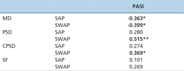
Demographic data are summarized in Table 1. There were no statistically significant intergroup differences in the age or gender of the patients. None of the study patients has systemic signs of psoriasis. The ophthalmologic examina-tions, including color vision tests and pupil reacexamina-tions, revealed unremarkable results in both groups. The best corrected visual acuity was 20/20 in both study groups and none of the study subjects presented with retinal and/or optic nerve pathology or ocular pathology. The visual field parameters are summarized in Table 2. No significant differences were observed between the two groups; how-ever, correlations were noted between disease severity (PASI) and the mean deviations of SAP and SWAP and between PASI and the CPSD and PSD of SWAP (r = -0.363, r = -0.399, r = 0.515, and r = 0.369, respectively) (Table 3).
& DISCUSSION
Psoriasis is an organ-specific autoimmune disease that is characterized by exacerbation and remission (1,4). Psoriasis activity increases with psychological stress, which has been explained by the common origin of keratinocytes and nervous cells during embryogenesis (3,8,9). Any part of the eye may be affected by psoriasis: the eyelids, con-junctiva, cornea, uvea, retina, optic nerve and lens may be involved in the disease process. Ocular symptoms are reported to occur during exacerbation of the disease (4,7). The current gold standard for the assessment of psoriasis severity is PASI, which evaluates the redness, thickness and scaliness of the lesions. PASI is a scoring system that takes into account the localization, extent and severity of the skin
Table 1 -Demographic data of patients in the experimental and control groups.
Control (n = 40) Experimental (n = 40) p-value
Gender Female 22 (55.0) 25 (62.5) 0.650
Male 18 (45.0) 15 (37.5)
Age 40.60¡11.17 39.03¡14.84 0.593
Disease duration (years) - 8.13¡7.39
-PASI - 22.05¡6.40
-The data are presented as n (%) or as the mean¡standard deviation.
PASI: Psoriasis Activity and Severity Index.
Table 2 -Visual parameters of patients in the
experimental and control groups obtained via standard achromatic perimetry and short wavelength automated perimetry.
Control (n = 40) Experimental (n = 40) p-value
MD SAP -3.30¡2.12 -3.73¡2.19 0.376
SWAP -7.75¡4.17 -7.80¡3.40 0.953
PSD SAP 3.21¡1.34 3.23¡1.66 0.953
SWAP 3.55¡0.97 3.48¡0.97 0.749
CPSD SAP 2.23¡1.62 2.42¡1.85 0.633
SWAP 2.00¡1.44 2.32¡1.32 0.313
SF SAP 1.97¡0.71 1.70¡0.66 0.075
SWAP 2.33¡0.72 2.05¡0.72 0.092 The data are presented as the mean¡standard deviation.
SAP: standard achromatic perimetry; SWAP: short wavelength automated perimetry; MD: mean deviation; PSD: pattern standard deviation; SF: short-term fluctuation; CPSD: corrected pattern standard deviation.
CLINICS 2015;70(1):14-17 Psoriasis and visual field
Demir HD et al.
signs and provides a single score for psoriasis severity ranging from 0 to 72 (12-14).
Abnormalities in the visual field may be observed in patients with normal central vision. In recent years, the sensitivity of SWAP was compared to that of SAP. SWAP was found to be more sensitive for detecting early glaucomatous and neuro-ophthalmologic visual field changes and was able to reveal functional abnormalities in the retina and optic nerve in patients with normal ocular structures (15-18).
To the best of our knowledge, the visual field has not yet been studied in psoriasis patients. We evaluated the visual field of the study group using both SAP and SWAP and we did not detect any statistically significant difference between patients and controls; however, we found a correlation between the visual field parameters and PASI. Our study group consisted of patients with moderate psoriasis, which might explain the statistically insignificant intergroup difference in the visual field parameters. When we examined the correlation between visual field para-meters and disease activity and severity, we found a negative correlation between the MD of both visual field analyses and PASI. Retinal sensitivity appears to decrease when psoriasis is exacerbated. We also found that the CPSD and PSD of SWAP were positively correlated with PASI, which may indicate localized defects in the visual field during exacerbation periods. SWAP appears to be more sensitive for detecting early functional changes in severe and active psoriasis.
Psoriasis is known as a skin disease and it has been shown to have a complex pathophysiology and a strong relation-ship with proinflammatory cytokines, including tumor necrosis factor-alpha (TNF-a), interferon-gamma (IFN-c)
and interleukins 6 and 8 (IL-6 and IL-8). Although one of the limitations of the current study is the lack of data regarding plasma proinflammatory cytokine levels of the study subjects, the role of cytokines has been extensively studied in patients with psoriasis. Serum TNF-a and IFN-c levels
were significantly higher in patients with active psoriasis and these high serum levels were positively correlated with the clinical severity and activity of the disease (19,20). The serum levels of these cytokines were reported to serve as a follow-up marker for monitoring disease severity (19,20). Overexpression of TNF-a has been demonstrated in the
plasma and skin lesions of psoriatic patients (1,5,6,19,20) and anti-TNF-a therapy has gained popularity in recent
years (1,6). TNF-a, an immunomediator and
proinflamma-tory cytokine, is a potent neurotoxic substance that induces apoptotic cell death (21-25). IFN-cplays an important role in
autoimmune and infectious diseases (26). Animal studies have shown that IFN-c induces apoptosis in the retinal
ganglion cell layer, resulting in photoreceptor loss and disturbance of the axonal transport system of the affected neurons (26,27). In addition, IL-8 induced cell death in cultured neurons, while IL-6 stimulated oxidative stress (28-31). The neurotoxic effects of these proinflammatory cytokines have been implicated in the pathogenesis of several central nervous system diseases, retinal ganglion cell damage and optic nerve crush (21,22,28).
The inflammatory response and likely the increased plasma levels of circulating proinflammatory cytokines, in active and severe psoriasis seem to affect retinal functions and decrease retinal sensitivity. Although the main target of psoriasis is the skin, based on the results of the current study, patients with psoriasis should undergo regular ophthalmological examinations to examine their retinal functions and identify potential ocular involvement. Additional studies with a larger number of participants should be performed to evaluate the retinal involvement and roles of these cytokines in psoriatic patients.
& AUTHOR CONTRIBUTIONS
Demir HD and Kalkan G collected data from the patients and wrote the manuscript. Kurt S wrote the manuscript. Gu¨nes¸ A revised the manuscript provided assistance to the manuscript writing and technical support. Sezer E collected the patient data. Erkorkmaz U¨ performed the data statistical evaluation.
& REFERENCES
1. Lowes MA, Bowcock AM, Krueger JG. Pathogenesis and therapy of psoriasis.
Nature. 2007;445(7130):866-73, http://dx.doi.org/10.1038/nature05663. 2. Coimbra S, Figueiredo A, Santos-Silva A. Brodalumab: an evidence-based
review of its potential in the treatment of moderate-to-severe psoriasis_ Core Evid. 2014;21(9):89-97, http://dx.doi.org/10.2147/CE.S33940. 3. Peter CM, van de Kerkhof, PEJ van Erp. Pathogenesis; in Peter CM. van
de Kerkhof (ed):Textbook of Psoriasis 2ndedt. Blackwell publishing: UK,
2003 pp 83-109.
4. Her Y, Lim JW, Han SH. Dry eye and tear film functions in patients with psoriasis. Jpn J Ophthalmol. 2013;57(4):341-6.
5. Rich SJ, Bello-Quintero CE. Advancements in the treatment of psoriasis: role of biologic agents.J Manag Care Pharm. 2004;10(4):318-25. 6. Schottelius AJ, Moldawer LL, Dinarello CA, Asadullah K, et al. Biology
of tumor necrosis factor-alpha- implications for psoriasis.Exp Dermatol. 2004;13(4):193-222, http://dx.doi.org/10.1111/j.0906-6705.2004.00205.x. 7. Rehal B, Modjtahedi BS, Morse LS, Schwab IR, Maibach HI. Ocular
psoriasis.J Am Acad Dermatol. 2011;65(6):1202-12, http://dx.doi.org/10. 1016/j.jaad.2010.10.032.
8. Grzybowski A, Grzybowski G, Druzdz A, Zaba R. Visual evoked potentials in patients with psoriasis vulgaris.Doc Ophthalmol. 2001;103(3):187-94, http://dx.doi.org/10.1023/A:1013059807998.
9. Perossini M, Turio E, Perossini T, Romagnoli M, et al. Pattern VEP alterations in psoriatic patients may indicate a sub clinic optic neuritis.
Doc Ophthalmol. 2005;110(2-3):203-7, http://dx.doi.org/10.1007/s10633-005-4830-1.
10. Shoeibi N, Taheri AR, Nikandish M, Omidtabrizi A, Khosravi N. Electrophysiologic evaluation of retinal function in patients with psoriasis and vitiligo. Doc Ophthalmol. 2014;128(2):131-6, http://dx. doi.org/10.1007/s10633-014-9425-2.
11. Menghini M, Duncan JL. Diagnosis and complementary examinations; Casaroli -Marano RP, Zarbin MA (eds): Cell-Based Therapy for Retinal Degenerative Disease. Dev Ophthalmol, Basel, Karger. 2014, pp:53-61. 12. Langley RG, Ellis CN. Evaluating psoriasis with Psoriasis Area and
Severity Index, Psoriasis Global Assessment, and Lattice System Physician’s Global Assessment. J Am Acad Dermatol. 2004;51(4):563-9, http://dx.doi.org/10.1016/j.jaad.2004.04.012.
13. Carlin CS, Feldman SR, Krueger JG, Menter A, Krueger GG. A 50% reduction in the Psoriasis Area and Severity Index (PASI 50) is a clinically significant endpoint in the assessment of psoriasis.J Am Acad
Table 3 -Correlation between visual field parameters and the psoriasis activity and severity index (PASI).
PASI
MD SAP -0.363*
SWAP -0.399*
PSD SAP 0.280
SWAP 0.515**
CPSD SAP 0.274
SWAP 0.369*
SF SAP 0.101
SWAP 0.269
*:p,0.05.
SAP: standard achromatic perimetry; SWAP: short wavelength automated perimetry; MD: mean deviation; PSD: pattern standard deviation; SF: short-term fluctuation; CPSD: corrected pattern standard deviation.
Psoriasis and visual field
Demir HD et al. CLINICS 2015;70(1):14-17
Dermatol. 2004;50(6):859-66, http://dx.doi.org/10.1016/j.jaad.2003.09. 014.
14. Scha¨fer I, Hacker J, Rustenbach SJ, Radtke M, Franzke N, Augustin M. Concordance of the Psoriasis Area and Severity Index (PASI) and patient-reported outcomes in psoriasis treatment.Eur J Dermatol. 2010; 20(1):62-7.
15. Wild JM. Short wavelenght automated perimetry.Acta Ophthalmol Scan. 2001;79(6):546-59, http://dx.doi.org/10.1034/j.1600-0420.2001.790602.x. 16. Heijl A. Ordering a test; in Heijl A (ed): Essential Perimetry 3thedt. Jena:
Germany: Carl Zeiss Meditec Inc., 2002. pp 25-43.
17. Keltner JL, Johnson CA. Short-wavelength automated perimetry in neuro-ophthalmologic disorders.Arch Ophthalmol. 1995;113(4):475-81, http:// dx.doi.org/10.1001/archopht.1995.01100040095033.
18. Fujimoto N, Kubota M, Saeki N, Adachi-Usami E. Use of blue-on-yellow perimetry for detection of sectoranopia.Eye. 2004;18(3):338-41, http:// dx.doi.org/10.1038/sj.eye.6700665.
19. Abdel-Hamid MF, Aly DG, Saad NE, Emam HM, Emam HM, Ayoub DF. Serum levels of interleukin-8, tumor necrosis factor-aandc-interferon in Egyptian psoriatic patients and correlation with disease severity.
J Dermatol. 2011;38(5):442-6, http://dx.doi.org/10.1111/j.1346-8138.2010. 01018.x.
20. Arican O, Aral M, Sasmaz S, Ciragil P. Serum levels of TNF-alpha, IFN-gamma, IL-6, IL-8, IL-12, IL-17, and IL-18 in patients with active psoriasis and correlation with disease severity.Mediators Inflamm. 2005;24(5):273-9, http://dx.doi.org/10.1155/MI.2005.273.
21. Tezel G, Li LY, Patil RV, Wax MB. TNF-alpha and TNF-alpha receptor-1 in the retina of normal and glaucomatous eyes.Invest Ophthalmol Vis Sci. 2001;42(8):1787-94.
22. Tezel G, Yang X, Yang J, Wax MB. Role of tumor necrosis factor receptor-1 in the death of retinal ganglion cells following optic nerve crush injury in mice.Brain Res. 2004;996(2):202-12, http://dx.doi.org/10.1016/j.brainres. 2003.10.029.
23. Wax MB, Tezel G. Immunoregulation of retinal ganglion cell fate in glaucoma.Exp Eye Res. 2009;88(4):825-30, http://dx.doi.org/10.1016/j. exer.2009.02.005.
24. Bai Y, Shi Z, Zhuo Y, Liu J, Malakhov A, Ko E, Burgess K, et al. In glaucoma the upregulated truncated TrkC.T1 receptor isoform in glia causes increased TNF-alpha production, leading to retinal ganglion cell death.Invest Ophthalmol Vis Sci. 2010;51(12):6639-51, http://dx.doi.org/ 10.1167/iovs.10-5431.
25. Levin LA, Gordon LK. Retinal ganglion cell disorders: types and treatments.Prog Retin Eye Res. 2002;21(5):465-84, http://dx.doi.org/10. 1016/S1350-9462(02)00012-5.
26. Egwuagu CE, Mahdi RM, Chan CC, Sztein J, Li W, Smith JA, et al. Expression of interferon-gamma in the lens exacerbates anterior uveitis and induces retinal degenerative changes in transgenic Lewis rats.Clin Immunol. 1999;91(2):196-205, http://dx.doi.org/10.1006/clim.1999.4701. 27. Geiger K, Howes E, Gallina M, Huang XJ, Travis GH, Sarvetnick N.
Transgenic mice expressing IFN-gamma in the retina develop inflamma-tion of the eye and photoreceptor loss. Invest Ophthalmol Vis Sci. 1994;35(6):2667-81.
28. Huang P, Zhang SS, Zhang C. The two sides of cytokine signaling and glaucomatous optic neuropathy. J Ocul Biol Dis Infor. 2009;2(2):78-83, http://dx.doi.org/10.1007/s12177-009-9026-6.
29. Kuchtey J, Rezaei KA, Jaru-Ampornpan P, Sternberg P Jr, Kuchtey RW. Multiplex cytokine analysis reveals elevated concentration of interleu-kin-8 in glaucomatous aqueous humor. Invest Ophthalmol Vis Sci. 2010;51(12):6441-7, http://dx.doi.org/10.1167/iovs.10-5216.
30. Thirumangalakudi L, Yin L, Rao HV, Grammas P. IL-8 induces expression of matrix metalloproteinases, cell cycle and pro-apoptotic proteins, and cell death in cultured neurons.J Alzheimers Dis. 2007;11 (3):305-11.
31. Wu WC, Hu DN, Gao HX, Chen M, Wang D, Rosen R, et al. Subtoxic levels hydrogen peroxide-induced production of interleukin-6 by retinal pigment epithelial cells.Mol Vis. 2010;16:1864-73.
CLINICS 2015;70(1):14-17 Psoriasis and visual field
Demir HD et al.

