Autonomic and Renal Alterations in the Offspring of
Sleep-Restricted Mothers During Late Pregnancy
Joyce R.S. Raimundo,ICassia T. Bergamaschi,IRuy R. Campos,IBeatriz D. Palma,II,IIISergio Tufik,II Guiomar N. GomesI,*
IEscola Paulista de Medicina – UNIFESP, Departamento de Fisiologia, Sa˜o Paulo/SP, Brazil.IIEscola Paulista de Medicina – UNIFESP, Departamento de
Psicobiologia, Sa˜o Paulo/SP, Brazil.IIICentro Universita´rio Sa˜o Camilo, Sa˜o Paulo/SP, Brazil.
OBJECTIVES:Considering that changes in the maternal environment may result in changes in progeny, the aim of this study was to investigate the influence of sleep restriction during the last week of pregnancy on renal function and autonomic responses in male descendants at an adult age.
METHODS:After confirmation of pregnancy, female Wistar rats were randomly assigned to either a control or a sleep restriction group. The sleep-restricted rats were subjected to sleep restriction using the multiple platforms method for over 20 hours per day between the 14thand 20thday of pregnancy. After delivery, the litters were
limited to 6 offspring that were designated as offspring from control and offspring from sleep-restricted mothers. Indirect measurements of systolic blood pressure (BPi), renal plasma flow, glomerular filtration rate, glomerular area and number of glomeruli per field were evaluated at three months of age. Direct measure-ments of cardiovascular function (heart rate and mean arterial pressure), cardiac sympathetic tone, cardiac parasympathetic tone, and baroreflex sensitivity were evaluated at four months of age.
RESULTS:The sleep-restricted offspring presented increases in BPi, glomerular filtration rate and glomerular area compared with the control offspring. The sleep-restricted offspring also showed higher basal heart rate, increased mean arterial pressure, increased sympathetic cardiac tone, decreased parasympathetic cardiac tone and reduced baroreflex sensitivity.
CONCLUSIONS:Our data suggest that reductions in sleep during the last week of pregnancy lead to alterations in cardiovascular autonomic regulation and renal morpho-functional changes in offspring, triggering increases in blood pressure.
KEYWORDS: Prenatal Exposure Delayed Effects; Hypertension; Kidney Disease; Sleep Restriction.
Raimundo JRS, Bergamaschi CT, Campos RR, Palma BD, Tufik S, Gomes GN. Autonomic and Renal Alterations in the Offspring of Sleep-Restricted Mothers During Late Pregnancy. Clinics. 2016;71(9):521-527
Received for publication onMarch 10, 2016;First review completed onMay 30, 2016;Accepted for publication onJune 7, 2016 *Corresponding author. E-mail: [email protected]
’ INTRODUCTION
During intrauterine development, fetal organs and tissues go through developmental periods designated as critical periods, in which cells undergo intense division (1). Altera-tions during these critical periods may cause fetal adaptaAltera-tions or ‘‘fetal programming’’that result in lifelong consequences related to metabolic and cardiovascular changes (2-4).
Sleep restriction (SR) seems to affect essential mechanisms required for the maintenance of homeostasis, resulting in disorders such as hypertension (5-7), glucose intolerance and increased production of various hormones such as cortico-sterone, growth hormone (GH) and adrenocorticotropic
hormone (ACTH), among others (8-11). The mechanisms underlying such alterations are not yet clear; however, increases in sympathetic nervous system activity and hypothalamic-hypophysis-adrenal axis activity appear to be related to the changes observed after SR (11,12). Studies performed in humans have shown that sleep deprivation of about 24–26 h is enough to alter arterial baroreflex function (7) and cardiac sympathetic modulation (13), increasing blood pressure values (14). These data support the notion that autonomic misbalance is related to the changes caused by SR. SR is a global phenomenon related to modern lifestyle that affects both men and women (15). During pregnancy, anatomical and physiological alterations are related to the onset of sleep disorders (16,17). Furthermore, SR associated with changes resulting from pregnancy may be harmful to both maternal and fetal health (17,18). Despite this, few studies have assessed the impact of SR during pregnancy on offspring. Alvarenga et al. (19) observed that the progeny of rats subjected to SR during pregnancy presented hormonal changes and prejudicial sexual responses in adulthood. Radhakrishnan et al. (20) showed that SR in late pregnancy
DOI:10.6061/clinics/2016(09)07
Copyright&2016CLINICS–This is an Open Access article distributed under the terms of the Creative Commons License (http://creativecommons.org/licenses/by/ 4.0/) which permits unrestricted use, distribution, and reproduction in any medium or format, provided the original work is properly cited.
caused anxiety-related behavioral alterations in young off-spring. Considering that renal development may be affected by insults during pregnancy (21), we studied the effects of SR both in late pregnancy and throughout pregnancy on renal morphology and function (21,22). The consequences of SR during the last week of pregnancy, a period critical for kidney development, were studied by Thomal et al. SR during this stage caused reductions in nephron number and augmented blood pressure in offspring (21). Lima et al. showed that SR throughout pregnancy did not produce clear renal morphological changes but did alter the sensitivity of the cardiac baroreflex response, suggesting that autonomic regulation of blood pressure was affected (22). The present study aimed to assess what effects SR at the end of preg-nancy has on kidney development and autonomic regulation of blood pressure.
’ MATERIALS AND METHODS
This study was evaluated and approved by the Ethical Research Committee of the Universidade Federal de São Paulo - UNIFESP (CEUA: 7647020614) and adhered to inter-national guidelines for the care of research animals.
Experimental Groups
Female (weighing 200-250 g) and male (weighing 300-350 g) three-month-old Wistar rats were used in this study. The animals (12 female and 6 male) could freely access food and water throughout the experimental protocol and were housed in a room with temperature and humidity control (21±2o
C, 60%) and a light/dark cycle of 12:12 h, with lights on at 07:00.
Pregnancy Confirmation
Two females spent the night with one male and vaginal discharge was collected the next morning. The presence of sperm was regarded as a positive result and considered day zero of pregnancy. The females were then randomly assigned to either the control mothers group or the SR mothers group.
Sleep Restriction Protocol
The SR technique was based on the muscle atony that accom-panies paradoxical sleep (23). Briefly, 10 narrow, circular platforms (6.5 cm in diameter) were placed inside a tiled tank (1234444 cm) filled with water to within 1 cm below the
upper border of the platforms. For the SR mothers, 2 to 6 rats were placed on the platforms in an arrangement that allowed them to move inside the tank and jump from one platform to the other. Two days before the beginning of the study, the animals were adapted to the water tank for a period of 1 h to avoid unnecessary falls into the water. The SR mothers were placed in the tank between the 14thand 20thday of pregnancy for 20 hours a day (from 2 pm until the next day at 10 am). After this period, they were returned to their home cages and could sleep freely. At 10 am on the 20thday of pregnancy, the SR mothers were placed back into their home cages for maintenance until spontaneous delivery and weaning of the offspring. The control group was housed in the same area in which the sleep deprivation took place.
Birth and Weaning
After birth, the animals were weighed, and six litters, with a proportion of four males to two females, stayed with their
mothers for 28 days. From the 28th of life, the offspring were separated from their mothers, and the male offspring were placed in collective cages containing four animals per cage. The females were not used in the present study. The offspring were distributed into two groups: a control mothers offspring (CO) group and a SR mothers offspring (SRO) group.
The four male offspring from each litter were used in different experimental procedures (i.e., analyses of renal function, cardiac baroreflex analysis, and double pharmaco-logical blockage) to avoid having siblings undergo the same evaluations. Initial renal function measurements were per-formed when the rats were three months old. Cardiovascular parameters were obtained sequentially. Because the renal experimental and analysis procedures used in this study are time-consuming, cardiovascular evaluations were performed when the rats were four months old.
Indirect Determination of Systolic Blood Pressure At two months of age, the offspring began adaptation to a tail plethysmography apparatus. Effective determination of indirect systolic blood pressure (BPi) was obtained at 3 three months of age. The animals were placed in acrylic cylinders with appropriate dimensions for the size of the animal while the tail remained exposed. The sphygmomanometer had a sensor connected to a register system (Monitor Ratpalp.b) and was adjusted to the proximal tail portion of the rat. Three measurements were performed in sequence; the mean of these measurements was considered the BPi.
Renal Function Studies
Clearance evaluations were performed when the animals were three months old. First, anesthesia was induced with sodium thiopental (90 mg/kg). Next, the trachea was cathe-terized to maintain adequate ventilation and the carotid artery and jugular vein were catheterized for infusions and for blood sampling, respectively. The bladder was catheterized for urine collection. Following this, the animals received an infusion solution (0.9% sodium chloride plus 3% mannitol) delivered at a constant rate. The animals received a primer solution (1 ml of saline containing inulin, 300 mg/kg and sodium para-aminohippurate (PAH) 6.66 mg/kg) and then a continuous infusion (saline containing inulin, 5 mg/min/kg and PAH, 1.33 mg/min/kg) at 0.1 ml/min. Inulin and PAH concentrations were measured by colorimetry in plasma and urine samples to estimate glomerular f iltration rate (GFR) and renal plasma flow (RPF). The urinary excretion of titra-table acid (TA) was measured by microtitration. The excreted amount of ammonium (EANH4) was evaluated by colori-metry as described previously (24). Sodium (Na+) and potas-sium (K+) concentrations were determined with a flame photometer, and after obtaining the concentrations of these ions in plasma and urine, the filtered load (FL), excreted load (EL) and fractional excretion (FE%) were calculated.
Renal Morphometric Analysis
connected to a microcomputer and a Nikon DS-Ri1 video camera. Twenty-five cortical fields (with an area of 277,000mm2) were analyzed; the glomeruli were counted and measured to determine their area and the results are expressed as the glomeruli per field andmm2, respectively (22,24).
Cardiovascular Parameters
Cardiovascular evaluation was performed when the animals were four months old. The animals were anesthetized with xylazine (4 mg/kg) and ketamine (100 mg/kg) and their femoral veins and arteries were catheterized for drug admin-istration and to obtain the following cardiovascular parameters: mean arterial pressure (MAP), systolic blood pressure (SBP), diastolic blood pressure (DBP) and heart rate (HR) (25). The ends of the catheters were externalized in the neck region of the animal, with the aid of a guide catheter. The functional experiments were performed 24 hours after surgical recovery. MAP, SAP, DAP and HR were recorded online using an analog-digital converter board (PowerLab AD Instruments).
Cardiac Baroreflex Analysis
To analyze the cardiac baroreceptor reflex in awake animals, increasing doses of phenylephrine (1-3 mg/kg, iv) (Sigma-Aldrich) were acutely administered to increase blood pressure and cause reflex bradycardia (25). To reduce blood pressure and induce reflex tachycardia, increasing doses of sodium nitroprusside (2, 5 and 7 mg/kg, iv) were administered. The cardiac baroreflex was evaluated by the mean index relating changes in HR to changes in MAP and expressed as beats per mmHg as previously described (25).
Double Pharmacological Blockage Analysis
Autonomic regulation of the heart was analyzed through recording changes in HR after selective pharmacological blockade of the parasympathetic and sympathetic nervous system using anti-cholinergic and beta-blocker drugs, respec-tively (26). The bradycardic response obtained afterb-adrenergic receptor blockade with atenolol (1 mg/kg, i.v.; Sigma-Aldrich Co, St Louis, MO, USA) was used to estimate sympathetic tone (AtenololD). The tachycardia response after muscarinic cholinergic receptor blockade with methyl atropine (3 mg/kg, i.v.; Sigma-Aldrich Co, St Louis, MO, USA) was used to esti-mate vagal tone (AtropineD). At the end of the experiment, hexamethonium bromide, a ganglion blocker that also inhibits the effects of noradrenalin on vessels, was slowly administered (1 mg/kg, iv; Sigma-Aldrich Co, St Louis, MO, USA). The sympathetic tone to the vessels was considered the difference between the minimum MAP obtained after hexamethonium blockade and the basal MAP (HexamethoniumD).
Spectral Analysis
For spectral analysis of HR and systolic pressure varia-bility, the beat-by-beat HR and systolic arterial pressure were recorded over a 10-min period in conscious rats. Fast Fourier transformation (FFT) was used to calculate the spectral densities of the frequency components of the HR and systolic pressure (26). The HR and systolic pressure data were converted every 100 ms with a cubic spline interpolation (10 Hz). The interpolated series were divided into half-overlapping sequential sets of 512 data points (51.2 s). The segments were inspected visually and nonstationary data were discarded. A Hanning window was used to attenuate the side effects. The power intensity was computed using a
direct FFT algorithm for discrete time series. The total power in the low frequency band (LF: 0.2-0.75 Hz) and high frequency band (HF: 0.75-3 Hz) was calculated. The LF/HF power ratio was calculated and used as an indicator of cardiac sympathovagal balance (27). The basal parameters of systolic arterial pressure and HR were registered 24 h after femoral catheterization and before the analysis of cardiac baroreflex or double pharmacological blockage.
Statistical Analysis
Data are expressed as the means±SE. Statistical analysis
was performed using the program Prism (GraphPad Software). The normality test, the Kolmogorov-Smirnov test and the comparative unpaired Student’s t-test were used. For
data that did not present a normal distribution, the Mann Whitney comparative test was used. Pp0.05 was
consid-ered significant.
’ RESULTS
Body Size Measurements
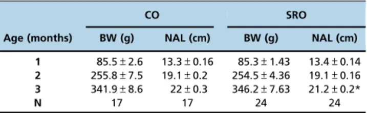
No significant differences were observed in body weight measured at 1, 2 and 3 months (Table 1). The naso-anal length (NAL) was measured monthly; at three months, the SRO group presented a decrease in NAL in comparison to the CO group.
Systolic Blood Pressure
At three months, the SRO group had significantly increased BPi values compared to the CO group (CO: 122.2±0.74; SRO:
144.6±0.94* mmHg).
Renal Function Analysis
GFR was significantly increased in the SRO group com-pared to the CO group (Table 2). However, RPF and urinary flow did not differ between the groups (V: CO: 0.11±0.02;
SRO: 0.10±0.01 ml/min/kg). Sodium, potassium and acid
excretion were similar in both groups.
Morphological Parameters for the Kidney
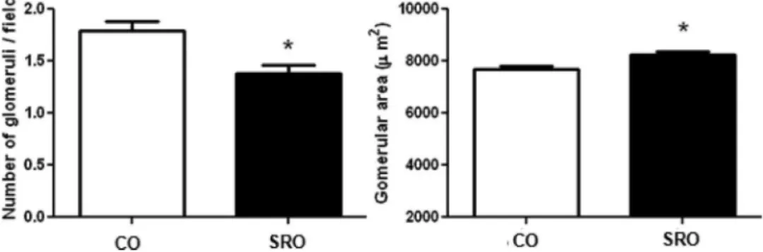
Under microscopic examination, the SRO rats presented a significant decrease in the glomeruli counted per field com-pared to the CO rats. Furthermore, the SRO rats also presented a significant increase in the glomerular area (Figure 1).
Cardiovascular Function
Direct measurements of cardiovascular parameters were performed at four months. The SRO group demonstrated significant increases in SAP, MAP and HR compared to the
Table 1-Body weight (BW) and naso-anal length (NAL) in the studied groups.
CO SRO
Age (months) BW (g) NAL (cm) BW (g) NAL (cm) 1 85.5±2.6 13.3±0.16 85.3±1.43 13.4±0.14 2 255.8±7.5 19.1±0.2 254.5±4.36 19.1±0.16 3 341.9±8.6 22±0.3 346.2±7.63 21.2±0.2*
N 17 17 24 24
Data are reported as the mean±SEM; N = number of animals in each group.
CO group. However, the DAP in the SRO rats was not significantly different from that in the CO rats (SAP: CO: 124±1.7; SRO: 139±3.02* mmHg; MAP: CO: 103±1.4; SRO:
110±2.7* mmHg; DAP: CO: 91±1.5; SRO: 96±2.9 mmHg;
HR: CO: 335±4.5; SRO: 354±7.5* bpm).
Cardiac Baroreceptor Reflex Sensitivity
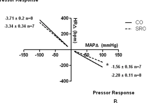
The SRO rats demonstrated significant impaired baroreflex sensitivity (bradycardic reflex response) for sudden eleva-tions in MAP. However, the tachycardic reflex response induced by sodium nitroprusside administration showed no difference between the groups (Figure 2).
Sympathetic and Parasympathetic Tone
The bradycardia observed following the administration of atenolol (Atenolol D) was significantly greater in the SRO rats compared to the CO rats. However, the tachycardic response observed after atropine administration (AtropinD) was not different between the SRO and CO groups (Table 3).
Balance of Sympatho-Vagal Tone
Table 4 shows the results of spectral analysis for the pulse interval (PI) and arterial pressure (AP). The LF values for the PI and AP were significantly higher in the SRO group compared to the CO group. The HF value for PI was decreased and the LF/HF ratio was increased in the SRO group, suggesting a sympatho-vagal misbalance with increased sympathetic modulation.
’ DISCUSSION
The present study revealed that SR during the last week of pregnancy reduced baroreflex response sensitivity, increased
sympathetic tone to the heart and caused sympatho-vagal misbalance in adult male offspring. This study also con-firmed that these offspring present elevated blood pressure values and renal morpho-functional changes, as described by Thomal et al. (21).
The participation of the kidneys in the development of hypertension has long been suggested and numerous mech-anisms have been proposed to confirm this hypothesis (28,29). In recent decades, the influence of inadequate intrauterine growth over nephron formation has also been associated with the development of hypertension (30-32).
Similarly to nutritional or growth restriction models (33,34), the SRO group exhibited a significantly reduced number of nephrons in comparison to the CO group and also presented glomerular hypertrophy; however, at the ages that the animals were studied, no decreases in GFR or in sodium excretion were observed, suggesting that renal adaptations in the SRO animals may be responsible for dislocation of the pressure-diuresis curve (35). In fact, increased GFR was observed in the SRO rats. Under normal conditions, GFR is maintained within narrow limits even when blood pressure changes due an auto-regulatory mechanism that acts by changing the resistance of glomerular arterioles. However, this mechanism may be weakened during hypertension and increased blood pressure in the glomerular capillaries may be responsible for enhancing the GFR (36). It is possible that these changes occurred in the SRO group; however, further experiments are necessary to corroborate this hypothesis.
To analyze whether changes in the central regulation of blood pressure contributed to the increased blood pressure in the SRO rats, we assessed autonomic cardiac function in this experimental model. Reduced baroreflex sensitivity, increased sympathetic tone to the heart and sympatho-vagal misbalance with an increase in sympathetic modulation were found in the adult SRO animals.
Impairment in the baroreflex response has been shown in diseases such as diabetes and hypertension (37,38). However, the role of baroreceptors in the cardiovascular system in chronic disease is not clear, especially considering that after approximately two days of hypertension the adap-tation of baroreceptors is completed (39). However, in parallel to baroreceptor adaptation, there is also a reduction in baroreflex sensitivity, which directly affects blood pressure regulation (40).
Reduced arterial baroreflex sensitivity may induce an increase in efferent sympathetic vasomotor activity because, under physiological conditions, the activity of aortic and carotid receptors results in an inhibitory effect on sympa-Table 2-Renal function analysis in the studied groups.
CO SRO
GFR (ml/min/Kg) 7.5±0.2 8.5±0.4*
RPF (ml/min/Kg) 21.8±1.6 22.8±1.9
TA mM/l 1.3±0.21 1.0±0.33
EANH4 mM/l 2.8±0.63 3.5±1.0
FE Na+% 0.75±0.16 0.51±0.15
FE K+% 28.6±2.7 25.6±2.6
N 9 8
Data are reported as the mean±SEM; N = number of animals/group. GFR = glomerular filtration rate, RPF = renal plasma flow, TA = titratable acid, EANH4 = excreted amount of ammonium, FE Na+= fractional excretion of sodium, FE K+= fractional excretion of potassium. CO = control mother offspring; SRO = sleep-restricted mother offspring. *pp0.05vs.CO group (Student´s T test and Mann Whitney test for RPF).
Figure 1 -Number of glomeruli/field and glomerular area of the studied groups.
thetic activity (40). Thus, in our experiments, the increase in sympathetic modulation found in the SRO rats may be related to a reduction in arterial baroreflex sensitivity, triggering sympatho-excitation and leading to blood pres-sure increase. However, the mechanisms underlying the baroreceptor dysfunction found in the SRO group remain unclear and require further investigation. Altered baroreflex sensitivity and sympathetic activity have also been observed in offspring subjected to prenatal exposure to dexamethasone (41,42), confirming the role of fetal exposure to corticosteroids in autonomic alterations in later life.
Interestingly, the progeny of rats subjected to nutritional restriction during pregnancy also exhibit cardiac baro-reflex impairment and increased expression of angiotensin II receptor type 1 (AT1) in brain regions responsible for blood pressure regulation (43). These changes are probably related to alterations in efferent autonomic baroreflex pathways. Increased AT1 receptor expression may be a consequence of
positive feedback due to increased Ang II in the central nervous system. This is because in rats, the chronic intracer-ebroventricular infusion of Ang II leads to increased expres-sion of AT1 receptors and AT1 mRNA in the brain (44). Other studies have associated increased hypothalamic expression of AT1 receptors with the use of glucocorticoids (dexamethasone) during pregnancy (45,46). In this experimental model, hyper-tension and decreased baroreflex sensitivity in offspring were also observed (41,42,47). Furthermore, the use of AT1 receptor inhibitors in late pregnancy attenuated baroreflex impair-ment and blood pressure increases in offspring (47), suggest-ing that changes in the expression of RAS components dursuggest-ing development can alter central structures involved in blood pressure regulation (47). Therefore, we cannot exclude the possibility that sleep deprivation during pregnancy may alter the expression of Ang II components within the brain, leading to cardiovascular alterations as previously reported (22,48)
Figure 2 -Cardiac baroreflex sensibility in the studied groups. (A) Depressor response induced by sodium nitroprusside. (B) Pressor response induced by phenylephrine.
CO = control mother offspring; SRO = sleep-restricted mother offspring. *pp0.05vs.CO group
Table 3-Evaluation of sympathetic and parasympathetic tone.
CO SRO
AtenololD(bpm) -15±3.27 -40±8.81*
AtropinD(bpm) 113±14.55 91±14.57
HexamethoniumD(mmHg) -38±3.86 -39±3.48
N 7 6
Data are reported as the mean±SEM; N = number of animals/group. CO = control mother offspring; SRO = sleep-restricted mother offspring. *pp0.05vs.CO group (Student´s T test and Mann Whitney test for HexamethoniumD).
Table 4-Results of spectral analysis.
CO SRO
LF PI (Hz) 14.08±0.98 25.8±2.1*
HF PI (Hz) 85.92±0.98 74.11±2.1*
LF/HF PI (Hz) 0.17±0.014 0.38±0.043*
LF AP (Hz) 4.48±0.65 7.82±1.09*
HF AP (Hz) 2.9±0.65 2.16±0.32
N 12 9
The alterations observed in the SRO group may be related to corticosterone expression. Several genes are modulated by glucocorticoids and fetal exposure to non-physiological concentrations of these hormones may result in the improper programming of these genes (49). During pregnancy, the enzyme 11-b-hydroxysteroid dehydrogenase (11bHSD) con-verts corticosterone (in rats) into non-active metabolites (49). However, in late pregnancy, the level of this enzyme is reduced and cortisol (or corticosterone) levels can increase during this period and reach the developing fetus (49).
The modulation of several hormones may be modified by SR or sleep deprivation (8,9). Increased secretion of ghrelin, ACTH, cortisol and GH after sleep deprivation was shown by Schussler et al. (9). During early pregnancy in mice, sleep deprivation caused a significant decrease in progesterone and an increase in corticosterone plasma concentration (50). Sleep deprivation in rats also increases the concentrations of corticosterone and norepinephrine in plasma and the secretion of hypothalamic hormones such as prepro-orexin (PPO) and neuropeptide Y (NPY) (10).
During critical periods of development, fetal exposure to non- physiological concentrations of hormones may promote hypothalamic dysfunction in offspring (51). This perinatal epigenetic programming phenomenon induced by hormonal changes during development was initially proposed by Günter Dörner. In this theory, hormones play a decisive role because they are influenced by the environment and also modulate neuroendocrine regulatory systems, which control all fundamental processes of life (52).
Thus, SR at the end of pregnancy influences the develop-ment of offspring through hormonal changes, causing adaptations that result in autonomic and kidney abnormal-ities that can be observed in the progeny during adulthood. However, additional research is needed to understand the mechanisms underlying the modifications observed in the present study.
The changes observed in offspring subjected to SR are probably related to increased plasma concentrations of corticos-terone. The levels of this hormone were not measured in the current study, although increased secretion of this hormone has been demonstrated in SR conditions (8-10,45). Thus, further experiments are needed to confirm this hypothesis.
The present study shows that SR during pregnancy is an additional risk factor for the development of hypertension in progeny. The elevation of blood pressure observed in our experimental model occurred through an increase in the sympathetic tone of the heart and a decrease in cardiac baroreflex control, suggesting that central nervous system changes were involved in fetal programming in this experi-mental model.
’ ACKNOWLEDGMENTS
Research support was provided by Fundac¸ão de Amparo à Pesquisa
(FAPESP- 2013-23622-7) and by Conselho Nacional de Desenvolvimento Científico e Tecnológico (CNPq). We thank American Journal Experts (AJE) for English language editing.
’ AUTHOR CONTRIBUTIONS
Gomes GN, Palma BD, Bergamaschi CT and Campos RR conceived and designed the experiments. Raimundo JR performed the experiments. Raimundo JR, Gomes GN, Bergamaschi CT and Campos RR analyzed the data. Gomes GN, Bergamaschi CT, Campos RR and Tufik S
contributed with the reagents, materials and analysis tools. Gomes GN, Bergamaschi CT, Campos RR and Raimundo JR wrote the manuscript.
’ REFERENCES
1. Widdowson EM, McCance RA. A review: new thoughts on growth. Pediatr Res. 1975;9(3):154-6, http://dx.doi.org/10.1203/00006450-19750 3000-00010.
2. Lucas A. Role of nutritional programming in determining adult morbidity. Arch Dis Child. 1994;71(4):288-90, http://dx.doi.org/10.1136/adc.71.4.288. 3. Barker DJ. In utero programming of chronic disease. Clin Sci (Lond).
1998;95(2):115-28, http://dx.doi.org/10.1042/CS19980019.
4. Barker DJ, Gluckman PD, Godfrey KM, Harding JE, Owens JA, Robinson JS. Fetal nutrition and cardiovascular disease in adult life. Lancet. 1993;341(8850):938-41, http://dx.doi.org/10.1016/0140-6736(93) 91224-A.
5. Gangwisch JE, Heymsfield SB, Boden-Albala B, Buijs RM, Kreier F, Pickering TG, et al. Short sleep duration as a risk factor for hypertension: analyses of the first National Health and Nutrition Examination Survey. Hypertension. 2006;47(5):833-9, http://dx.doi.org/10.1161/01.HYP.0000 217362.34748.e0.
6. Dean E, Bloom A, Cirillo M, Hong Q, Jawl B, Jukes J, et al. Association between habitual sleep duration and blood pressure and clinical impli-cations: a systematic review. Blood Press. 2012;21(1):45-57, http://dx.doi. org/10.3109/08037051.2011.596320.
7. Ogawa Y, Kanbayashi T, Saito Y, Takahashi Y, Kitajima T, Takahashi K, et al. Total sleep deprivation elevates blood pressure through arterial baroreflex resetting: a study with microneurographic technique. Sleep. 2003;26(8):986-9.
8. Machado RB, Tufik S, Suchecki D. Chronic stress during paradoxical sleep deprivation increases paradoxical sleep rebound: association with pro-lactin plasma levels and brain serotonin content. Psychoneuroendo-crinology. 2008;33(9):1211-24, http://dx.doi.org/10.1016/j.psyneuen. 2008.06.007.
9. Schüssler P, Yassouridis A, Uhr M, Kluge M, Weikel J, Holsboer F, et al. Growth hormone-releasing hormone and corticotropin-releasing hormone enhance non-rapid-eye-movement sleep after sleep deprivation. Am J Physiol Endocrinol Metab. 2006;291(3):E549-56, http://dx.doi.org/10.1152/ ajpendo.00641.2005.
10. Martins PJ, Marques MS, Tufik S, D’Almeida V. Orexin activation
pre-cedes increased NPY expression, hyperphagia, and metabolic changes in response to sleep deprivation. Am J Physiol Endocrinol Metab. 2010; 298(3):E726-34, http://dx.doi.org/10.1152/ajpendo.00660.2009. 11. Aldabal L, Bahammam AS. Metabolic, endocrine, and immune
con-sequences of sleep deprivation. Open Respir Med J. 2011;5:31-43, http:// dx.doi.org/10.2174/1874306401105010031.
12. Meerlo P, Sgoifo A, Suchecki D. Restricted and disrupted sleep: effects on autonomic function, neuroendocrine stress systems and stress respon-sivity. Sleep Med Rev. 2008;12(3):197-210, http://dx.doi.org/10.1016/ j.smrv.2007.07.007.
13. Tobaldini E, Cogliati C, Fiorelli EM, Nunziata V, Wu MA, Prado M, et al. One night on-call: sleep deprivation affects cardiac autonomic control and inflammation in physicians. Eur J Intern Med. 2013;24(7):664-70, http:// dx.doi.org/10.1016/j.ejim.2013.03.011.
14. Carter JR, Durocher JJ, Larson RA, DellaValla JP, Yang H. Sympathetic neural responses to 24-hour sleep deprivation in humans: sex differences. Am J Physiol Heart Circ Physiol. 2012;302(10):H1991-7, http://dx.doi. org/10.1152/ajpheart.01132.2011.
15. (CDC) CfDCaP. Effect of short sleep duration on daily activities--United States, 2005-2008. MMWR Morb Mortal Wkly Rep. 2011;60(8):239-42. 16. Lopes EA, Carvalho LB, Seguro PB, Mattar R, Silva AB, Prado LB, et al.
Sleep disorders in pregnancy. Arq Neuropsiquiatr. 2004;62(2A):217-21, http://dx.doi.org/10.1590/S0004-282X2004000200005.
17. Pires GN, Andersen ML, Giovenardi M, Tufik S. Sleep impairment during pregnancy: possible implications on mother-infant relationship. Med Hypotheses. 2010;75(6):578-82, http://dx.doi.org/10.1016/j.mehy. 2010.07.036.
18. Chang JJ, Pien GW, Duntley SP, Macones GA. Sleep deprivation during pregnancy and maternal and fetal outcomes: is there a relationship? Sleep Med Rev. 2010;14(2):107-14, http://dx.doi.org/10.1016/j.smrv.2009. 05.001.
19. Alvarenga TA, Aguiar MF, Mazaro-Costa R, Tufik S, Andersen ML. Effects of sleep deprivation during pregnancy on the reproductive capability of the offspring. Fertil Steril. 2013;100(6):1752-7, http://dx.doi.org/10.1016/ j.fertnstert.2013.08.014.
20. Radhakrishnan A, Aswathy BS, Kumar VM, Gulia KK. Sleep deprivation during late pregnancy produces hyperactivity and increased risk-taking behavior in offspring. Brain Res. 2015;1596:88-98, http://dx.doi.org/ 10.1016/j.brainres.2014.11.021.
22. Lima IL, Rodrigues AF, Bergamaschi CT, Campos RR, Hirata AE, Tufik S, et al. Chronic sleep restriction during pregnancy--repercussion on cardi-ovascular and renal functioning of male offspring. PLoS One. 2014;9(11): e113075, http://dx.doi.org/10.1371/journal.pone.0113075.
23. Jouvet D, Vimont P, Delorme F, Jouvet M. Study of selective deprivation of the paradoxal sleep phase in the cat. C R Seances Soc Biol Fil. 1964;158:756-9.
24. Rocco L, Gil FZ, da Fonseca Pletiskaitz TM, de Fátima Cavanal M, Gomes GN. Effect of sodium overload on renal function of offspring from dia-betic mothers. Pediatr Nephrol. 2008;23(11):2053-60, http://dx.doi.org/ 10.1007/s00467-008-0884-0.
25. Nishi EE, Oliveira-Sales EB, Bergamaschi CT, Oliveira TG, Boim MA, Campos RR. Chronic antioxidant treatment improves arterial renovas-cular hypertension and oxidative stress markers in the kidney in Wistar rats. Am J Hypertens. 2010;23(5):473-80, http://dx.doi.org/10.1038/ ajh.2010.11.
26. Fazan R, de Oliveira M, Oliveira JA, Salgado HC, Garcia-Cairasco N. Changes in autonomic control of the cardiovascular system in the Wistar audiogenic rat (WAR) strain. Epilepsy Behav. 2011;22(4):666-70, http:// dx.doi.org/10.1016/j.yebeh.2011.09.010.
27. Montano N, Ruscone TG, Porta A, Lombardi F, Pagani M, Malliani A. Power spectrum analysis of heart rate variability to assess the changes in sympathovagal balance during graded orthostatic tilt. Circulation. 1994;90(4):1826-31. Epub 1994/10/01, http://dx.doi.org/10.1161/01.CIR. 90.4.1826.
28. Johnson RJ, Feig DI, Nakagawa T, Sanchez-Lozada LG, Rodriguez-Iturbe B. Pathogenesis of essential hypertension: historical paradigms and modern insights. J Hypertens. 2008;26(3):381-91, http://dx.doi.org/10.1097/HJH. 0b013e3282f29876.
29. Marín R, Gorostidi M, Fernández-Vega F, Alvarez-Navascués R. Systemic and glomerular hypertension and progression of chronic renal disease: the dilemma of nephrosclerosis. Kidney Int Suppl. 2005(99):S52-6. 30. Brenner BM, Garcia DL, Anderson S. Glomeruli and blood pressure. Less
of one, more the other? Am J Hypertens. 1988;1(4 Pt 1):335-47, http://dx. doi.org/10.1093/ajh/1.4.335.
31. Luyckx VA, Brenner BM. Low birth weight, nephron number, and kidney disease. Kidney Int Suppl. 2005(97):S68-77, http://dx.doi.org/10.1111/ j.1523-1755.2005.09712.x.
32. Singh RR, Denton KM. Role of the kidney in the fetal programming of adult cardiovascular disease: an update. Curr Opin Pharmacol. 2015; 21:53-9, http://dx.doi.org/10.1016/j.coph.2014.12.010.
33. Merlet-Bénichou C, Gilbert T, Muffat-Joly M, Lelièvre-Pégorier M, Leroy B. Intrauterine growth retardation leads to a permanent nephron deficit in the rat. Pediatr Nephrol. 1994;8(2):175-80, http://dx.doi.org/10.1007/BF00865473. 34. Alves GM, Barão MA, Odo LN, Nascimento Gomes G, Franco Md MoC, Nigro D, et al. L-Arginine effects on blood pressure and renal function of intrauterine restricted rats. Pediatr Nephrol. 2002;17(10):856-62, http:// dx.doi.org/10.1007/s00467-002-0941-z.
35. Hall JE, Mizelle HL, Hildebrandt DA, Brands MW. Abnormal pressure natriuresis. A cause or a consequence of hypertension? Hypertension. 1990;15(6 Pt 1):547-59, http://dx.doi.org/10.1161/01.HYP.15.6.547. 36. Ofstad J, Iversen BM. Glomerular and tubular damage in normotensive
and hypertensive rats. Am J Physiol Renal Physiol. 2005;288(4):F665-72, http://dx.doi.org/10.1152/ajprenal.00226.2004.
37. Dall’Ago P, Silva VO, De Angelis KL, Irigoyen MC, Fazan R, Salgado HC.
Reflex control of arterial pressure and heart rate in short-term
streptozotocin diabetic rats. Braz J Med Biol Res. 2002;35(7):843-9, http:// dx.doi.org/10.1590/S0100-879X2002000700013.
38. Pagani M, Lucini D. Autonomic dysregulation in essential hypertension: insight from heart rate and arterial pressure variability. Auton Neurosci. 2001;90(1-2):76-82, http://dx.doi.org/10.1016/S1566-0702(01)00270-3. 39. Salgado HC, Krieger EM. Reversibility of baroreceptor adaptation in
chronic hypertension. Clin Sci Mol Med Suppl. 1973;45 Suppl 1:123s-6, http://dx.doi.org/10.1042/cs045123s.
40. Fahim M. Role of the arterial baroreceptor reflex in controlling circulation. Current Science. 1998:443-50.
41. Segar JL, Bedell KA, Smith OJ. Glucocorticoid modulation of cardiovas-cular and autonomic function in preterm lambs: role of ANG II. Am J Physiol Regul Integr Comp Physiol. 2001;280(3):R646-54.
42. Shaltout HA, Chappell MC, Rose JC, Diz DI. Exaggerated sympathetic mediated responses to behavioral or pharmacological challenges follow-ing antenatal betamethasone exposure. Am J Physiol Endocrinol Metab. 2011;300(6):E979-85, http://dx.doi.org/10.1152/ajpendo.00636.2010. 43. Pladys P, Lahaie I, Cambonie G, Thibault G, Lê NL, Abran D, et al. Role of
brain and peripheral angiotensin II in hypertension and altered arterial baroreflex programmed during fetal life in rat. Pediatr Res. 2004;55(6): 1042-9, http://dx.doi.org/10.1203/01.PDR.0000127012.37315.36. 44. Porter JP. Chronic intracerebroventricular infusion of angiotensin II
increases brain AT1 receptor expression in young rats. Brain Res Dev Brain Res. 1999;112(2):293-5, http://dx.doi.org/10.1016/S0165-3806(98) 00182-5.
45. Aguilera G, Kiss A, Luo X. Increased expression of type 1 angiotensin II receptors in the hypothalamic paraventricular nucleus following stress and glucocorticoid administration. J Neuroendocrinol. 1995;7(10):775-83, http://dx.doi.org/10.1111/j.1365-2826.1995.tb00714.x.
46. Dodic M, Abouantoun T, O’Connor A, Wintour EM, Moritz KM.
Pro-gramming effects of short prenatal exposure to dexamethasone in sheep. Hypertension. 2002;40(5):729-34, http://dx.doi.org/10.1161/01.HYP.0000 036455.62159.7E.
47. Shaltout HA, Rose JC, Figueroa JP, Chappell MC, Diz DI, Averill DB. Acute AT(1)-receptor blockade reverses the hemodynamic and baroreflex impairment in adult sheep exposed to antenatal betamethasone. Am J Physiol Heart Circ Physiol. 2010;299(2):H541-7, http://dx.doi.org/10.1152/ ajpheart.00100.2010.
48. Perry JC, Bergamaschi CT, Campos RR, Andersen ML, Casarini DE, Tufik S. Differential sympathetic activation induced by intermittent hypoxia and sleep loss in rats: Action of angiotensin (1-7). Auton Neurosci. 2011;160(1-2):32-6, http://dx.doi.org/10.1016/j.autneu.2010.11.006. 49. Mark PJ, Augustus S, Lewis JL, Hewitt DP, Waddell BJ. Changes in the
placental glucocorticoid barrier during rat pregnancy: impact on placental corticosterone levels and regulation by progesterone. Biol Reprod. 2009; 80(6):1209-15, http://dx.doi.org/10.1095/biolreprod.108.073650.
50. Calegare BF, Fernandes L, Tufik S, D’Almeida V. Biochemical, biometrical
and behavioral changes in male offspring of sleep-deprived mice. Psy-choneuroendocrinology. 2010;35(5):775-84, http://dx.doi.org/10.1016/ j.psyneuen.2009.11.004.
51. Plagemann A, Harder T, Janert U, Rake A, Rittel F, Rohde W, et al. Malformations of hypothalamic nuclei in hyperinsulinemic offspring of rats with gestational diabetes. Dev Neurosci. 1999;21(1):58-67, http://dx. doi.org/10.1159/000017367.