Introduction
Cardiovascular disease (CVD) is the name of a larger construct, which involves diseases of the heart, brain vasculature and blood vessels1. Several data indicate that CVD is the major cause of
dis-ability and death worldwide1,2. To date, CVD seems to be responsible
for 30% of the annual deaths in low and middle income countries2.
Projections for the next years do not indicate a better scenario, since is expected that this number will increase exponentially2.
Among the variety of CVD risk factors, physical inactivity is highlighted as a phenomenon strongly associated with CVD development, regardless of body mass index2. The effects of
physical activity (PA) (e.g., walking, climbing stairs) on CVD risk factors are widely known, which explains its popularity with healthy individuals who want to avoid CVD2. In turn, physical
exercise (PE), which concerns planned and structured body movement aimed to improve one or more physical capacities, has been widely suggested as a powerful non-pharmacological tool by different international associations, in order to prevent and counteract the deleterious effects of CVD in the organic system, due its capacity to offer larger effects in comparison with PA2,3,4,5,6.
In fact, several studies, including systematic reviews and meta-analytic data, indicate that PE is capable of leading to changes in the pathophysiological course of different CVD, such as hypertension (HTN), coronary artery disease/myocardial
infarction (MI) and heart failure (HF)7,8,9,10. Although the clinical
effects of PE on the different CVD are already known, the mecha-nisms associated with such changes must still be elucidated.
It is widely acknowledged that experiments with humans have limited capacity to contribute to the investigation of the mechanisms associated with the effects of PE on CVD and, usually, inferences are limited to systemic analyses (i.e., plasma and/or serum).
In this sense, studies using animal models have emerged as an effective tool to explore the several mechanisms triggered by PE in different organs and tissues, as well as in the whole organic system. Regarding animal models in the CVD context, experiments have been performed with different types of animals, including species that spontaneously developed the disease due to genetic factors and animals that underwent surgical procedures.
In fact, HTN, for example, is commonly studied in Spontaneous Hypertensive Rats (SHR), since the pathogenesis of HTN in this species is multifactorial, as in humans. However, if necessary, researches can also study HTN triggered by speciic alterations in the renal system (i.e., one kidney one clip [1K1C], two kidneys one clip [2K1C] and two kidneys two clip [2K2C]), using pharmacological approaches (i.e., L-NAME [Nω – nitro-L-arginine methyl ester], DOCA [deoxycorticosterone acetate]) and associated with diseases, such as obesity, and physiologi-cal conditions, such as menopause (i.e., ovariectomized), for
Mini-Review
Exercise training on cardiovascular diseases: Role of
animal models in the elucidation of the mechanisms
Bruno Rodrigues Daniele Jardim Feriani Bruno Bavaresco Gambassi
Universidade Estadual de Campinas, Campinas, SP, Brasil
Maria Claudia Irigoyen
Instituto do Coração, São Paulo, SP, Brasil
Kátia De Angelis
Universidade Nove de Julho, São Paulo, SP, Brasil
Hélio José Coelho-Júnior
Universidade Estadual de Campinas, Campinas, SP, Brasil
Abstract — Cardiovascular diseases, which include hypertension, coronary artery disease/myocardial infarction and heart failure, are one of the major causes of disability and death worldwide. On the other hand, physical exercise acts in the prevention and treatment of these conditions. In fact, several experiments performed in human beings have demonstrated the eficiency of physical exercise to alter clinical signals observed in these diseases, such as high blood pressure and exercise intolerance. However, even if human studies demonstrated the clinical eficiency of physical exercise, most extensive mechanisms responsible for this phenomenon still have to be elucidated. In this sense, studies using animal models seem to be a good option to demonstrate such mechanisms. Therefore, the aims of the present study are describing the main pathophysiological characteristics of the animal models used in the study of cardiovascular diseases, as well as the main mechanisms associated with the beneits of physical exercise.
example. Moreover, such possibilities are not exclusively related to HTN, and several possibilities are available in the context of MI (e.g., Left anterior descending coronary artery ligation [CAL], Ischemia-Reperfusion model [IRM] and MI-induced by isoproterenol) and HF (e.g., hyperadrenergic activity, CAL, doxo-rubicin, left coronary artery microembolization with polystyrene). The mechanisms responsible for the beneicial effects of PE on CVD have been studied in some of the aforementioned models, and much has been discovered. To contribute with this Special Issue, denominated: Animal Studies: Contributions to Exercise Physiology, we aimed to provide a brief description of the mechanisms and clinical aspects observed in the animal models that are most used to study the effects of PE on CVD.
This knowledge is important not only for undergraduate stu-dents, but also for graduate and post-graduate stustu-dents, as well as for researches that require an overview of the main animal models used in the context of PE and CVD.
Hypertension
HTN is one of the most prevalent diseases in adult life11. In older
people, for example, the prevalence of HTN is elevated, reaching values above 60% in both sexes11,12. The main concern about this
disease is its poor prognosis because patients with high blood pressure (BP) show increased risk for stroke (i.e., hemorrhagic and ischemic) and MI11,12. Moreover, a recent report from the
World Health Organization (WHO) established HTN as the main risk factor for death worldwide11,12.
SHR have been widely used in scientiic experiments, mainly because it is considered analogous with essential HTN in human by several authors13,14,15,16,17. This animal model of HTN was created
by mating Wistar rats that showed the highest BP levels. After 20 generations, the animals began to develop spontaneous HTN in early adulthood13. In these animals, BP increases exponentially
with aging, which occur mainly due to elevated vascular peripheral resistance (VPR) rather than to modiied cardiac output (CO)15,18.
As in humans, the pathogenesis of HTN in SHR seems to be multifactorial, since these animals show morphological and functional alterations on the different physiological elements that compose BP control, such as heart, kidney, blood vessels and autonomic control. Interestingly, these alterations have dis-similar time-courses, and factors associated with elevated BP in SHR are observed from the irst weeks of life.
Data are inconclusive about HTN condition in young SHR (ySHR) (~4 weeks old), once during this age the animals present a high oscillation in BP values (as demonstrated by data using direct and indirect measurements) with some evidence indicat-ing HTN19,20 — BP values next to 150 mmHg — and others
not21,22. Therefore, during this time of life animals are generally
denominated as pre-hypertensive. However, a signiicant number of evidence have been indicating that, even in the absence of alterations on BP measurements, morphological alterations on different vascular beds of the cardiovascular system and in the kidney, are observed in ySHR21,22,23,24.
In the kidney, for example, increased cross-sectional area (CSA) of the intima and adventitia layer, concomitantly with
increased wall lumen, are found in the renal arteries of ySHR in comparison with age-matched normotensive control23. Data are
also observed in other vascular beds (i.e., aorta, mesenteric), and evidence indicate elevated intima (IT), media (MT) wall thick-ness (WT), media-lumen ratio (M/L), media cross-sectional area (MCSA) and hypertrophy of smooth muscle cells in ySHR21,22,24,25.
Such alterations increase linearly and progressively with aging, and results of modiied vascular structure in 8-week and 12-week old SHR (adult SHR [aSHR]) are larger than in ySHR and age-matched WKY control22. In this sense, results from analyses in the
kidney of aSHR demonstrated increased indications of renal injury and abnormalities in comparison with normotensive animals26.
Nevertheless, experiments in the thoracic aorta of aSHR identiied elevated mRNA expression of α-actin (+9-fold), elastin (+6-fold) and collagen I and III (+11-fold) in comparison with normotensives27. Moreover, as observed in ySHR, the content of connective tissue, elastic ibers, and ibrils, as well as the CSA, were higher in the aorta of aSHR in comparison with age- matched control 27.
Regarding functional alterations, aSHR show an endothelium phenotypic proile close to what is generally observed in endothelial dysfunction, characterized by increased reactive oxygen species (ROS) synthesis, oxidative stress activity and decreased antioxidant activity, culminating in a substantial impairment on endothelium-dependent dilation28,29,30. In fact, endothelium-dependent dilation is
decreased in the vessels of aSHR in comparison with WKY28,29,30.
Participation of the endothelium in this phenomenon seems to be clear when pre-treatment with L-Nitro-Arginine Methyl Ester (L-NAME) — a nitric oxide (NO) inhibitor — abolishes differ-ences between normotensive and hypertensive animals30.
Noteworthy, NO is a molecule synthesized by the vascular endothelium is response to several stimuli (e.g., shear stress, acetyl-choline [ACh]) and it is the main action that occurs in the smooth muscle cells, causing vasodilation by altering calcium kinetics, through activation of soluble guanylate cyclase (sGC)/ cyclic guanosine monophosphate (cGMP) (sGC/cGMP) pathway31,32.
Moreover, this free radical seems to be important to mediate the effects of other vasodilatory molecules, such as angiotensin (ANG) (1-7)33,34. Interestingly, vasodilatory response to ANG (1-7) is
decreased in the aorta of aSHR, and impairment on NO pathway activity is a possible hypothesis to explain this phenomenon29.
As aforementioned, increased oxidative stress activity is generally observed during endothelial dysfunction. In aSHR, this phenomenon is not observed only in the heart, lung and kidney — organs associated with cardiovascular control — but also in the aorta and erythrocytes35,36. On the other hand, superoxide
dismutase (SOD), catalase (CAT) and glutathione peroxidase (GPx) — antioxidant enzymes that contribute to the control of oxidative stress —, as well as the total antioxidant activity, are found decreased35,36.
Oxidative stress decreases NO mainly due to anion superoxide (O2-) actions37,38,39,40,41. After superoxide anion (O2-) synthesis,
by uncoupled endothelial NO synthase (eNOS), NAD(P)H oxidase and mitochondria, this molecule reacts quickly with NO, creating peroxynitrite (ONOO-)37,38,39,40,41, which, in turn,
it is possible to observe that decrease in NO bioavailability is a retro-pathway, which, if not stopped, can cause serious damages to the homeostasis of the organic system, mainly on endothelial function.
One interesting issue about SHR animals concerns the asso-ciation between autonomic control and vascular function. In the mesenteric vessels, for example, ySHR show higher sensitivity to noradrenaline (NA), demonstrated by early vasoconstrictor response, than WKY rats, which is associated with higher inlux of calcium and lower barorelex sensitivity (gain) (BrS)22,42,43.
This phenomenon is exacerbated by age, since aSHR dem-onstrate not only higher sensibility to NA infusion and electrical stimulus, but also decreased vasodilator response after ACh infusion in mesenteric and carotid vessels22,23,43,44,45, as well as
BrS in conscious and nonconscious rats47,48,49. Furthermore,
evidence have been found that morphological and functional alterations on blood vessels, such as arterial stiffness, decreased endothelium-dependent dilation, and oxidative stress are associ-ated with BrS impairment35. Thus, data indicate that vascular
and morphological alterations in blood vessels are associated with cardiac autonomic control.
In this regard, aSHR showed increased cardiac and periph-eral sympathetic activity19,26,44,47,48 and blocked of α
1-receptor
with prazosin inhibit in ~95% of the vasoconstrictor response to electrical stimulation, causing absence of differences in relation to the response of control animals23. Several evidence
have showed that aSHR presents reduced heart rate variability (HRV) and increased systolic AP variability (SAP), followed by alterations in analyses of the frequency domain, indicating alteration in the sympathovagal balance26. Moreover, this
phe-nomenon is not just an effect of increased sympathetic activity discharge from the central nervous system to the periphery, but an environmental complex proving vasoconstrictor response.
This idea seems to be clear in the experiment of Reja et al. (2002)49, who observed elevated α
1A-receptor (α1A-R),
concomi-tantly with decreased α2A-receptor (α2A-R), gene expression levels
on neural system — central (i.e., ventromedial hypothalamus [VHM] and rostral ventrolateral medulla oblongata [RVLM]) and peripheral (i.e., spinal cord) — and on peripheral organs associated with BP control, as myocardium and adrenal medulla tissue studying aSHR 50. Furthermore, this overexpression of
α1A-R on the central and peripheral nervous system and on the
peripheral organs are positively correlated with BP in SHR49.
Yet, administration of phenoxybenzamine — a α-adrenergic receptor blocker — causes signiicant decrease on BP and HR in a dose-dependent manner in comparison with normotensive control rats50. High drug levels caused total absence of differences of the
aforementioned parameters between aSHR and control groups50.
Authors also tested the effects of β-adrenergic receptor blocker, propranolol, in cardiovascular parameters. Results indicate that inhibition of β-adrenergic receptor leads to signiicant decrease on HR in aSHR to levels similar to control group50.
Additionally to the morphological and functional alterations in the vascular endothelium, in organs associated with BP control (e.g., heart, kidney), and in the autonomic control of the cardio-vascular system, an upregulation of some molecular pathways in the brain areas responsible for BP control are found in SHR.
As in the peripheral tissues, brain renin-angiotensin system (RAS) acts regulating cardiovascular control51,52. Importantly,
during HTN the blood-brain barrier (BBB) is markedly disrupted in areas associated with autonomic cardiac control — such as RVLM, nucleus tractus solitarius (NTS) and paraventricular nucleus (PVN) —, which allows for extravasation of peripheral ANGII to these areas, as demonstrated trough infusion of luo -rescently labeled ANGII in SHR117,118. Therefore, it is possible
that elevated ANGII in the brain is, partially, explained by the peripheral activity of RAS117,118.
Regarding the pathway, in summary, after the cleavage of renin by the substrate angiotensinogen (Aogen), the decapeptide angiotensin I (ANG I) will be formed51,52. From the activity of
the angiotensin-converting enzyme (ACE), ANG I is cleaved in an octapeptide, called angiotensin II (ANG II), which seems to be the neuropeptide responsible for RAS negative effects on cardiovascular control through its binding with AT1R51,52.
Seminal data demonstrated that inhibition of the activity of RAS components in the brain by direct ventricular infusion of saralasin — a ANGII antagonist —, or even by antisense treatment, with consequently impair AT1R and Aogen, leads to decrease on BP values of aSHR in a dose-dependent manner16,53. Recent studies
have conirmed these evidences, showing that the expression of RAS components, as ACE, AT1R, Aogen, are increased in brain areas responsible for cardiovascular control (e.g., RVLM, NTS), which, in turn, is associated with BP values34,54.
Interestingly, BP modulation by RAS does not seem to occur alone, but in conjunction with the activity of ROS and inlam -matory elements, such as proinlam-matory cytokines, adhesion molecules, and transcription factors (nuclear factor-κB [NFκB])55.
In fact, blockade of NFκB in the PVN leads to lowering of mean BP, as well as decreased mRNA expression of proinlammatory cytokines, such as tumor necrosis factor-α (TNF – α), interleukin-1β (IL-interleukin-1β) and IL-6 (IL-6), and ROS (e.g., O2
-,ONOO-)56. In
ad-dition, analysis of data from experiments on aSHR indicate that ROS and proinlammatory elements are elevated in similar brain areas, and times, supporting the idea of an integrated complex of cardiac control between these pathways in SHR34,47,48,54.
After elevation of ROS by RAS, O2 – and other ROS can
lead to phosphorylation of the extracellular signal–regulated kinases ½ ( ERK ½), — a signaling protein kinase — which promptly activates NFκB trough the inhibition of Iκβα, an inhibitory anchoring protein, allowing the translocation of NFκB to nucleus, increasing the synthesis of ROS and
PICs39,47,57. In aSHR, mRNA expression of NAD(P)H oxidase
and the activity of ROS are elevated in the PVN, followed by elevated phosphorylation of ERK ½ and IKKβ, in addition to NFκB binding to the DNA33,58. Moreover, data demonstrated
that increased ROS, ERK ½ and NFκB activity is associated with increased levels of PICs, as well as decreased levels of anti-inlammatory cytokines (i.e., interleukin-10 [IL-10]) in aSHR33. Considered together, these data indicate that, in the
brain of aSHR, a molecular complex formed by RAS, ROS and PICs acts on cardiovascular control.
control59,60. In the experiment of Takagishi et al.60, for example,
exogenous IL-6 administrated by microinjection in the NTS causes ~35% inhibition of baroreceptor bradycardia relex gain in a dose-dependent manner60.
Moreover — despite the exacerbation of activity of hyper-tensive elements in the brain areas of SHR — some pathways, such as ANG 1-7/Mas receptor and anti-inlammatory cytokines (i.e., IL-10), that could act as counter-regulatory agents, con-sequently decreasing BP values, show lower expression in the brain of aSHR in comparison with normotensive age-matched control33,34. ANG (1-7), for example, is formed by the cleavage
of ANG II by ACE 2 and, in the brain areas of cardiovascular control, acts through Mas receptors in the tonic and relex control of BP52,61. Yet, evidence indicates that ANG (1-7) control of BP
can occur due to its inluence on BrS52,61.
The effects and mechanisms of PE on hypertension: evidences from SHR
PE is considered a beneicial non-pharmacological therapy to induce decrease on BP values in hypertensive patients, being considered one of the most important changes in lifestyle of these patients3,62. Several evidence indicate that aerobic PE is an
effective stimulus to induce acute (hypotension post-exercise) and chronic decrease on BP values in humans3,63,64,65. Nevertheless,
recent reviews (i.e., descriptive and meta-analyses) have described the potential of resistance training to contribute to this control58,66.
Experiments have demonstrated lower BP values in trained aSHR in comparison with sedentary aSHR after treadmill, swimming and resistance exercise26,33,34,47,48,54,67. A recent
meta-analysis, which analyzed 17 studies in SHR, conirmed these data and demonstrated that PE is capable of leading to signiicant decrease on BP values17.
Morphological and functional alterations on organs associ-ated with BP control (e.g., heart, kidney) and on blood vessels are one of the most evident alterations triggered by PE in aSHR. In the aorta, for example, low to moderate aerobic physical training performed 5 days per week, one hour per day, over three months caused decrease on smooth cells volume, elastic components and connective tissue in the aorta27. These effects
are followed by signiicant decrease in the mRNA expression of α-actin, elastin, and collagen I and III27. Results were not
dif-ferent in the heart since collagen content and cardiac load were decreased, and myocardial performance index (MPI) and left ventricular chamber diameter were increased in aSHR submitted to moderate aerobic exercise during 10 weeks68. Yet, swimming
exercise decreased the number of sclerosis glomerular index in the kidney of SHR26.
Morphological alterations are generally followed by functional changes caused by oxidative stress and characterized by decreased endothelium-dependent dilation. PE seems to be an eficient stimulus to counter-regulate this phenotype. In fact, after PE, the total antioxidant activity was increased in the heart, kidney, aorta, and erythrocytes of aSHR35,36. Concomitantly, total oxidant activity
was decreased in the lung, as well as lipid peroxidation (LPO) in the aorta35,36. Such alterations on pro and antioxidant molecules
seem to be sensible, and after 10 weeks of detraining total oxidant activity is again increased in the kidney and in the liver36.
Functionally, PE improves ACh-mediated vasodilation and low-mediated dilation in aSHR, restoring endothelial function in hypertensive animals28,30. PE also increases the vasodilator
response to ANG (1-7) and improves the expression of Mas receptor in the aorta29. Interestingly, this response seems to be
endothelium-dependent, since in endothelium-denuded vessels from trained SHR the vasodilator effect induced by ANG (1- 7) was abolished. Moreover, NO and Mas receptor inhibition impairs the effects of PE29.
Improvements on autonomic cardiac control have been extensively studied and suggested as one of the main beneits in response to PE in hypertensive patients. In turn, use of SHR models have contributed to a better understanding of this phe-nomenon and Krieger et al.69 already indicated this change as
one of the main physiological adaptations in response to PE. As in humans, several evidence using different types of exer-cise (i.e., treadmill running, swimming) have indicated that PE is capable of improving HRV and SAPV, which is accompanied by improvement in sympathovagal balance in aSHR26,47,48,68.
Furthermore, authors reported that the aforementioned changes are associated with total restore of BrS and vagal tonus, as well as sympathoinhibiton26,47,48. Regarding BrS, the effects of PE
have been cited since the late 1990s69. In a seminal experiment,
for example, Brum et al.70 observed that after 13 weeks of
low-intensity aerobic PE the AP range for triggering baroreceptor activation and the relation between the barorelex discharge and changes on SAP of aSHR were increased in comparison with sedentary animals70.
Interestingly, changes in cardiac and peripheral autonomic control after PE seem to occur through the afferent baroreceptor modulation68. In fact, experiments have indicated that aSHR submitted to sinoaortic denervation presented no alteration on BrS, HRV and SAP after PE protocol68.
Results from experiments on aSHR have contributed to understanding the effects of PE on the expression and activ-ity of hypertensive elements in the brain areas responsible for cardiovascular control. Nevertheless, evidence were not limited to this speciic issue, also demonstrating a possible correlation with cardiovascular function.
In fact, data demonstrate that low to moderate and moderate PE can cause signiicant decrease on RAS in the brain areas responsible for cardiovascular control of SHR. Data indicate lower mRNA expression of Aogen, ACE and AT1R in the NTS and RVLM of trained aSHR in comparison with sedentary
aSHR34,54. On the other hand, ANG (1-7) pathway components
(i.e., ACE2 and Mas receptor) showed elevated expression in the RVLM after exercise34. Similarly, ROS generation, NAD(P)
H subunits (i.e., p47phox, gp91phox) activity, phosphorylated
ERK ½ and IKKβ, NFκB translocation and proinlammatory cytokines synthesis (i.e., IL-6 and TNF-α) were lower in the PVN, RVLM of aSHR33,34,47,48. Noteworthy, such alterations
In an interesting experiment, Masson et al.47 described the
time-course changes in the expression of the aforementioned pathways in the PVN of aSHR47. During 8 weeks, animals were
submitted to low-to-moderate intensity PE, which occurred 5 days per week, 1 hour per day. To evaluate the time-course, evaluations occurred before the start and during the 1st, 2nd,
4th and 8th weeks of the PE program. Results showed that PE
was capable of causing signiicant decrease on ROS, ERK1/2 phosphorylation, NFκB translocation and proinlammatory cytokines synthesis in the irst two weeks of exercise47. These
results remained during the next weeks of training and were accompanied by an increase on autonomic cardiac control47.
In addition, experiments have demonstrated that PE can change the pattern of neurotransmitters release in PVN, as demonstrated by Jia et al.33, who submitted aSHR to 16 weeks
of moderate-intensity aerobic exercise (60% of maximal aero-bic velocity, 5 days per week, 60 min per day). After exercise, authors observed lower levels of excitatory neurotransmitters — glutamate and NA — and high levels of inhibitory neurotrans-mitters — GABA — in the PVN of SHR33.
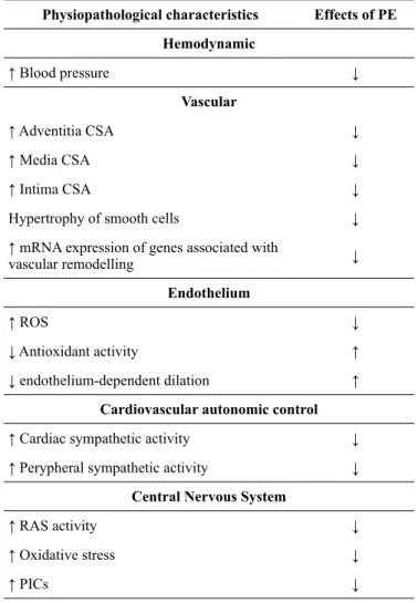
Table 1 shows a summary of the pathophysiological elements present in SHR, as well as the effects of PE.
Table 1. Physiopathological characteristics observed in SHR and the effects of PE
Physiopathological characteristics Effects of PE
Hemodynamic
↑ Blood pressure ↓
Vascular
↑ Adventitia CSA ↓
↑ Media CSA ↓
↑ Intima CSA ↓
Hypertrophy of smooth cells ↓
↑ mRNA expression of genes associated with
vascular remodelling ↓
Endothelium
↑ ROS ↓
↓ Antioxidant activity ↑
↓ endothelium-dependent dilation ↑
Cardiovascular autonomic control
↑ Cardiac sympathetic activity ↓
↑ Perypheral sympathetic activity ↓
Central Nervous System
↑ RAS activity ↓
↑ Oxidative stress ↓
↑ PICs ↓
CSA: Cross-sectional area; PE: Physical exercise; PICs: Proinlammatory cytokines; SHR: Spontaneuos hypertensive rats; RAS: Renin-angiotensin system; ROS: Reactive oxygen species.
Myocardial infarction
As aforementioned, CVDs are the leading cause of mortality worldwide1. Among them, coronary artery disease (CAD) stands
out due to its high risk of death11.
CAD has, as the main characteristic, the formation of ath-erosclerotic plaque in medium and large arteries. In summary, atherosclerotic plaque starts to build up due to endothelial dysfunction, which increases the permeability of the arterial intima layer to plasma lipoproteins — such as low-density lipoprotein (LDL), favoring the retention of such elements in the subendothelial space, when, later, in association with in-lammatory markers (e.g., macrophages), the lipoproteins will undergo oxidation, forming the oxidized LDL (oxLDL)71. This
phenomenon is followed by a myriad of inlammatory events, which have the formation of foam cells through phagocytosis of oxLDL by macrophages as the inal event of the pathway71.
It should be noted that the formation of oxLDL is associated with the quantum of available LDL in the plasma. Therefore, a higher number of LDL in the plasma will lead to increased formation of oxLDL and, consequently, of foam cells71.
Once developed, atherosclerotic plaque is composed of a cholesterol-rich lipid and a collagen-rich ibrous cap. The lipid content of the atherosclerotic plaque is the element responsible for its integrity, since disruption of this structure leads to forma-tion of thrombus, which, if in contact with coronary circulaforma-tion, can impair myocardial blood low, thus causing ischemia and, possibly, MI71. Therefore, MI is deined as myocardial cell death
due to prolonged ischemia72.
Studies using animal models are conducted to provide better understanding concerning the effects of PE pre and post MI. The most widely used protocol of MI in animals, mainly rodents, is occlusion of left anterior coronary artery. The MI surgery is conducted with the rat anesthetized with ketamine (80mg/kg) and xylazine (12mg/kg). After intubation, animals are positive-pressure ventilated with room air at 2.5mL, 65 strokes/minute with a pressure-cycled rodent ventilator. To induce MI, a 2-cm left lateral thoracotomy is performed in the third intercostal space, and the left anterior descending coronary artery is oc-cluded with a nylon (6.0) suture at approximately 1 mm from its origin below the tip of the left atrium. It is important to mention that studies generally use a sham group, which is also submitted to the same procedures, except for myocardial ischemia, which was not induced in this case73,74.
The effects and mechanisms of PE on MI: evidence from the surgical MI model
PE is a non-pharmacological therapy widely recommended for CVD patients, which has been demonstrated to be effective in improving endothelial function76, BrS, autonomic function77,
as well as reducing tissue and systemic inlammatory state78.
Such improvements can be identiied in human beings with tools already validated. Moreover, these evaluations are relatively easy and non-invasive if performed by an experienced evaluator, since, sometimes, just blood is necessary. However, in order to thoroughly evaluate the molecular, cellular and physiological mechanisms involved in these improvements, experimental models are necessary.
In fact, guidelines for rehabilitation programs in MI pa-tients recommend that low-intensity PE starts approximately 1 month after MI5. However, animal studies have shown that if
PE starts as soon as possible after MI this can further improve heart function, increasing maximum stroke volume, ejection fraction and attenuating the deterioration of LV contractility. These beneicial effects may be associated with PE-induced proliferation of cardiomyocytes, angiogenesis, attenuation of apoptosis in cardiomyocytes, due to improvement of myoila -ments and management of intracellular calcium (Ca2+)79.
It is known that — during and after MI — neurohumoral changes occur in order to minimize the consequences of re-duced ventricular function and, consequently, cardiac output. On the other hand, chronically, autonomic imbalance is usually followed by abnormalities in cardiorespiratory relex control, leading to impairment of BrS and function, and increased acti-vation of ergorelex and chemorelex. In turn, evidence shows that PE allows for the improvement of autonomic function and subsequent reduction of mortality in humans77.
In this sense, in order to identify the mechanisms associated with improvement in autonomic function after PE in MI, Jorge et al.80 tested the effects of early aerobic exercise training on LV
and autonomic function, hemodynamics, tissue blood low, and mortality rate after MI in rats. Results from PE demonstrated that the intervention induced improvement of cardiac function (i.e., systolic and diastolic), followed by normalization of he-modynamic and regional blood low, as well as improvement of autonomic control of peripheral circulation (i.e., BrS and increase on pulse interval [PI]) and cardiac function. Furthermore, the authors observed increased SERCA2 and VEGF mRNA ex-pression in LV. However, these beneits resulted in signiicant reduction in mortality rate in trained animals. According to the authors, the fact that early training restored autonomic control of circulation – represented by BrS and HRV – suggests that training may not only increase relex responses mediated by the parasympathetic nervous system, but also suppress the inluence of the sympathetic nervous system on ischemic heart disease. Moreover, elevated SERCA2 and VEGF mRNA expression suggests that these improvements are associated with alterations in intracellular calcium handling and blood supply.
Nerve growth factor (NGF) inducing cardiac sympathetic nerve sprouting is another characteristic observed post-MI. This phenomenon causes substantial sustained increase in sympathetic
activity, resulting in downregulation and desensitization of β1 and β2-adrenergic receptor (β1-AR and β2-AR, respectively), and in upregulation of β3-AR.
On the other hand, Chen et al.79, assuming that several
evi-dences indicate that PE decreases sympathetic activity after MI, investigated whether such phenomenon occurred by inhibition of sympathetic nerve sprouting and restoring of β3-AR/β1-AR ratio.
Results from the aforementioned study showed that PE in-hibits cardiac sympathetic nerve sprouting and restores β3-AR/ β1-AR balance after MI; which seems to occur due to increase in mRNA expression of β3-AR. Moreover, authors observed increased activation of NO synthase 1 (NOS1) and NOS2 in the heart of animals submitted to PE, indicating possible modulation of β3-AR through NO pathway. Therefore, considered together, these results indicate that the protective effect of PE in MI can be modulated by β3-AR/NO pathway79.
In fact, it is known that after MI, β3-AR is upregulated and activated due to high availability of NA. Dissimilar from the other β-receptor subunits, β3 seems to play a protective role after MI, acting as a counterregulatory mechanism during sympathetic overstimulation. Activation of β3-AR induces NO production, which, in turn, is associated with NOS1 activity. NOS1 signaling leads to increased cardiac calcium cycling, followed by enhanced cardiac contraction and accelerated relaxation79.
Thus, the authors suggested that the beneicial effects of β3-AR stimulation after PE are associated with the activation of NOS2 and NOS1, and the normalization of β-AR balance.
Regarding menopause, its main characteristic is the loss of the cardioprotective effects of estrogen, including on autonomic function. In this sense, Flores et al.81 investigated the effects
of PE in MI-rats with ovarian hormone deprivation. Results demonstrated PE-induced improvement in cardiopulmonary BrS, which was correlated with improvement in the autonomic control, represented by increased vagal tone in trained animals.
These data are conirmed by Rondon et al.82, who observed
that the improvement in BrS induced by exercise training in infarcted rats is due, in part, to increased aortic depressor nerve activity, concomitantly with improving cardiac vagal modulation.
Studies have not been exclusively developed in order to verify the effects of PE after MI, but also its cardioprotective effects. In the experiment of Bozi et al.83 and Rodrigues et
al.74, for example, the authors conducted a study in which rats
performed aerobic exercise training for 8 weeks prior to MI surgery. After MI event, trained rats showed a smaller infarct extension and sympathetic activity, as well as increased BrS, and parasympathetic modulation compared with sedentary in-farcted animals. Additionally, Rodrigues et al.74 observed that
improvements in autonomic balance and in parasympathetic modulation were strongly correlated with structural, systolic, diastolic and global LV function.
A recent study aimed to explore the effects of prior PE on the inlammatory aspects associated with MI. In fact, Santos et al.84 evaluated rats exercised prior MI and observed that exercise
regulation and inlammatory processes, acting as a suppressor of the inlammatory state.
In the control group, negative correlation between TNF-α and NF-κB was observed. However, these results were not observed in the trained group, which seems to be mediated by PPAR-α activation. Therefore, these data demonstrated that previously exercised animals had lower levels of local inlammatory mark -ers and less myocardial apoptosis, which seemed to be related to the presence of PPAR-α.
It is important to mention that due to signiicant functional loss in MI patients, mainly due to exacerbated muscle atrophy, resistance exercise was recommended as a complementary type of PE in relation to aerobic exercise. In this sense, Grans et al.116
evaluatedthe effects of dynamic resistance training on cardiac and hemodynamic function, as well as cardiovascular autonomic control after MI in rats. Results demonstrated that resistance exercise did not improve cardiac function. On the other hand, PE improved exercise tolerance and prevented additional loss in cardiovascular autonomic modulation.
Interestingly, Barboza et al.85 conducted one of the few
stud-ies that aimed to understand the effects of detraining on cardiac function, BrS, and mortality rate. To this end, MI rats were sub-mitted to PE for 3 months with subsequent 1 month of detraining. The authors observed that PE reduced the infarcted area, concomi-tantly with improvement on systolic and diastolic functions, on BrS and reduction on mortality rate. Moreover, the detraining period was
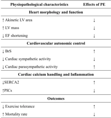
not enough to reverse the beneicial outcomes resulting from PE. Table 2 presents a summary of the physiopathological ele-ments present in MI rats, as well as the effects of PE.
Table 2. Physiopathological characteristics observed after MI in rats and the effects of PE
Physiopathological characteristics Effects of PE
Heart morphology and function
↑ Akinetic LV area ↓
↑ LV mass ↓
↓ EF shortening ↓
Cardiovascular autonomic control
↓ BrS ↑
↓ Cardiac sympathetic activity ↓
↓ Cardiac parasympathetic activity ↑
Cardiac calcium handling and Inlammation
↓SERCA2 ↑
↑PICs ↓
Outcomes
↓ Exercise tolerance ↑
↑ Mortality rate ↓
BrS: Barorelex sensitivity; EF: Ejection fraction; LV: Left ventricular; PE: Physical exercise; SERCA2: sarcoplasmic reticulum Ca2+-ATPase; PICs: Proinlammatory cytokines.
Heart Failure
Heart failure (HF) is a complex clinical condition that occurs in response to ventricular dysfunction due to structural and func-tional alterations in the heart, which lead to decreased capacity of the heart to pump blood to itself and to the periphery5,6,86. To
counteract such alterations, in an attempt to regulate CO, neurohu-moral compensatory mechanisms (e.g., the sympathetic nervous system [SNS]) are increased during HF87. Although, initially, this
phenomenon seems advantageous, chronic activation of the SNS has a toxic effect on the organic system of HF patients87.
In this sense, in order to study the relation between hyper-adrenergic activity and HF, α2A/α 2C adrenergic receptor (AR)
knockout (KO) mice was developed by mating two heterozygous C57B16 mice: a α2A-ARKO and a α2c-ARKO
88. During the irst
months of life (1st to 4th month), these animals present no evident
signals of HF, although muscular and cardiac abnormalities may be observed in this period88.
In fact, exercise intolerance, pulmonary edema associated with ventricular dysfunction (e.g., lower fractional shortening), and cardiac remodeling (e.g., cardiac hypertrophy) are signiicantly highlighted from the 5th month of life89,90,91. The development of
HF reaches the peak during the 7th month when it is proposed
that these animals developed a severe HF phenotype91,92,93. In
addition to the high mortality rate found in α2A/α 2CaARKO mice,
the animals present rest tachycardia and increased plasma NA levels due to increased adrenergic activity92,93,94,95.
Cardinal manifestations in HF patients involve limited muscu-lar and cardiac functioning, leading the patient to poor prognosis6.
Regarding muscular functioning, skeletal myopathy consists of intrinsic alterations in skeletal muscle observed during HF, which are indicated to be responsible for exercise intolerance and early fatigue96,97. This condition is one of the main features present
in cardiac cachexia syndrome and is strongly associated with poor outcomes96. In addition to the alterations in skeletal muscle
structure and function, skeletal myopathy is associated with a shift toward fast twitch ibers, oxidative stress, local and systemic inlammatory state (i.e., elevated TNF-α), and muscle metabolic dysfunction (i.e., mitochondrial respiration, energy transfer system and pH regulation) in response to stress93,96,98,99,100,101.
This phenotype is not well established in 3-month old α2A/α2Ca ARKO mice
90,92,102. However, from the 5th month of life,
these animals present suggestible muscle proile of skeletal myopathy due to decreased motor performance (i.e., Rotard test), increased oxidative stress (i.e., lipid hydroperoxidation and protein carbonylation), gastrocnemius capillary rarefaction, muscle atrophy of type I and type II ibers and, due to all these factors, exercise intolerance89,91,93,95,101,102.
Catabolic (i.e., ubiquitin-proteasome system [UPS]) and ana-bolic muscle pathways (i.e., insulin growth factor-1 [IGF- 1]) are not exclusively associate with muscle mass homeostasis during aging and stroke57, to name a few, but also seem to be present in
skeletal myopathy of HF93,101. Observations in α
2A/α2Ca ARKO mice
with established congestive HF (i.e., 7 months of age) showed that these animals present decreased IGF-1 protein content, and phos-phorylated AKTSer473, 4E-BP1Thr37/46, p70S6KThr389 and GSK3βSer9
soleus muscle93. Moreover, evidences allow to infer that
cata-bolic pathways are activate by oxidative stress and inlammatory state99,101. Besides muscular functioning disorders, several levels
of alterations — functional and structural — are observed in the heart of HF patients. In relation to cardiac function, is knowledge that HF associated with hyperadrenergic activity is followed by calcium (Ca2+) cardiac kinetics impairment.
It is important to mention that Ca2+ has a crucial role in car-diac excitation-contraction coupling (ECC), since this molecule regulates muscle contraction acting as a critical intermediary between the electrical stimulus and the coupling of actin. Here, it is described an overview of this phenomenon, while more detailed and extensive reviews were performed by several authors103,104,105.
Initially, to generate cardiac systole, the action potential propagates trough the membrane of the cardiomyocyte leading to its depolarization (from ~ – 90mV to ~+20mV) and, conse -quently, opening of the voltage-gated sodium (Na+) channels, mainly Nay1.5, allowing Na+ inlux103,105. The crescent increase
on ion Na+ concentration alters the membrane voltage, until reaching the threshold to the opening of the L-type voltage-gated Ca2+ channels — in this case, Cav1.2105. Subsequent to
the increase in cytosolic Ca2+ bioavailability (~10-fold), the ryanodine receptors 2 (RyR2) — the predominant subtype of RyR in the cardiac sarcoplasmic reticulum (SR) — are activated by a Ca2+-dependent mechanism, leading to Ca2+ release in the junctional zone — a space between cardiac sarcollema and SR —, which, subsequently, migrates to the cytosol, binding in its site on the troponin C, allowing for muscle contraction through actin-myosin interaction103,104,105. During cardiac diastole, a
decrease in Ca2+ bioavailability is necessary to cause cardiac muscle relaxation104,105. This process is dependent on proteins
involved in transsarcolemmal lux and sarcoplasmic reuptake of Ca2+103,105. Regarding transsarcolemmal lux, Na+/Ca2+ exchanger (NCX) is one of the main cellular mechanisms responsible for the clearance of Ca2+ in the cardiomyocyte103. NCX is located in
the cardiac cell membrane and — through the electrochemical gradient — exchanges one Ca2+ ion to the extracellular milieu at the same time that uptakes three Na+ ions103,105. The sarco/
endoplasmic reticulum Ca2+-ATPase protein 2a, henceforth de-nominated as SERCA2a, is another important cardiac structure with key role in calcium handling103. During cardiac systole,
SERCA2a remains inhibited by dephosphorylated phosphol-amban (PLN)103,105. However, after its phosphorylation by PKA
and Ca2+/calmodulin-dependent protein kinase (CaMKII), PLN allows SERCA to sequester Ca2+ from cytosol, contributing to cardiac relaxation103,104,105. It is important to mention that other
structures, such as plasma membrane Ca2+ ATPase (PMCA) and the mitochondrial uniporter seem to contribute, in a lower magnitude, to Ca2+ clearance during cardiac diastole103.
The failing heart is characterized by marked contractile (i.e., systolic and diastolic) dysfunction and high prevalence of arrhythmias, which has been considered, at least in part, as a result of decreased SR Ca2+ handling104,105,106. In fact, in vitro
experiments with human HF cardiac cells demonstrated smaller amplitude of Ca2+ transient, followed by lower SR Ca2+ content and load, as well as slow decline of Ca2+ during action potential depolarizing in comparison with healthy hearts107.
Moreover, detailed analysis adds data to the aforementioned and mentions several other alterations on cardiac Ca2+ transient, such as decreased Ca2+ sequestration during cardiac diastole, decreased SR Ca2+ stores during cardiac systole, elevated Ca2+ availability during cardiac diastole and elevated SR Ca2+ leak — which is the inappropriate Ca2+ release during diastole104,105.
Interestingly, authors have suggested the key role of SERCA2a and RyR2 in this phenotype104,105.
Regarding cardiac SERCA2a, experiments have demonstrated a decrease of 57% on its activity in HF hearts107. This
phenom-enon seems to be strongly associated with decreased cardiac Ca2+ uptake during diastole, concomitantly with increased inhibition on PLN104. In turn, in the experiment of AI et al.106, the authors
observed decreased RyR2 mRNA and protein expression in the LV of HF rabbits106. However, during chronic hyperadrenergic
state, which is present in HF, activation of cardiac β-receptors leads to decrease on calstabin 2 and increase on PKA phosphory-lation at Ser 2808 causing “leaky” of RyR2 and, consequently, diastolic SR Ca2+ leak104.
SERCA2a, NCX and SERCA2a/NCX are decreased by 26% and 34%, respectively, in the heart of α2A/α 2CaARKO mice
92,94.
Concomitantly, it is possible to observe increase on NCX94. In
rela-tion to RyR2, its expression is not changed in α2A/α2Ca ARKO mice 92.
Due to several compensatory stimuli, including hyperad-renergic activity, HF patients may present cardiac remodeling, which involves the combination of several mechanisms. Cardiac hypertrophy is the major pathophysiological response to stress in HF. Initially, the hypertrophic response is beneicial, since it minimizes parietal stress and maintains contractile performance. However, over time, this response becomes harmful, aggravating HF. Nevertheless, such response is usually related to detrimental changes in the components of extracellular matrix, reduced myocardial vascularization, and ibrosis87,108,109.
Cardiac ibrosis is caused by excessive accumulation of collagen in the heart during pathological remodeling. As a result of ibrosis, electrical conduction is impaired and the risk of arrhythmias is increased110,111. The development of ibrosis
alters the normal operation of the extracellular matrix and may lead to systolic and diastolic dysfunctions. The ibrotic tissue also contains myoibroblasts with contractile properties, par -ticipating in the collagen regulation. In response to paracrine and autocrine components, such as circulating hormones, me-chanic stress, and proinlammatory cytokines, and segregation of ibrillar collagen precursors, as well as signaling molecules responsible for the interaction between parenchymal cells and extracellular matrix111.
Another mechanism involved in cardiac remodeling is oxida-tive stress, which may impair the contractile function — through changes in the proteins that participate in the excitation-con-traction coupling — and activate signaling kinases hypertrophy, activate matrix metalloproteinases, and trigger apoptosis112.
Caspase substrates in the heart comprise, for example, troponin, tropomyosin, α-actin, and myosin chains113.
Apoptosis may occur by the extrinsic or intrinsic pathways. Briely, in the extrinsic pathway, a death binder (such as FasL or TNF-α) activates a death receptor, triggering the death-inducing signaling complex, activating caspase-8, which, in turn, acti-vates caspase-3, causing apoptosis. In the intrinsic pathway, mitochondria are essential to mediate the apoptotic process. The mitochondria release cytochrome c into the cytosol, causing the formation of an activation complex — the apoptosome — containing apoptotic protein activating factor-1 and caspase-9, leading to activation of other caspases, such as caspase-3114.
In 5–7-month old α2A/α 2CaARKO mice, it is possible to
ob-serve increased heart height, LV mass, cardiomyocyte width and CSA, as well as cardiac ibrosis and collagen volume, suggesting a phenotype generally observed in remodeled hearths89,91,94,95,102. In
the study of Pereira et al.90, quantitative morphometric analyses
indicated that cardiomyocyte width and cardiac collagen were, respectively, 28% and 55% increased in α2A/α 2CaARKO mice
in comparison with age-matched control90.
Nevertheless, the failing heart of α2A/α 2CaARKO mice show,
associated with structural alterations, decreased fractional shortening (FS) and increased LV dilation linked to increased left ventricular end-systolic dimension (LVSED) and left ven-tricular end-diastolic dimension (LVEDD), characterizing LV dysfunction phenotype89,94,95,102.
The effects and mechanisms of PE on HF: evidence
from α2A/α 2CARKO
Physical inactivity contributes to the progression of HF, whereas PE has been widely recommended as a non-pharmacological therapy capable of counteracting the deleterious effects of HF on the organic system4,97,109.
Evidence have demonstrate the effectiveness of moderate intensity PE (i.e., 60% of the maximal workload, 1 hour per day, 5 days per week, for 8 weeks) to increase exercise tolerance in α2A/α 2CaARKO mice to levels similar to those observed in
age-matched WT90,93,94,95,96,97,101. Of interest, PE does not seem to act
just by reversing the deleterious effects of HF in muscle func-tioning in the established pathology, but evidence has indicated its action in preventing the development of such effects92,102.
In fact, in the experiments of Medeiros et al.92,115,
low-to-moderate swimming trained 3-month old α2A/α 2CaARKO
mice (5 days per week, 60 minutes per day, during 8 weeks) presented preserved exercise tolerance during the development of HF, in comparison with age-matched control92,111. Moreover,
it is important to mention that inhibition of the development of exercise intolerance seems to be an adaptation exclusively in response to PE since data have demonstrated that treatment with β-blocker — carvedilol — did not alter exercise tolerance in α2A/α 2CaARKO mice
102.
Such improvements in exercise tolerance after PE are prob-ably associated with changes in the catabolic proile present in α2A/α 2CaARKO, such as capillary rarefaction
93,102 . Recently,
in the experiment of Bacurau et al.93, HF mice submitted to
low-to-moderate aerobic exercise presented elevated exer-cise tolerance and motor performance, as well as attenuated soleus muscle mass atrophy in comparison with age-matched control93. Authors also demonstrated that attenuated muscular
atrophy could be associated with changes on regulating muscle mass pathways, since PE elevated the protein content of the anabolic arm (i.e., IGF-1, PI3K, phosphorylated AKTSer473,
4E-BP1Thr37/46, p70S6KThr389) and decreased the catabolic arm
(i.e., proteasome activity)93.
Functional and structural parameters in α2A/α 2C ARKO also
seem to be responsive to PE109. In fact, experiments have
demon-strated the effectiveness of PE to lead to decreased Ca2+ decay, concomitantly with the peak of Ca2+ transient increase in the cardiomyocytes of α2A/α 2CaARKO
95. PE increases the balance
between Ca2+ reuptake by SERCA2a and Ca2+ clearance by
NCX92. These alterations on Ca2+ after PE are associated with
improvement on cardiac function91,95.
Regarding molecular mechanisms, PE increases SERCA2a and SERCA2A/NCX ratio toward control group levels92,94. This
phenomenon is an isolated product of increase on SERCA2a since NCX levels are found decreased in the heart of α2A/α 2CaARKO
mice92,94. Furthermore, it is possible to observe an increase in
the phosphorylation of PLN at Ser16 and Thr17 after PE92,94,95.
Therefore, considered together, these data indicate that PE leads to signiicant phosphorylation of PLN at Ser16 and Trh17 removing its inhibitory effect on SERCA2a, thus contributing to enhanced Ca2+ transient observed in trained animals95.
In turn, morphological alterations generally observed in remodeled hearts are impaired in trained α2A/α 2C ARKO, since
heart weight, cardiomyocyte width, LV mass, collagen content and cardiomyocyte CSA are decreased in these animals in com-parison with sedentary age-matched control89,95. Considering
these data it is possible to indicate that PE has an anti-remodeling effect on the heart of α2A/α 2C ARKO mice
89.
Moreover, some studies have aimed to describe the mechanisms associated with the anti-remodeling effect of PE. Experiments demonstrated that PE is capable of trigger-ing signiicant decrease on the translocation to the nucleus of elements strongly associated with cardiac remodeling, such as calcineurin and its downstream targets — NFATe3 and GATA-4 — in the heart of α2A/α 2C ARKO
89. It is important mention that,
in the heart of sedentary mice, both results were increased and associated with elevated β-MHC expression, suggesting a key role of this pathway in cardiac hypertrophy89. However, other
factors indirectly associated with cardiac remodeling (i.e., RAS system) also demonstrated responsiveness to PE, since ANGII and ACE activity were decreased, while ACE2 expression was increased, after PE90.
Such post-PE improvements on cardiac Ca2+ handling, remodel-ing, and skeletal myopathy occur in conjunction with improving
FS in α2A/α 2C ARKO to levels similar to those observed in control
non-KO mice89,91,95,102. As in skeletal myopathy, data indicate that
PE can also act as a preventive tool, since 3-month old animals — without evident signals of HR — submitted to swimming exercise demonstrated preserved FS in comparison with sedentary mice92.
Exercise and hypertension. Med Sci Sports Exerc, United States. 2004; 533-553.
4. Piepoli MF, Corrà U, Adamopoulos S, Benzer W, Bjarnason-Wehrens B, Cupples M, et al.. Secondary prevention in the clini-cal management of patients with cardiovascular diseases. Core components, standards and outcome measures for referral and delivery. Eur J Prev Cardiol. 2012.
5. SBC, Sociedade Brasileira de Cardiologia. Diretriz Sul-Americana de Prevenção e Reabilitação Cardiovascular. Arq Bras Cardiol, 2014; 103 (1).
6. Haskell WL, Lee IM, Pat RR, Powell KE, Blair SN, Franklin BA, et al. Physical activity and public health: updated recommendation for adults from the American College of Sports Medicine and the American Heart Association. Circulation. 2007; 116 (9). 7. Cornelissen VA, Neil AS. Exercise training for blood pressure:
a systematic review and meta-analysis.J Am Heart Assoc. 2013; 2 (1).
8. Cornelissen VA, Fagard RH, Coeckelberghs E, Vanhees L.. Impact of resistance training on blood pressure and other cardio-vascular risk factors a meta-analysis of randomized, controlled trials. Hypertension. 2011; 58 (5): 950-958.
9. Lawler PR, Filion KB, Eisenberg MJ. Eficacy of exercise-based cardiac rehabilitation post–myocardial infarction: a systematic review and meta-analysis of randomized controlled trials. Am Heart J. 2011; 162 (4): 571-584.
10. Ostman C, Jewiss D, Smart NA. The Effect of Exercise Training Intensity on Quality of Life in Heart Failure Patients: A Systematic Review and Meta-Analysis. Cardiology. 2016; 136 (2): 79-89. 11. Go AS; Mozaffarian D, Roger VL, Benjamin EJ, Berry JD, Borden
WB et al. Heart disease and stroke statistics – 2013 update: a report from the American Heart Association. Circulation. 2013; 127: e6-e245.
12. World Health Organization. World health statistics, 2009. 13. Okamoto K, Kyuzo A. Development of a strain of spontaneously
hypertensive rats. Japanese Jpn Circ J. 1963; 27 (3): 282-293. 14. Okamoto K, Kyuzo A. Participation of neural factor in the
patho-genesis of hypertension in the spontaneously hypertensive rat. Jpn Circ J; 1967 8(2), 168-180.
15. Albrecht I. The hemodynamics in female spontaneously hyper-tensive rats (SHR). Jpn Circ J. 1974; 38 (8): 651-654.
16. Phillips MI, Mann JF, Haebara H, Hoffman WE, Dietz R, Schelling P, et al. Lowering of hypertension by central saralasin in the absence of plasma renin. Nature. 1977; 270: 445-447. 17. Schlüter KD, Schreckenberg R, Rebelo RMC. Interaction between
exercise and hypertension in spontaneously hypertensive rats: a meta-analysis of experimental studies. Hypertens Res. 2010; 33 (11): 1155-1161.
18. Evenwel RT, Kasbergen CM, Struyker Boudier HAJ. Central and regional hemodynamics and plasma volume distribution during the development of spontaneous hypertension in rats. Clin. Exp. Hypert. 1983.
19. Okamoto K, Tabei R, Fukushima M, Nosaka S, Yamori Y, Ichijima K, et al. Further observations of the development of a strain of spontaneously hypertensive rats. Jpn Circ J. 1966; 30 (6): 703-716. 20. Weiss L, Lundgren Y, Folkow B. Effects of Prolonged Treatment
with Adrenergic β□receptor Antagonists on Blood Pressure,
Conclusions
In conclusion, when the ethical principles of animal experimenta-tion are considered, the use of animals in Physical Educaexperimenta-tion as a ield of knowledge has gained great prominence, particularly in understanding the pathophysiological mechanisms of CVD and the effects of PE on parameters that are unquantiiable in humans. Moreover, considering the data observed in this review, it is possible to infer that animal models of CVD seem to be an eficient and reliable tool to study the mechanisms responsible for the effects of PE on CVD.
References
1. WHO, World Health Organization. Global status report on non-communicable diseases 2010.World Health Organization, 2011. 2. Fuster V, Bridget BK, editors. Promoting cardiovascular health
in the developing world: a critical challenge to achieve global health. National Academies Press, 2010.
3. Pescatello LS, Franklin BA, Fagard R, Farquhar WB, Kelley GA, Ray CA. American College of Sports Medicine position stand.
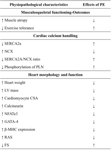
Table 3. Physiopathological characteristics observed in α2A/α
2CaARKO mice and the effects of PE
Physiopathological characteristics Effects of PE
Musculosqueletal functioning-Outcomes
↑ Muscle atropy ↓
↓ Exercise tolerance ↑
Cardiac calcium handling
↓ SERCA2a ↑
↑ NCX ↓
↓ SERCA2A/NCX ratio ↑
↓ Phosphorylation of PLN ↑
Heart morphology and function
↑ Heart weight ↓
↑ LV mass ↓
↑ Cardiomyocyte CSA ↓
↑ Calcineurin ↓
↑ NFATe3 ↓
↑ GATA-4 ↓
↑ β-MHC expression ↓
↑ RAS ↓
↓ FS ↑
Cardiovascular Design and Reactivity in Spontaneously Hypertensive Rats (SHR). Acta Physiol Scand. 1974; 91 (4): 447-457. 21. Adams AM, Bobik A, Korner PI. Differential development of
vascular and cardiac hypertrophy in genetic hypertension. Relation to sympathetic function. Hypertension. 1989; 14 (2): 191-202. 22. Rizzoni D, Castellano M, Porten E, Bettoni G, Muiesan ML,
Agabiti-Rosei E.. Vascular structural and functional alterations before and after the development of hypertension in SHR. Am J Hypertens. 1994; 7 (2):
193-23. Smeda JS, RM Lee, Forrest JB. Structural and reactivity altera-tions of the renal vasculature of spontaneously hypertensive rats prior to and during established hypertension. Circ Res.1988; 63 (3): 518-533.
24. Wellens D, Borgers M, Verheyen A. Structural basis for resetting of baroreceptor regulation in spontaneously hypertensive rats (SHR). Experientia. 1973; 29 (10): 1268-1271.
25. Levy D. Prognostic implications of echocardiographically deter-mined left ventricular mass in the Framingham Heart Study. N Engl J Med. 1990; 322 (22): 1561-1566.
26. Neto OB, Abade DT, Júnior MM, Mota GR, Orsatti FL, Silva RCR, et al. Exercise training improves cardiovascular autonomic activity and attenuates renal damage in spontaneously hyperten-sive rats. J Sports Sci Med. 2013; 12: 52-59.
27. Jordão MT, Ladd FC, Coppi AA, Chopard RP, Michelini LC. Exercise training restores hypertension-induced changes in the elastic tissue of the thoracic aorta. J Vasc Res. 2011; 48 (6): 513-524.
28. Gündüz F, Koçer G, Ülker S, Meiselman HJ, Baskurt OK, Sentürk
ÜK. Exercise training enhances low-mediated dilation in sponta -neously hypertensive rats. Physiol Res. 2011; 60 (4).
29. Silva DMR, Gomes-Filho A, Olivon VC, Santos TM, Becker LK, Santos RA, et al. Swimming training improves the vasodilator effect of angiotensin-(1–7) in the aorta of spontaneously hyper-tensive rat. J Appl Physiol. 2011; 111 (5): 1272-1277.
30. Guerrero J, Catros S, Derkaoui SM, Lalande C, Siadous R, Bareille R, et al. Cell interactions between human progenitor-derived endothelial cells and human mesenchymal stem cells in a three-dimensional macroporous polysaccharide-based scaffold promote osteogenesis. Acta Biomaterialia 2013; 9 (9): 8200-8213. 31. Asano RY, Browne RAV, Sotero RDC, Sames MM, Moraes
JFCND, Campbell, CSG, et al. Cycling above rather than below lactate threshold is more effective for nitric oxide release and post-exercise blood pressure reduction in individuals with type-2 diabetes. Motriz: J. Phys Ed. 2013; 19 (3): 633-640.
32. Bauer V, Ružena S. Nitric oxide—the endothelium-derived relaxing factor and its role in endothelial functions. Gen Physiol Biophys. 2010; 29 (4): 319-340.
33. Jia LL, Kang YM, Wang FX, Li HB, Zhang Y, Yu XJ, et al. Exercise training attenuates hypertension and cardiac hypertrophy by modulating neurotransmitters and cytokines in hypothalamic paraventricular nucleus. PloS one. 2014; 9 (1): e85481. 34. Ren CZ, Yang YH, Sun JC, Wu ZT, Zhang RW, Shen D, et al.
Exercise Training Improves the Altered Renin-Angiotensin System in the Rostral Ventrolateral Medulla of Hypertensive Rats. Oxid Med Cell Longev. 2016.
35. Bertagnolli M, Campos C, Schenkel PC, de Oliveira VL,
De Angelis K, Belló – Klein A, et al. Barorelex sensitivity
improvement is associated with decreased oxidative stress in trained spontaneously hypertensive rat. J Hypertens. 2006; 24 (12): 2437-43.
36. Kilic-Erkek O, Kilic-Toprak E, Caliskan S, Ekbic Y, Akbudak IH, Kucukatay V, et al. Detraining reverses exercise-induced improvement in blood pressure associated with decrements of oxidative stress in various tissues in spontaneously hypertensive rats. Mol Cell Biochem. 2016; 412 (1): 209-219.
37. Brieger K, Schiavone S, Miller JR, Krause KH. Reactive oxygen species: from health to disease. Swiss Med Wkly. 2012; 142. 38. Csiszar A, Wang M, Lakatta EG, Ungvari Z. Inlammation and
endothelial dysfunction during aging: role of NF-κB. J Appl
Physiol. 2008; 105 (4): 1333-1341.
39. El Assar M, Ângulo J, Rodríguez-Mañas L. Oxidative stress and
vascular inlammation in aging. Free Radic Biol Med. 2013; 65:
380-401.
40. Herrera MD, Mingorance C, Rodríguez-Rodríguez R, de Sotomayor MA. Endothelial dysfunction and aging: an update. Ageing Res Rev. 2010; 9 (2): 142-152.
41. Van Der Loo B, Labugger R, Skepper JN, Bachschmid M, Kilo J, Powell JM, et al. Enhanced peroxynitrite formation is associ-ated with vascular aging. J Exp Med. 2000; 192 (12): 1731-1744. 42. Head GA., Adams MA. Time course of changes in baroreceptor
relex control of heart rate in conscious SHR and WKY: contri -bution of the cardiac vagus and sympathetic nerves. Clin Exp Pharmacol Physiol. 1988; 15 (4): 289-292.
43. Mulvany MJ, Nyborg N. An increased calcium sensitivity of mesenteric resistance vessels in young and adult spontaneously hypertensive rats. Br J Pharmamcol. 1980; 71 (2): 585-596. 44. Iriuchijma J. Sympathetic discharge rate in spontaneously
hyper-tensive rats. Jap Heart J. 1973; 14: 350-356
45. Tesfamariam B, Halpern W. Endothelium-dependent and en-dothelium-independent vasodilation in resistance arteries from hypertensive rats. Hypertension. 1988; 11 (5): 440-444. 46. Head GA, Michael AA. Time course of changes in baroreceptor
relex control of heart rate in conscious SHR and WKY: contri -bution of the cardiac vagus and sympathetic nerves. Clin Exp Pharmacol Physiol. 1988; 15 (4): 289-292.
47. Masson G S, Costa TS, Yshii L, Fernandes DC, Soares PPS, Laurindo FR,et al. Time-dependent effects of training on cardio-vascular control in spontaneously hypertensive rats: role for brain
oxidative stress and inlammation and barorelex sensitivity. PloS
one. 2014; 9 (5): e94927.
48. Kishi, T, Hirooka Y, Katsuki M, Ogawa K, Shinohara K, Isegawa K, et al. Exercise training causes sympathoinhibition through anti-oxidant effect in the rostral ventrolateral medulla of hypertensive rats. Clin Exp Hypertens. 2012; 34.4: 278-283.
49. Reja V, Goodchild AK, Pilowsky PM. Catecholamine-related gene expression correlates with blood pressures in SHR. Hypertension. 2002; 40 (3): 342-347.
50. Numao Y, Iriuchijima J. Effects of alpha and beta blockers on hemodynamics of SHR. Jap Heart J. 1974; 15 (2): 166-172. 51. Bohlen und Halbach OV. The renin-angiotensin system in the
mammalian central nervous system. Curr Protein Pept Sci . 2005. 6 (4): 355-371.