Dendrimers
Jean-Sebastian Mandeville1, Phillipe Bourassa1, Thekkumkattil John Thomas2,3, Heidar-Ali Tajmir-Riahi1*
1De´partement de Chimie-Biologie, Universite´ du Que´bec a` Trois-Rivie`res, Trois-Rivie`res, Que´bec, Canada,2Department of Medicine, University of Medicine and Dentistry of New Jersey-Robert Wood Johnson Medical School, New Brunswick, New Jersey, United States of America,3The Cancer Institute of New Jersey, University of Medicine and Dentistry of New Jersey, Robert Wood Johnson Medical School, New Brunswick, New Jersey, United States of America
Abstract
Biogenic polyamines are essential for cell growth and differentiation, while polyamine analogues exert antitumor activity in multiple experimental model systems, including breast and lung cancer. Dendrimers are widely used for drug deliveryin vitro and in vivo. We report the bindings of biogenic polyamines, spermine (spm), and spermidine (spmd), and their
synthetic analogues, 3,7,11,15-tetrazaheptadecane.4HCl (BE-333) and 3,7,11,15,19-pentazahenicosane.5HCl (BE-3333) to dendrimers of different compositions, mPEG-PAMAM (G3), mPEG-PAMAM (G4) and PAMAM (G4). FTIR and UV-visible spectroscopic methods as well as molecular modeling were used to analyze polyamine binding mode, the binding constant and the effects of polyamine complexation on dendrimer stability and conformation. Structural analysis showed that polyamines bound dendrimers through both hydrophobic and hydrophilic contacts with overall binding constants of
Kspm-mPEG-G3= 7.66104M21,Kspm-mPEG-PAMAM-G4= 4.66104M21, Kspm-PAMAM-G4= 6.66104M21,Kspmd-mPEG-G3= 1.06105M21, Kspmd-mPEG-PAMAM-G4= 5.56104M21, Kspmd-PAMAM-G4= 9.26104M21, KBE-333-mPEG-G3= 4.26104M21, KBe-333-mPEG-PAMAM-G4=
3.26104M21, KBE-333-PAMAM-G4= 3.66104M21, KBE-3333-mPEG-G3= 2.26104M21, KBe-3333-mPEG-PAMAM-G4= 2.46104M21,
KBE-3333-PAMAM-G4= 2.36104M21. Biogenic polyamines showed stronger affinity toward dendrimers than those of synthetic
polyamines, while weaker interaction was observed as polyamine cationic charges increased. The free binding energies calculated from docking studies were:23.2 (spermine),23.5 (spermidine) and23.03 (BE-3333) kcal/mol, with the following order of binding affinity: spermidine-PAMAM-G-4.spermine-PAMMAM-G4.BE-3333-PAMAM-G4 consistent with spectro-scopic data. Our results suggest that dendrimers can act as carrier vehicles for delivering antitumor polyamine analogues to target tissues.
Citation:Mandeville J-S, Bourassa P, Thomas TJ, Tajmir-Riahi H-A (2012) Biogenic and Synthetic Polyamines Bind Cationic Dendrimers. PLoS ONE 7(4): e36087. doi:10.1371/journal.pone.0036087
Editor:Laurent Kreplak, Dalhousie University, Canada
ReceivedFebruary 21, 2012;AcceptedMarch 26, 2012;PublishedApril 27, 2012
Copyright:ß2012 Mandeville et al. This is an open-access article distributed under the terms of the Creative Commons Attribution License, which permits
unrestricted use, distribution, and reproduction in any medium, provided the original author and source are credited.
Funding:The authors have no funding or support to report.
Competing Interests:The authors have declared that no competing interests exist. * E-mail: [email protected]
Introduction
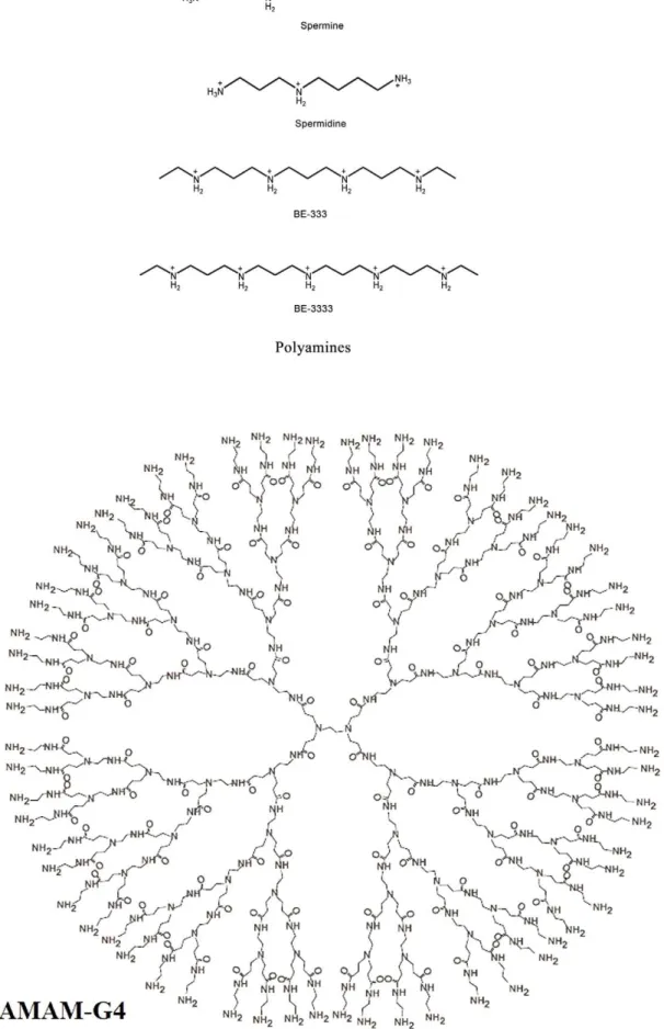
Polyamine analogues (Fig. 1) exert antitumor activity in multiple experimental model systems, including breast and lung cancer models and they are being used in clinical trials [1–5]. Synthetic polyamines can mimic some of the self-regulatory functions of biogenic polyamines but are unable to substitute for natural polyamines in their growth promoting role [6–13]. Natural polyamines are ubiquitous cellular cations and are involved in cell growth and differentiation (14). They are capable of modulating gene expression and enzyme activities, activation of DNA synthesis, and facilitating protein-DNA interactions [13–20]. Even though interactions of biogenic and synthetic polyamines with DNA and RNA are well characterized [21–25], little is known about their interaction with therapeutically important synthetic polymers, such as dendrimers [26].
Synthetic polymers with a specific shape and size play important roles in the development of modern drug and gene delivery systems [27–29]. Dendrimers are unique synthetic macromole-cules of nanometer dimensions with a highly branched structure and globular shape [29,30]. Among dendrimers, polyamidoamine
Figure 1. Chemical structures of polyamines and PAMAM-G4 dendrimer.
combination with other substrates produces conjugated molecules, that combine the properties of both the substrate and the polymer. However, conjugate formation can alter the binding affinity of dendrimers in general since a part of the functional pendant groups are removed by conjugation.
In this report, we present the results of spectroscopic and molecular docking experiments on the interaction of biogenic and synthetic polyamines with dendrimers of different composition, PAMAM (G4), m-PEG-PAMAM (G3) and m-PEG-PAMAM (G4), in aqueous solution, using a constant polymer concentration and different drugs concentrations. Structural data regarding polyamine binding modes as well as the stability of polyamine-dendrimer complexes are presented in this report.
Materials and Methods
Materials
Spermine.4HCl and spermidine.3HCl were purchased from Sigma Chemical Company and used as supplied. Polyamine
analogues, BE-333 and BE-3333, were synthesized in the laboratory of Dr. Akira Shirahata (Josai University, Saitama, Japan). PAMAM-G4 (MW 14214 g/mol) was purchased from Aldrich Chemical Co and used as supplied. mPEG-PAMAM-G3 (MW 13423 g/mol) and mPEG-PAMAM-G4 (MW 19214 g/mol) were synthesized according to published methods [35,41,42]. mPEG block has a molecular weight of 5000 g/mol. Other chemicals were of reagent grade and used without further purification.
Preparation of stock solutions
Dendrimer solution (1 mM) were prepared in distilled water and diluted to various concentrations in Tris-HCl buffer. Polyamine solutions (1 mM) were prepared in water and diluted in Tris-HCl buffer. The pH of stock solutions was kept at 760.2.
FTIR spectroscopic measurements
Infrared spectra were recorded on a FTIR spectrometer (Impact 420 model), equipped with deuterated triglycine sulphate (DTGS)
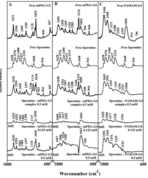
Figure 2. FTIR spectra and difference spectra (diff.) in the region of 1800-600 cm21 of hydrated films (pH 7.4) for free mPEG-PAMAM-G3 (A), mPEG-PAMAM-G4 (B) PAMAM-G4 (C) (0.5 mM) and their spermine complexes obtained at different spermine concentrations (indicated on the figure).
detector and KBr beam splitter, using AgBr windows. Polyamine solutions were added drop-wise to dendrimer solutions, with constant stirring to ensure the formation of homogeneous solutions and to reach target polyamine concentrations of 0.125, 0.25, and 0.5 mM and a final dendrimer concentration of 0.5 mM. Spectra were collected after 2 h incubation of polyamine and polymer solution at room temperature, using hydrated films [42]. Interferograms were accumulated over the spectral range of 4000-600 cm21, with a nominal resolution of 4 cm21 and 100 scans. The difference spectra [(dendrimer+polyamine solution)2
(dendrimer solution)] were generated, using dendrimer bands at 843 (mPEG-PAMAM-G3), 841 (mPEG-PAMAM-G4) and 1037 cm21 (PAMAM-G4). These vibrations are related to the polymers C-C stretching and semi ring skeletal modes [43,44] that show no spectral changes (intensity or shifting) upon polyamine-dendrimer complex formation, and cancelled on spectral subtrac-tion.
UV-Visible spectroscopy
The UV-Vis spectra were recorded on a Perkin-Elmer Lambda spectrophotometer with a slit of 2 nm and scan speed of 400 nm min21. Quartz cuvettes of 1 cm were used. The absorbance measurements were performed at pH 7.0 by keeping the concentration of dendrimer constant (0.10 mM), while increasing polyamine concentrations (0.005 mM to 0.10 mM).
The binding constants were obtained according to the method described by Connors [45]. It is assumed that the interaction between the ligand L and the substrate S is 1:1; for this reason a single complex SL (1:1) is formed. It was also assumed that the sites (and all the binding sites) are independent and all species obeyed the Beer’s law. A wavelength is selected at which the molar absorptivities eS (molar absorptivity of the substrate) and e11
(molar absorptivity of the complex) are different. In the absence of ligands and light path length (b) of 1 cm and at total substrate concentration St, the solution absorbance is given by the following
equation:
Figure 3. FTIR spectra and difference spectra (diff.) in the region of 1800-600 cm21 of hydrated films (pH 7.4) for free mPEG-PAMAM-G3 (A), mPEG-PAMAM-G4 (B) PAMAM-G4 (C) (0.5 mM) and their spermidine complexes obtained at different spermidine concentrations (indicated on the figure).
Ao~eSbSt ð1Þ
At total concentration Ltof a ligand, the absorbance of a solution
containing the same total substrate concentration is:
AL~eSb S½ zeLb L½ ze11b SL½ ð2Þ
where [S] is the concentration of the uncomplexed substrate, [L] the concentration of the uncomplexed ligand and [SL] is the concentration of the complex) which, combined with the mass balance on S and L, gives
AL~eSbStzeLbLtzDe11b SL½ ð3Þ
whereDe11=e112eS2eL (eLmolar absorptivity of the ligand). By
measuring the solution absorbance against a reference containing ligand at the same total concentration Lt, the measured
absorbance becomes
A~eSbStzDe11b SL½ ð4Þ
Combining equation (4) with the stability constant definition K11= [SL]/[S][L], gives
DA~K11De11b S½ ½ L ð5Þ
where DA = A2Ao . From the mass balance expression
St= [S]+[SL] we get [S] = St/(1+K11[L]), which is equation (5),
giving equation (6) at the relationship between the observed absorbance change per centimeter and the system variables and parameters.
DA
b ~
StK11De11½ L
1zK11½ L ð6Þ
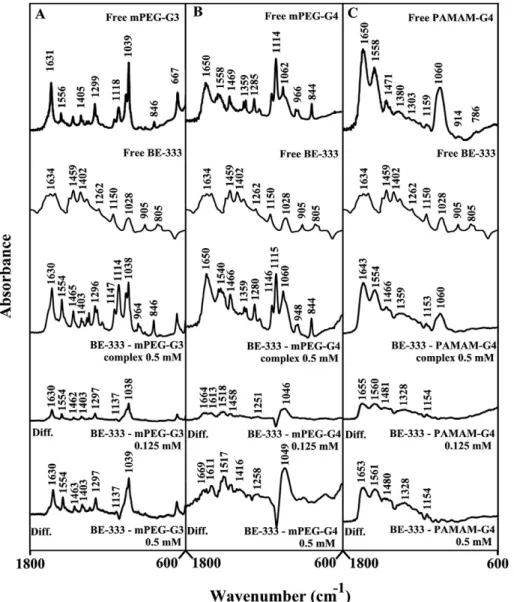
Figure 4. FTIR spectra and difference spectra (diff.) in the region of 1800-600 cm21 of hydrated films (pH 7.4) for free mPEG-PAMAM-G3 (A), mPEG-PAMAM-G4 (B) PAMAM-G4 (C) (0.5 mM) and their BE-333 complexes obtained at different BE-333 concentrations (indicated on the figure).
Equation (6) is the binding isotherm, which shows the hyperbolic dependence on free ligand concentration.
The double-reciprocal form of plotting the rectangular hyperbola1
y~ f d:
1
xz e
d, is based on the linearization of equation
(6) according to the following equation,
b DA~
1
StK11De11½ L z
1
StDe11
ð7Þ
Thus the double reciprocal plot of 1/DA versus 1/[L] is linear
and the binding constant can be estimated from the following equation
K11~intercept
slope ð8Þ
Molecular modeling
The PAMAM-G4 and polyamine structures were generated using the ChemOffice Ultra 6.0 software suite. The polyamine was then automatically docked to the rough PAMAM-G4 structure using ArgusLab 4.0.1 (ArgusLab 4.0.1, Mark A. Thompson, Planaria Software LLC, Seattle,WA, http://www.arguslab.com). The docked polyamine-PAMAM-G4 structures were optimized by means of molecular dynamics using the MM+force field available in HyperChem Pro 7.0. The heat time and run time for the simulations were 2 ps and 28 ps respectively with a step size of 0.001 ps. The temperature was initially set at 1 K and gradually increased to 300 K during the heat time by increments of 30 K. In all the simulations, equilibrium (achieving constant temperature near the selected final value) was reached after approximately 20 ps. The free binding energies of the optimized PAMAM-G4– polyamine complex structures were calculated using the Ascore scoring function provided in the ArgusLab software.
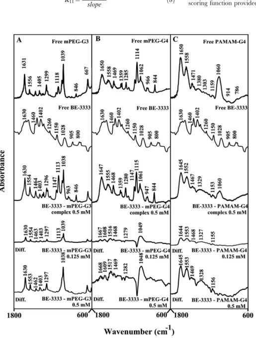
Figure 5. FTIR spectra and difference spectra (diff.) in the region of 1800-600 cm21 of hydrated films (pH 7.4) for free mPEG-PAMAM-G3 (A), mPEG-PAMAM-G4 (B) PAMAM-G4 (C) (0.5 mM) and their BE-3333 complexes obtained at different BE-3333 concentrations (indicated on the figure).
Results
FTIR spectral analysis of polyamine-dendrimer complexes
Figure 2 shows the infrared spectra and difference spectra of dendrimers complexed with spermine. Spectral shifting was observed for the polymer C = O, C-N, C-O stretching and NH bending [43,44] due to drug hydrophilic interactions with polymer polar groups. The major infrared bands at 1631 (C = O stretch and NH bending), 1556 (C-N stretch), 1405, 1299 (C-O), 1118, 1061 and 1039 cm21 (C-O and C-C stretch), in the infrared spectra of the free mPEG-PAMAM-G3 exhibited shifting and intensity increases upon spermine binding (Figs. 2A). Similarly, the major infrared bands of the free mPEG-PAMAM-G4 at 1650, 1558, 1469, 1359, 1285, 1114 and 1062 cm21showed shifting and intensity changes upon complex formation with spermine (Fig. 2 B). The infrared bands of the free PAMAM-G4 at 1650, 1558, 1471, 1380, 1159 and 1060 cm21 also shifted upon spermine interaction (Fig. 2C). The observed spectral shifting was accompanied with gradual increase in intensity of the above vibrational frequencies in the difference spectra [(dendrimer
+-spermine solution)2(dendrimer solution)] of drug-polymer
com-plexes (Fig. 2A, 2B and 2C, diffs). The spectral changes observed are attributed to the hydrophilic interactions of polyamine polar groups with dendrimer NH2, C-O and C-N groups. The
hydrophilic interaction is more pronounced at high spermine concentrations as evidenced by an increase in the intensity of several positive bands, centered at 1650-1000 cm21 in the difference spectra of polyamine-dendrimer complexes (Fig. 2A, 2B and 2C, compare diffs 0.125 and 0.5 mM).
Spermidine-polymer complex formation produced major spec-tral changes of the dendrimer infrared vibrational frequencies (Fig. 3). The spectral changes were observed mainly for the C = O, C-N, C-O stretching and NH bending modes [43,44] in the region of 1650-1000 cm21 of the infrared spectra of
mPEG-PAMAM-G3, mPEG-PAMAM-G4 and PAMAM-G4, upon complex formation with spermidine (Fig. 3A, 3B and 3C). The spectral shifting was associated with an increase in intensity of these vibrations in the difference spectra of spermidine-dendrimer complexes (Fig. 3A, 3B and 3C, diffs). More perturbations of polymer spectra occurred at high polyamine concentrations, as the intensity of the positive features increased as a result of spermidine-polymer complex formation (Fig. 3A, 3B and 3C. Compare diffs of
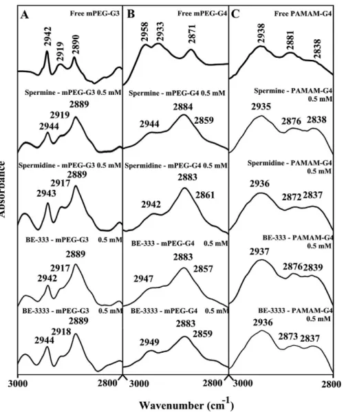
Figure 6. FTIR spectra in the region of 3300-2800 cm21of hydrated films (pH 7.4) for free mPEG-PAMAM-G3 (A), mPEG-PAMAM-G4
(B) and PAMAM-G4 (C) and their polyamine complexes obtained with 0.5 mM polymer and polyamine concentrations.
0.125 mM and 0.50 mM). The observed spectral changes are attributed to the hydrophilic contacts of drug OH groups with dendrimer NH2, C-O and C-N groups.
BE-333-dendrimer complexation caused minor spectral changes (shifting and intensity) at low polyamine analogue concentration, while major spectral changes occurred at high polyamine concentrations (Fig. 4A, 4B and 4C, 0.125 and 0.5 mM). The observed spectral changes (shifting and intensity increases) are due
to BE-333-polymer complex formationviadendrimer C-O, C-N and NH2and the polyamine NH2groups.
The infrared spectra of BE-3333-dendrimer complexes present-ed in Fig. 5 showpresent-ed minor spectral changes at low polyamine concentration and major shifting and intensity changes at high BE-3333 concentrations (Fig. 5A, 5B, 5C, compare 0.125 and 0.5 mM). The observed spectral changes are due to polyamine-polymer interaction via dendrimer C-O, C-N and NH and polyamine NH2groups (hydrophilic contacts).
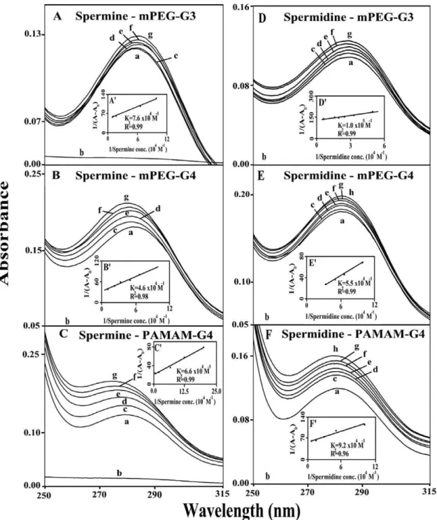
Figure 7. UV-visible spectra of mPEG-PAMAM-G3, mPEG-PAMAM-G4 and PAMAM-G4 and their complexes with spermine and spermidine with free dendrimer at 100mM and complexes c-g at 5, 10, 20, 40 and 80mM. (A, B and C) for spermine and c-g at 5, 10, 20, 40, and 80mM for spermidine.mPEG-G3 (D), c-h c-g at 5, 10, 20, 40, 80 and 100mM for mPEG-G4 (E) and spermidine-PAMAM-G4 (F). Plots of 1/(A2A0) vs (1/ polyamine concentration) and binding constant (K) for spermine (A9, B9and C9) and spermidine (D9, E9and F9).
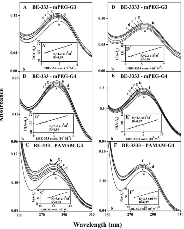
Figure 8. UV-visible spectra of mPEG-PAMAM-G3, mPEG-PAMAM-G4 and PAMAM-G4 and their complexes with 333 and BE-3333 with free dendrimer at 100mM and complexes c-h at 5, 10, 20, 40, 80 and 100mM. (A, B and C); c-h at 5, 10, 20, 40, 80 and 100mM for BE-3333-mPEG-G3 (D), c-i at at 5, 10, 20, 40, 60, 80 and 100mM BE-3333-mPEG-G4 (E) and c-f at 5, 10, 20 and 40mM ( F).
Hydrophobic contacts
The effect of polyamine-polymer complex formation on dendrimer antisymmetric and symmetric CH2 stretching
vibra-tions in the region of 3000-2800 cm21was investigated by infrared spectroscopy [43.44]. From Fig. 6A, the antisymmetric and symmetric CH2 bands of the free mPEG-PAMAM-G3 and its
polyamine complexes were assigned as follows: free mPEG-PAMAM-G3 at 2942, 2912 and 2890 cm21, spermine-mPEG-PAMAM-G3 at 2944 and 2889 cm21; spermidine-mPEG-PA-MAM-G3 at 2943, 2917 and 2889 cm21; BE-333-mPEG-PAMAM-G3 at 2942, 2917 and 2889 cm21; and BE-3333-mPEG-PAMAM-G3 at 2944, 2918 and 2889 cm21. In Fig. 6B, the antisymmetric and symmetric CH2bands of the free
mPEG-PAMAM-G4 and its polyamine complexes were observed as follows: mPEG-PAMAM-G4, 2958, 2933 and 2817 cm21; spermine-mPEG-PAMAM-G4, 2944, 2984 and 2859 cm21;
spermidine-mPEG-PAMAM-G4, 2942, 2983 and 2861 cm21;
BE-333-mPEG-PAMAM-G4, 2947, 2983 and 2857 cm21;
BE-3333-mPEG-PAMAM-G4, 2949, 2983 and 2859 cm21. Similarly,
Fig. 6C shows the CH2stretching vibrations of the free
PAMAM-G4 and its complexes as follows: PAMAM-PAMAM-G4, 2938, 2881 and 2838 cm21; spermine-PAMAM-G4, 2935 and 2876 and 2838 cm21; spermidine-PAMAM-G4, 2936, 2872 and 2837 cm21; BE-333-PAMAM-G4, 2937, 2876 and 2839 cm21; and BE-3333-PAMAM-G4, 2936, 2873 and 2837 cm21. The observed spectral shifting for polymer CH2vibrations is indicative
of some degree of hydrophobic interactions for polyamine-dendrimer complexes. This is due to the hydrophobic contacts
viapolyamine hydrophobic parts (aliphatic CH2 groups) and the
interior hydrophobic cavities present in dendrimers.
UV-Visible spectra and stability of polyamine-dendrimer complexex
The UV spectra of polyamine-dendrimer complexes are presented in Figures 7 (spermine and spermidine) and 8 (BE-333 and BE-3333). There is clear evidence that as polyamine complex formation occurred, major intensity increases of the dendrimer UV band, centered at 260–290 nm, also occurred [46,47]. The spectral changes are more pronounced in the case of biogenic spermine and spermidine than those of polyamine analogues BE-333 and BE-BE-3333 (Figs. 7 and 8).
The polyamine-dendrimer binding constants, obtained (accord-ing to the method described in experimental section (us(accord-ing plots of 1/(A2A0) vs (1/polyamine concentrations), showed one binding
constant for each polyamine-polymer complex formation (Figs. 7 and 8 and Table 1). The calculated binding constants are:
Kspm-mPEG-G3= 7.66104M21,Kspm-mPEG-PAMAM-G4= 4.6610 4
M21, Kspm-PAMAM-G4= 6.6610
4
M21, Kspmd-mPEG-G3= 1.06105M21, Kspmd-mPEG-PAMAM-G4= 5.56104M21, Kspmd-PAMAM-G4= 9.26
104M21,KBE-333-mPEG-G3= 4.26104M21,KBe-333–mPEG-PAMAM-G4=
3.26104M21, KBE-333-PAMAM-G4= 3.66104M21,KBE-3333-mPEG-G3=
2.26104 M21, KB e - 3 3 3 3 –m P EG- P A M A M -G4= 2.46104M21,
KBE-3333-PAMAM-G4= 2.36104M21(Table 1). The binding affinity
of biogenic polyamines toward dendrimers was stronger than that of synthetic polyamines, while weaker interaction was observed as polyamine cationic charge increased (Table 1). The reason why biogenic polyamine-dendrimers are more stable than those of the synthetic polyamines can be due to other factors such as the primary amines ((NH3+) in biogenic polyamines, that possess a higher density of positive charge than the secondary ones ((NH2+), in synthetic polyamines and also the presence of more hydrophobic contacts in the biogenic polyamine-polymer complexes.
Docking studies
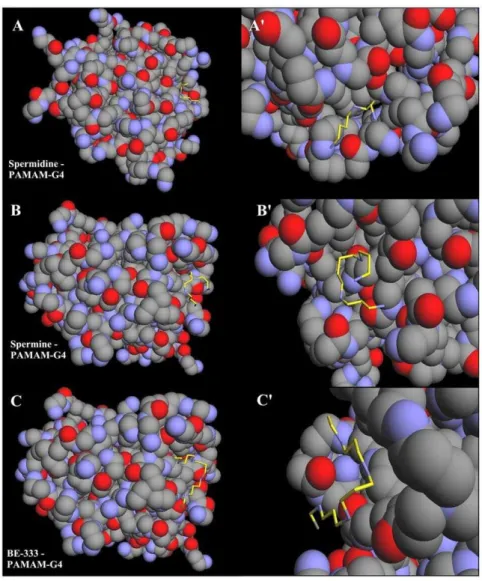
Our results from FTIR and UV-visible spectroscopic methods were complemented with molecular dynamic simulations in which the polyamines spermine, spermidine and BE-333 were automat-ically docked to PAMAM-G4 and the resulting structures were optimized using the MM+force field to determine the preferred conformations of the polyamine-polymer complexes. The simula-tion results are shown in Fig. 9 and Table 2. The models showed that polyamines are located on the surface of dendrimers and in cavities of PAMAM-G4 polymer (Fig. 9). The free binding energies calculated from docking studies were as follows: spermine, (3.2; spermidine, (3.5 and BE-333, (3.03 kcal/mol, with the following order of binding affinity: spermidine-PAMAM-G-4.spermine-PAMMAM-G4.BE-333-PAMAM-G4. These re-sults are consistent with the data obtained from spectroscopic studies (Tables 1 and 2).
Discussion
Several synthetic macromolecules have been developed as drug and gene delivery vehicles [27,28,30,32]. An ideal drug carrier vehicle must be biochemically inert and non-toxic, while protecting the payload (drug) from dissociation until it reaches the target site, and capable of releasing the drug at target site. Among synthetic polymers, dendrimers are unique macromole-cules with nanometer dimensions, a highly branched structure and globular shape. These macromolecules have uniform size and are mono-disperse, with modifiable surface functionality as well as internal cavities. They contain several binding sites for hydropho-bic, hydrophilic, cationic and anionic drugs (Fig. 1) Dendrimers are capable of binding and transporting DNA, RNA and drug molecules with high efficiency [30,32]. Dendrimers can be used as a containers to encapsulate drug molecules and carry them to different targets in vivo [35,48,49]. It has been shown that dendrimers with a hydrophobic interior and hydrophilic chain ends are capable of solubilizing hydrophobic compounds in aqueous solutions [49,50,51]. Attempts have been made to design different dendrimers as drug carriers [36]. For example, anticancer drug, 5-fluorouracil (5-FU) was attached to dendrimers with cyclic core [36]. Dendrimers having poly(ethylene glycol)
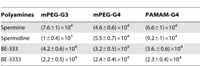
Table 1.Binding constants of polyamine-dendrimers (KM21).
Polyamines mPEG-G3 mPEG-G4 PAMAM-G4
Spermine (7.661)6104 (4.6
60.6)6104 (6.6 61)6104
Spermidine (160.4)6105 (5.560.7)6104 (9.261)6104
BE-333 (4.260.6)6104 (3.260.5)6104 (3.6.60.6)6104
BE-3333 (2.260.5)6104 (2.4
60.4)6104 (2.3 60.4)6104
doi:10.1371/journal.pone.0036087.t001
Table 2.Free binding energy of the docked polyamine-PAMAM complexes.
Complex DGbinding(kcal/mol)
Spermidine – PAMAM-G4 23.50
Spermine – PAMAM-G4 23.20
BE-333 – PAMAM-G4 23.03
grafts have been used to encapsulate antitumor drugs adriamycin and methotrexate [37]. The complexation of dendrimers with anti-inflammatrory drug flurbiprofen was studied in vitro and in vivo, while drug biodistribution in different organs has been monitored [39]. Gene delivery targeted to brain has been attempted using transferring-conjugated polyethyleneglycol-mod-ified polyamidoamine dendrimer [40]. The purpose of our investigation was to analyze the interaction of dendrimers with biogenic and synthetic polyamines in order to test the feasibility of these nanocarrier molecules for polyamine-based drug delivery. Infrared spectroscopic data in the region of 1700-1000 cm21, where most of the polymer in-plane vibrations related to C = O, C-N, NH and C-O modes are located, exhibit spectral changes (shifting and intensity variations) upon polyamine-polymer com-plex formation. These changes are more profound at high polyamine concentrations. There was clear evidence that the hydrophilic polyamine entity induced more perturbations of polymer hydrophilic group vibrational frequencies, with the following order of spectral changes: spermidine.spermine.
BE-333.BE-3333 (Figs. 2, 3, 4, 5). This can be expected since polyamines with several positively charged NH and NH2groups
show more affinity for dendrimer terminal groups than those of the hydrophobic groups located in polymer interior cavities. Molecular modeling also showed polyamine binding with the dendrimer with spermidine-PAMAM more stable than spermine-and BE-333-PAMAM complexes.
In conclusion, we find that synthetic and natural polyamines are capable of binding to different dendrimers. The binding affinity is relatively low to enable them to act as polyamine delivery vehicles, especially polyamine analogues under investigation as cancer chemotherapeutic agents.
Author Contributions
Conceived and designed the experiments: JSM PB TJT HATR. Performed the experiments: JSM PB TJT. Analyzed the data: JSM PB. Contributed reagents/materials/analysis tools: TJ. Wrote the paper: HATR. Figure 9. Optimized polyamine-PAMAM-G4 docking structures.The polyamines are shown in yellow color. (A) shows whole PAMAM-G4 in spheres with spermine and (A9) shows the zoom on the binding site represented in sticks. (B) shows whole PAMAM-G4 in spheres with spermidine and (B9) shows the binding site represented in sticks. (C) whole PAMAM-G4 in spheres with BE-333 and (C9) shows the binding site in represented in sticks.
References
1. Huang Y, Keen JC, Pledgie A, Marton LJ, Zhu T, et al. (2006) Polyamine analogues down-regulate estrogen receptoraexpression in human breast cancer cells. J Biol Chem 281: 19055–19063.
2. Huang Y, Keen JC, Hager E, Smith R, Hacker A, et al. (2004) Regulation of polyamine analogue cytotoxicity by c-jun in human MDA-MB-435 cancer cells. Mol Cancer Res 2: 81–88.
3. Casero RA, Woster PM (2001) Terminally alkylated polyamine analogues as chemotherapeutic agents. J Med Chem 44: 1–26.
4. Gerner EW, Meyskens FL (2004) Polyamines and cancer: old molecules, new understanding. Nat Rev Cancer 4: 781–792.
5. Bergeron RJ, Neims AT, McManis JS, Hawthorne TR, Vinson JRT, et al. (1988) Synthetic polyaminesanalogues as antineoplastics. J Med Chem 31: 1183–1190.
6. Bergeron RJ, McManis JS, Liu CZ, Frng Y, Weimar WR, et al. (1994) Antiproliferative properties of polyamine analogues: a structure-activity study. J Med Chem 37: 3464–3476.
7. Davidson NE, Mank AR, Prestigiacomo LJ, Bergeron JR, Casero RA (1993) Growth inhabitation of hormone-responsive and resistant human breast cancer cells in culture byN1,N12
-bis (ethyl)spermine. Cancer Res 53: 2071–2075. 8. Thomas T, Balabhadrapathruni S, Gallo MA, Thomas TJ (2002) Development
of polyamine analogues as cancer therapeutic agents. Oncol Res 13: 123–135. 9. Faaland CA, Thomas TJ, Balabhadrapathruni S, Langer T, Mian S, et al. (2000) Molecular correlates of the action of bis(ethyl)polyamines in breast cancer cell growth inhibition and apoptosis. Biochem Cell Biol 78: 415–426.
10. Thomas T, Thomas TJ (2003) Polyamine metabolism and cancer. J Cell Mol Med 7: 113–126.
11. McCloskey DE, Pegg AE (2003) Properties of the spermidine/spermine N1
-acetyltransferase mutant L156F that decreases cellular sensitivity to the polyamine analogue N1, N11
-bis(ethyl)norspermine. J Biol Chem 278: 13881–13887.
12. Ha HC, Woster PA, Yager JD, Castro RA (1997) The role of polyamine catabolism in polyamine analogue-induced programmed cell death. Proc Natl Acad. Sci USA 94: 11557–11562.
13. Ha HC, Sirisoma NS, Kuppusamy P, Zweier LJ, Woster PM, et al. (1998) The natural polyamines spermine functions directly as a free radical scavenger. Proc Natl Acad Sci U S A 95: 11140–11145.
14. Tabor CW, Tabor H (1984) Polyamines. Annu Rev Biochem 53: 749–790. 15. Pegg AE (1988) Polyamine metabolism, and its importance in neoplastic growth
and a target of chemotherapy. Cancer Res 48: 759–774.
16. Heby O (1981) Role of polyamines in the control of cell proliferation and differentiation. Differentiation 19: 1–20.
17. Tkachenko AG, Nesterova LY (2003) Polyamines as modulators of gene expression under oxidative stress inEscherichia coli. Biochemistry (Moscow) 68: 850–856.
18. Marton LJ, Pegg AE (1995) Polyamines as targets for therapeutic intervention. Annu Rev Pharmacol Toxicol 35: 55–91.
19. Cohen SS (1998) A Guide to the polyamines, Oxford Uni. Press, New York. 20. Thomas T, Thomas TJ (2001) Polyamine in cell growth and death. Cell Mol
Life Sci 58: 244–258.
21. Ahmed Ouameur A, Tajmir-Riahi HA (2004) Structural analysis of DNA interactions with biogenic polyamines and cobalt (III) hexamine studied by Fourier transform infrared and capillary electrophoresis. J Biol Chem 279: 42041–42050.
22. Ahmed Ouameur A, Bourassa P, Tajmir-Riahi HA (2010) Probing tRNA interaction with biogenic polyamines. RNA 16: 1968–1979.
23. N’soukpoe´-Kossi CN, Ahmed Ouameur A, Thomas T, Shirahata A, Thomas TJ, et al. (2008) DNA interaction with antitumor polyamine analogues: Acompar-ison with biogenic polyamines. Biomacromolecules 9: 2712–2718.
24. N’soukpoe´-Kossi CN, Ahmed Ouameur A, Thomas T, Thomas TJ, Tajmir-Riahi HA (2009) Interaction tRNA with antitumor polyamine analogues. Biochem Cell Biol 87: 621–630.
25. D’Agostino L, di Pietro M, Di Luccia A (2005) Nuclear aggregates of polyamines are supramolecular structures that play a crucial role in genomic DNA protection and conformation. FEBS J 272: 3777–3787.
26. Hardy JG, Mauri A, David K, Smith K, Gabrielson NP, et al. (2006) Dendrons with spermine surface groups as potential building blocks for nonviral vectors in gene therapy. Bioconjugate Chem 17: 172–178.
27. Galeazzi S, Hermans TM, Paolino M, Anzini M, Mennuni L, et al. (2010) Multivalent supramolecular dendriemr-based drugs. Biomacromolecules 11: 182–186.
28. Hu J, Fang M, Cheng Y, Zhang J, Wu Q, et al. (2010) Host-guest chemistry of dendrimer-drug complexes. 4. An in depth look into the binding/encapsulation of guanosine monophosphate by dendrimers. J Phys Chem B 114: 7148–7157. 29. Maiti PK, Caing T, Wang G, Goddard WA (2004) Structure of PAMAM
dendrimers: Generations 1 through 11. Macromolecules 37: 6236–6254. 30. Patri AK, Majoros IJ, Baker JR (2002) Dendritic polymer macromolecular
carriers for drug delivery. Curr Opinion Chem Biol 6: 466–471.
31. Tomilia DA (2005) Birth of a new macromolecular architecture: dendrimers as quantized building blocks for nanoscale synthetic polymer chemistry. Prog Polymer Sci 30: 294–324.
32. Klajnert B, Bryszewska M (2007) Dendrimers as delivery systems in gene therapy; in : New developments in mutation research Valon CL, ed. Chapter 9. pp 217–240.
33. Lee CC, Mackay JA, Frechet JMJ, Szoka FC (2005) Designing dendrimers for biological applications. Nature Biotech 23: 1517–1526.
34. Klajnert B, Bryszewska M (2007) in: Dendrimers in Medeicines Klajnert B, Bryszewska M, eds. Nova Science publisher, Inc., New York.
35. Kojima C, Kono K, Maruyama K, Takagishi T (2000) Synthesis of pollyamidoamine dendriemrs having poly(ethylene glycol) grafts and their ability to encapsulate anticancer drugs. Bioconjugate Chem 11: 910–917. 36. Zhuo RX, Du B, Lu ZR (1999) In vitro release of 5-flurouracil with cyclic core
dendritic polymer. J Control Release 57: 249–257.
37. Kono K, Liu M, Frechet JMJ (1999) Design of dendritic macromolecules containing folate or methotrexate residues. Bioconjugate Chem 10: 1115–1121. 38. Jansen JFGA, Meijer EW, de Brabander-van den Berg EMM (1995) The dendritic box: shape-selective liberation of encapsulated guests. J Am Chem Soc 117: 4417–4418.
39. Malik N, Wiwattanapatapee R, Klopsch R, Lorenz K, Frey H, et al. (2000) Dendrimers: relationship between structure and biocompatibility in vitro, and preliminary studies on the biodistribution of 125I-labeled poly (amido amine) dendrimers in vivo. J Control Release 65: 133–148.
40. Jevprascsphant R, Penny J, Jalal R, Atwood D, McKcown N, et al. (2000) The influence of surface modifications on the cytotoxicity of PAMAM dendrimers. Int J Pharm 252: 263–266.
41. Iyer J, Fleming K, Hammond PT, Paula TH (1998) Synthesis and solution properties of new linear–dendritic diblock copolymers. Macromolecules 31: 8757–8765.
42. Froehlich E, Mandeville JS, Weinert CM, Kreplak L, Tajmir-Riahi HA (2011) Bundling and aggregation of DNA by cationic dendrimers. Biomacromolecules 12: 511–517.
43. Popescu MC, Filip D, Vasile C, Cruz C, Rueff JM, et al. (2006) Characterization by Fourier transform infrared spectroscopy (FT-IR) and 2D IR correlation spectroscopy of PAMAM dendrimer. J Phys Chem B 110: 14198–14211. 44. Singh P, Gupta U, Asthana A, Jain NK (2008) Folate and folate#PEG#
PAMAM dendrimers: synthesis, characterization, and targeted anticancer drug delivery potential in tumor bearing mice. Bioconjugate Chem 19: 2239–2252. 45. Connors K. Binding constants: The measurement of molecular complex
stability, John Wiley & Sons, New York.
46. Dubeau S, Bourassa P, Thomas TJ, Tajmir-Riahi HA (2010) Biogenic and synthetic polyamines bind bovine serum albumin. Biomacromolecules 11: 1507–1515.
47. Esseminea J, Hasnia I, Carpentiera R, Thomas TJ, Tajmir-Riahi HA (2011) Binding of biogenic and synthetic polyamines to beta-lactoglobulin. Intl J Biol Macromol 49: 201209.
48. Jansen JFGA, Meijer EW, de Brabander-van den Berg EMM (1994) Encapsulation of guest molecules into a dendritic box. Science 266: 1226–1229. 49. Hawke CJ, Wooley KL, Fre´chet JMJ (1999) Unimolecular micells and globular amphiphiles;dendritic macromolecules as novel recyclable solubilization agents. J Chem Soc Perkin Trans. pp 1287–1297.
50. Twyman LJ, Beezer AE, Esfand R, Dardy MJ, Michell JC (1999) The synthesis of water soluble dendrimers and their application as possible drug delivery systems. Tetrahedron Lett 40: 1743–1746.