and
RBSP3 (CTDSPL)
in Cancer
Vladimir I. Kashuba1,2, Tatiana V. Pavlova1,3, Elvira V. Grigorieva1,4, Alexey Kutsenko1,3, Surya Pavan Yenamandra1, Jingfeng Li1, Fuli Wang1, Alexei I. Protopopov1, Veronika I. Zabarovska1, Vera
Senchenko1,3, Klas Haraldson1, Tatiana Eshchenko1,4, Julia Kobliakova1, Olga Vorontsova1, Igor Kuzmin5,6, Eleonora Braga7, Vladimir M. Blinov8, Lev L. Kisselev3, Yi-Xin Zeng9, Ingemar Ernberg1, Michael I. Lerman5*, George Klein1, Eugene R. Zabarovsky1,3*
1Department of Microbiology, Tumor and Cell Biology, Karolinska Institute, Stockholm, Sweden,2Institute of Molecular Biology and Genetics, Ukrainian Academy of Sciences, Kiev, Ukraine,3Engelhardt Institute of Molecular Biology, RAS, Moscow, Russia,4Institute of Molecular Biology and Biophysics SD RAMS, Novosibirsk, Russia, 5Cancer-Causing Genes Section, Laboratory of Immunobiology, National Cancer Institute, Frederick, Maryland, United States of America,6Basic Research Program, SAIC-Frederick, Inc., SAIC-Frederick, Maryland, United States of America,7Russian State Genetics Center, Moscow, Russia,8State Research Center of Virology and Biotechnology ‘‘Vector’’, Novosibirsk, Russia,9Department of Experimental Research, Cancer Center, Sun Yat-sen University, GuangZhou, People’s Republic of China
Abstract
Background: Many different genetic alterations are observed in cancer cells. Individual cancer genes display point mutations such as base changes, insertions and deletions that initiate and promote cancer growth and spread. Somatic hypermutation is a powerful mechanism for generation of different mutations. It was shown previously that somatic hypermutability of proto-oncogenes can induce development of lymphomas.
Methodology/Principal Findings: We found an exceptionally high incidence of single-base mutations in the tumor suppressor genesRASSF1andRBSP3 (CTDSPL)both located in 3p21.3 regions, LUCA and AP20 respectively. These regions contain clusters of tumor suppressor genes involved in multiple cancer types such as lung, kidney, breast, cervical, head and neck, nasopharyngeal, prostate and other carcinomas. Altogether in 144 sequenced RASSF1A clones (exons 1–2), 129 mutations were detected (mutation frequency, MF = 0.23 per 100 bp) and in 98 clones of exons 3–5 we found 146 mutations (MF = 0.29). In 85 sequenced RBSP3 clones, 89 mutations were found (MF = 0.10). The mutations were not cytidine-specific, as would be expected from alterations generated by AID/APOBEC family enzymes, and appearedde novo
during cell proliferation. They diminished the ability of corresponding transgenes to suppress cell and tumor growth implying a loss of function. These high levels of somatic mutations were found both in cancer biopsies and cancer cell lines.
Conclusions/Significance:This is the first report of high frequencies of somatic mutations inRASSF1andRBSP3in different cancers suggesting it may underlay the mutator phenotype of cancer. Somatic hypermutations in tumor suppressor genes involved in major human malignancies offer a novel insight in cancer development, progression and spread.
Citation:Kashuba VI, Pavlova TV, Grigorieva EV, Kutsenko A, Yenamandra SP, et al. (2009) High Mutability of the Tumor Suppressor GenesRASSF1andRBSP3
(CTDSPL)in Cancer. PLoS ONE 4(5): e5231. doi:10.1371/journal.pone.0005231
Editor:Maria G. Masucci, Karolinska Institutet, Sweden
ReceivedDecember 9, 2008;AcceptedMarch 18, 2009;PublishedMay 29, 2009
Copyright:ß2009 Kashuba et al. This is an open-access article distributed under the terms of the Creative Commons Attribution License, which permits unrestricted use, distribution, and reproduction in any medium, provided the original author and source are credited.
Funding:This work was supported by research grants from the Swedish Cancer Society, the Swedish Research Council, the Swedish Foundation for International Cooperation in Research and Higher Education (STINT), the Swedish Institute, the Royal Swedish Academy of Sciences, INTAS and Karolinska Institute. A.K. would like to acknowledge the Swedish Research Council for the Project grant for young researchers. EB was supported with the Russian Foundation for Basic Research (Grant 07-04-00097-a). IK and MIL were funded by the Intramural Research Program of the NIH, National Cancer Institute, Center for Cancer Research. VNS and LLK were supported by Russian Foundation for Basic Research and by Ministry of Education and Science (grant for support of leading Russian scientific schools). The funders had no role in study design, data collection and analysis, decision to publish, or preparation of the manuscript.
Competing Interests:The authors have declared that no competing interests exist. * E-mail: lermanmi@gmail.com (MIL); eugzab@ki.se (ERZ)
Introduction
We have performed a comprehensive deletion survey of 3p on more than 400 of lung, renal, breast, cervical and ovarian carcinomas (major epithelial cancers) using a defined set of markers, combining conventional LOH with quantitative real-time PCR (QPCR), comparative genomic and NotI microarrays hybridisa-tions [1,2,3,4,5]. We identified two most frequently affected 3p21.3 regions, LUCA (LUng CAncer) at the centromeric and AP20 at the telomeric border of 3p21.3. Aberrations of either region were
detected in more than 90% of the studied tumors suggesting they harbor multiple tumor suppressor genes (TSG) [5,6,7].
(RCC) and in about 40% of non-small cell lung carcinomas (NSCLC). The gene is able to suppress growth of lung and renal cancer cells in culture and tumor formation in mice [6]. In addition, occasional missense mutations in RASSF1A have been reported.RASSF1A codes for 340 amino acids. The amino acid sequence ofRASSF1A contains a predicted diacylglycerol (DAG) binding domain and a Ras association domain. RASSF1A can induce cell-cycle arrest by engaging the Rb-family cell cycle checkpoint [9]. These and other results strongly suggest that RASSF1A is an important human tumor suppressor protein acting at different levels of tumor progression [6].
Another gene isRBSP3also calledHYA22andCTDSPL. It exists in two splice forms (A, 265 amino acids and B, 276 amino acids) that map to AP20 region and belongs to a gene family of small C-terminal domain phosphatases that may control the RNA polymerase II transcription machinery [10]. Expression of the gene was greatly decreased in several SCLC and NSCLC cell lines.RBSP3showed growth suppression with regulated transgenes in culture and suppression of tumor formation in SCID mice. It was demonstrated that transient expression of both A and B forms resulted in drastic reduction of phosphorylated form of RB protein presumably leading to a block of the cell cycle at the G1/S boundary. After this finding the gene was renamed (RB protein serine phosphatase from chromosome 3). All these features are consistent with classical characteristics of a TSG.
Interestingly, bothRASSF1andRBSP3could collaborate in cell cycle arrest: the former by inhibiting cyclin D1 [9] and the latter by dephosphorylating RB [10]. This supports the hypothesis that TSGs in these two regions could act synergistically [4,5]. Moreover two other TSGs from these regions could cause increasing mutation frequencies in tumors (MLH1 from AP20
andG21/NPRL2from LUCA) [11,12,13].
It is well known that cancer is the result of genetic and epigenetic changes and point mutations is one of the most important mechanisms for the development of cancer [14,15].
Previously, others and we detected numerous single-base changes/mutations in RASSF1A that were believed to be SNPs [8,16,17]. Moreover, RBSP3 mutations were detected in all 14 tumors of different origins expressing the gene [10].
To study the apparently high mutation frequencies of TSG(s) in these regions of 3p21.3, we performed a comprehensive mutation analysis of RASSF1A [18,19] and RBSP3/HYA22 [10] in several cancers. Here we show that exceptionally frequent single-base mutations occur in these genes in multiple cancer types. The mutations were not cytidine-specific as would be expected if generated by AID [20] or other APOBEC family [21,22] enzymes. These mutations were not due to RNA editing and appearedde novoduring cell divisions.
Results
Bioinformatics analysis of EST cDNA clones reveals high mutation frequency ofRASSF1andRBSP3
First we examined publicly available EST sequence data for
RASSF1A and RBSP3(forRASSF1A Accession No. NM_007182;
RBSP3A, Accession No. AJ575644, and for RBSP3B, Accession
No. AJ575645). Sequences with homology below the threshold (see Materials and Methods) i.e. containing multiple distinct mismatches to the annotated genes and unknown nucleotides (N) were not considered. Sequences close to the end of reads were also excluded. The data presented in Table 1 show that theRASSF1A
and RBSP3genes were mutated at extremely high rates. For the
RASSF1A we considered only 17 clones (mutation frequency per
100 bp, MF = 0.22). Six of them were obtained from cancer tissues Table
1. Bioinformatic analysis of mutation frequency in the RASSF1 and RBSP3 genes. Gene, length* Type of tissue Number of ESTs Total length of ESTs, Kbp Number of mutations Mutation frequency, per 100 bp total nonsynonymo us + nonsense + frameshift total
nonsynonymous +nonsense +frameshift
and all of them contained mutations (MF = 0.42). Eleven sequences were from normal tissues (four clones with one mutation) and MF = 0.1, i.e. mutation frequencies were statistically significantly different (P = 0.025).
Eighty one per cent of the RBSP3 sequences (63 out of 79) contained mutations/mismatches. MF forRBSP3ESTs was 0.63. Again it was much higher in clones isolated from cancer (MF = 1.05) than from normal tissues (MF = 0.45). This difference was also significant (P,0.001). The difference was even more pronounced for mutations changing amino acid sequences (MF 0.72 versus 0.24) and similar for RASSF1A clones (MF 0.33 versus 0.1).
The number of available (and mutated) EST sequences was significantly higher for bothRASSF1AandRBSP3, but due to the very stringent criteria many were excluded from analysis.
Importantly, we have also detected hypermutations in other exons ofRASSF1A, shared withRASSF1C(recently shown to be a TSG with a different tissue specificity thanRASSF1A, see [23]. MF for the exons 3–6 was 0.43 and for the mutations changing amino acids MF = 0.25 and thereforeRASSF1Cis also hypermutated.
A similar bioinformatic analysis was done for the insulin gene (333 bp, complete ORF). No mutations were detected in more than 1000 sequenced clones isolated from cell lines and somatic tissues. In 20 available p16/INK4a (exons 1–3, 447 bp) clones sequenced from cancer and normal cells we found no mutations and in 6 clones forGPR14(1170 bp) only 1 mutation was found in cancer cells (MF = 0.01). In our experiments described below (see next Section and Section ‘‘Different mutations frequencies in other genes’’) in 31 sequencedGPR14clones no mutations were found indicating that this mutation is rather rare. The mutation frequency for GPR14 was statistically significantly different as compared both to the RASSF1A and RBSP3 (P = 0.01).
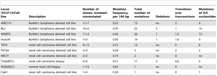
Frequent mutations inRASSF1Ain human carcinomas, cancer and haematopoietic cell lines
During analysis of RASSF1A we have isolated several mutant clones including one double mutant [16]. This high frequency of mutations was surprising since for RASSF1Aand other candidate genes in the AP20 and LUCA regions the mutation frequencies were reported to be low to none [6,19]. At the same time many polymorphisms were recorded forRASSF1Aand in many cases it was not clear whether it was a real single nucleotide polymorphism or somatic mutation in cancer cells because control normal cells were not available [8]. Importantly, in all these studies single-strand conformation polymorphism (SSCP) and direct sequencing from PCR products was used. The admixture of stroma, blood vessels, lymphocytes and other normal cells would hamper detection of mutations using these methods (see M/M). Tumor heterogeneity creates additional problems for recognizing mutations. Therefore we decided to re-investigate the mutational status ofRASSF1Ain multiple tumor types including primary tumors and cancer cell lines. First, RASSF1A cDNA was isolated from an RCC biopsy (T356) and the surrounding normally looking kidney parenchyma (N356). Several cDNA clones were sequenced. In six clones derived from normal kidney parenchyma, no mutations were found. However of seven clones from the tumor tissue, mutations were detected in three (P = 0.14). All were A to G substitutions. To exclude RNA editing, genomic DNA from the same patient was isolated and the first two exons (DAG domain) ofRASSF1Awere amplified by PCR. Several clones derived from normal and tumor tissue were then sequenced: all six clones from the tumor biopsy showed mutations while of the fourteen analyzed clones from normal tissue only one was mutated (P,0.001). The observed mutations in the cDNA from tumor cells were not created by RNA editing because the mutations were detected also on genomic DNA
level. Normal cell contamination and high expression ofRASSF1A in normal cells, compared to cancer cells, can explain the different ratios between mutated and normalRASSF1Aclones from cDNA and genomic DNA. Most surprising was the fact that with the exception of two genomic clones from the tumor biopsy with deletion of C at position 254 (Accession No. NM_007182), all other detected mutations were in different positions.
As a control we amplified GPR14from the same patient and sequenced 10 clones from cancer and from the surrounding normal tissue. No mutations were found proving that high mutation rate is specific for theRASSF1Agene.
To check whether different mutations in the same tumor occurred due to the tumor heterogeneity or some other mechanism(s), we isolated and sequenced RASSF1A clones (only exon 1 and 2; 391 bp) from genomic DNA of four RCC cell lines. In TK164 all three and in KRC/Y (2+2) all four sequenced clones contained mutations. In TK10, among 22 clones, 9 were mutated. Importantly, the majority of clones contained different mutations. Only one clone was sequenced from Caki1, and it was mutated.
We also sequenced this gene from genomic DNA of four lymphoid cell lines (BL2 and RAMOS are Burkitt cell lines, and IARC171 and MutuIII are lymphoblastoid cell lines) and the results were very similar to the RCC cell lines (Table 2). Altogether, among 84 sequenced clones 55 contained mutations that in most cases differed. MF inRASSF1Ain these experiments was between 0.14 and 0.70.
In all further experiments, we analyzed genomic DNA (exon 1 and 2 for RASSF1A and the whole RBSP3 transgene in pETE vector) if not specially mentioned.
Mutations inRASSF1Acan be generatedde novo To distinguish between the possibility that different mutated
RASSF1A genes were mutated at once (‘‘burst of mutations’’) or
were constantly generated over time, we performed experiments with single cells. In this experiment BL2 cells, (which previously showed the highest rate of mutation: 10 clones with 25 mutations), were diluted and plated into wells with an expected frequency of 0.3 cells per well. Three randomly selected wells (designated as BL2-cl.1, 2 and 3) containing single cells were expanded and further analyzed. DNA was isolated from these clones after 10 days (approximately 10 divisions, 103cells).
The results were as follows: for BL2-cl.1, five of 10 sequenced clones were mutated (mutation frequency per 100 bp (MF), was 0.14), for BL2-cl.2, five of 13 clones (MF = 0.15; two clones contained T43T mutations with codon changed from ACA to ACG) and for BL2-cl.3, three of 17 clones were mutated (MF = 0.07; two clones contained N70G mutations). Altogether 16 single base pair mutations were detected, all were transitions and only five of them showed mutated G or C. This experiment clearly shows that mutations in the RASSF1A locus could be generatedde novoduring cell proliferation.
The complete list of 129 mutations (111 mutations were different) found in exons 1 and 2 ofRASSF1Ain all experiments is shown in Table S1A. See also Table 2 and 3 and Figure 1A. Altogether 144 clones were sequenced (56,3 KB) and the average frequency of mutations was 0.23/100 bp for transcribed sequences and 0.17/100 bp for coding sequences. Among them, there were four nucleotide changes that occurred in non-coding 59, three stop (nonsense) and five frameshift (deletions) mutations. Of the remaining 127 mutations, 82 were missense and 35 synonymous.
RBSP3is also hypermutated in various cancers
ovarian carcinoma (OC) biopsies that all expressed RBSP3, we detected mutations in the RBSP3cDNA in all six cases [10; see Table S2A].
To test whether the hypermutation feature is a characteristic only of the RASSF1A gene or a more general phenomenon, we similarly analyzed the recently identified multiple tumor suppres-sor geneRBSP3located in AP20, 3p21.3 telomeric region [10].
Using RT-PCR, cDNA was isolated from two of each RCC, BC and OC biopsies and the SCLC cell line N417. Multiple clones were sequenced. Results, presented in Table S2A, Table 3 and Figure 1B, showed that almost all isolated clones suffered mutations. As reverse transcriptase used in RT-PCR has a significantly higher error rate than other polymerases used in PCR, we attempted to reproduce the observed high mutation rate at the genomic DNA level, as in the case with RASSF1A. Unfortunately, it was difficult to perform this experiment on the genomic RBSP3 due to the large size of the gene (more than 120 kb), numerous small exons (at least 9), and high GC content (reaching 100% in some regions). However this problem was solved using clonedRBSP3in SCID mice.
RBSP3revealed high mutability in SCID mice on genomic level
SCLC cell line ACC-LC5 and RCC cell line KRC/Y were transfected with RBSP3A and RBSP3B splicing isoforms in the pETE vector and stable cell clones were isolated. Four of these clones (AHA1 and AHB1 for ACC-LC5 and KHA4 and KHB9 for KRC/Y) were inoculated into SCID mice (see M/M).
Cell clones KHA4 and KHB9, containingRBSP3AorRBSP3B were grownin vitroin parallel with tumors in SCID mice. After 8
weeks DNA was isolated from grown tumors and cell lines, and the
RBSP3AandBgenes were amplified by PCR from pETE vector
and cloned. Again multiple clones were sequenced and results of the experiment are shown in Table 4 and Table S2B. Only 30% of
RBSP3KHA4 and KHB9 plasmid clones were mutatedin vitro, as
compared to 85% mutated clones after growth in SCID mice. This difference according to Fischer test is statistically significant (P,0.001).
In summary, inRBSP3experiments we identified 89 mutations among which 79 were individually distinct (see Tables S2A and S2B). The average frequency of mutations was 0.10/100 bp for transcribed sequences. This frequency is more than 0.11/100 bp for coding sequences (see Table 3 and Figure 1B). Among them, seven nucleotide changes occurred in non-coding regions and five were frameshift (deletions) mutations. Of the remaining 77 mutations, 68 were missense and 9 synonymous.
Thus, the mutation frequency was 2.5 fold less than for the first two exons of theRASSF1A(see above). The significant difference in mutation frequencies could be accounted for by differences in nucleotide composition of the genes, or it could reflect intrinsic differences in the hypermutation rates of the genes. It could also be important that for theRBSP3the whole gene was sequenced while for theRASSF1Aonly its 59end.
RASSF1AandRBSP3amplified by PCR fromE.coli DNA don’t show high frequency of mutations
We have performed PCR amplification of E. coli DNA containing plasmids (i.e. total DNA isolated fromE.colicontaining mixture of genomic and plasmid DNA) with these two genes. For each gene ten and four ng of DNA was used. Unfortunately lower Table 2.Mutations inRASSF1Aexon 1and 2 in different cell types.
Locus RASSF1A/Cell
line Description
Number of clones, mutated+
nonmutated
Mutation frequency, per 100 bp
Total number of
mutations Deletions
Transitions over transversions
Mutations of G/C nucleotides
IARC171 Burkitt’s lymphoma derived cell line 11+7 0.23 15 no 2 9
BL2 Burkitt’s lymphoma derived cell line 10+0 0.70 25 3 1 16
RAMOS Burkitt’s lymphoma derived cell line 11+2 0.56 26 1 1.2 15
mutuIII Burkitt’s lymphoma derived cell line 7+0 0.56 14 1 1.6 9
TK10 renal cell carcinoma derived cell line 9+13 0.15 12 no 0 8
TK164 renal cell carcinoma derived cell line 3+0 0.28 3 no 2 2
KRC/Y renal cell carcinoma derived cell line 2+2 0.14 2 no 0 no
T356(RCC) renal cell carcinoma biopsy 6+0 0.51 11 3 0.6 5
N356(RCC) normal renal cell biopsy 1+13 0.02 1 no 0 no
Caki1 renal cell carcinoma derived cell line 1+0 0.28 1 no 0 1
doi:10.1371/journal.pone.0005231.t002
Table 3.Experimental mutations frequency in theRASSF1AandRBSP3genes.
Gene, length
Number of sequenced clones
Total length/coding
sequences, Kbp Number of mutations
Mutation frequency, per 100 bp
total In coding region total In coding region
RASSF1, exons 1–2 144 56.3/51.4 129 89 0.23 0.17
RASSF1, exons 3–5 98 50.6 146 145 0.29 0.29
RBSP3, exons 1–8 85 85.3/70.6 89 79 0.10 0.11
amount ofE. coliDNA didn’t produce sufficient amount of PCR products for further cloning. Ten clones in each experiment were sequenced and no mutations were detected. These results indicate that the observed hypermutation rate of RASSF1A and RBSP3 cannot be explained by PCR polymerase errors.
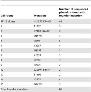
Search for founder mutations inRASSF1Ain single cell clones
The main idea of this experiment was the following. If a mutation originates in the cell and not in the tubein vitrothen in the cell population grown from one cell some fraction (depending on the number of alleles present in the single cell) of plasmid clones should contain the same (i.e. a founder) mutation. To perform this
experiment we isolated 15 single cell KRC/Y clones as described in the section ‘‘Mutations generated de novo’’. In this case we grew the cells for three weeks to obtain more DNA and generate more mutated clones. KRC/Y cells were used instead of BL2 cells as it was easier to detect that we have one cell in the well. However, we of course cannot exclude that in some of the 15 selected wells there were more than one cell.RASSF1Aexons 3–5 were tested in this experiment (see M/M) as they were more easily isolated than exons 1 and 2 and contained more sequence information (516 nt vs. 391 nt). Moreover, according to EST sequence data this part of
RASSF1A has higher MF. From each PCR reaction 10 plasmid
clones were selected and DNA was isolated. However, due to different technical problems (no or rearranged insert, bad quality DNA or sequencing, etc.) usually only six-seven plasmid clones were further analysed. Totally 98 plasmid clones were sequenced (Table 5). One founder mutation was detected in all cell clones and in 46 (47%) of plasmid clones. It was a change of A to G (nt26735, Accession No. AC002481) just at the border of intron 2 and exon 3. This mutation destroyed the splice acceptor site AG/G and thus inactivated the gene. As this founder mutation appeared in all single cell clones most probably it originated before we started to do this experiment. Forty other founder mutations specific for each cell clone were also detected (see Table 5 and Table S1B). Interestingly in one case it was possible to construct a tree showing how founder mutations were accumulated. First it was only one mutation than two and then additional independent mutations (Figure 2).
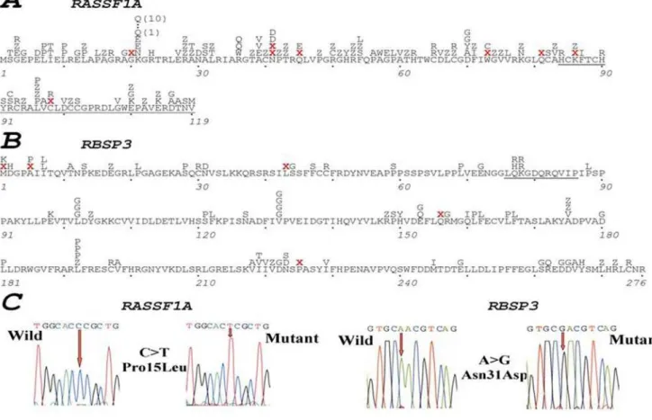
Figure 1. Mutations inRASSF1AandRBSP3in natural and experimental tumors.Position of mutations detected inRASSF1AandRBSP3is
shown in A and B respectively. Examples of mutations are shown in C. ForRASSF1Aonly mutations in coding sequences of exons 1 and 2 are shown. Mutations in the whole coding part of RBSP3 are shown. Red ‘‘X’’ marks stop nonsense mutations or deletions. ‘‘Z’’ designates synonymous mutations.
doi:10.1371/journal.pone.0005231.g001
Table 4.Mutation frequency of theRBSP3AandRBSP3B
genesin vitroandin vivoin the gene inactivation test.
Gene/cell clone In vitro In vivo
tested mutated tested mutated
RBSP3A/KHA4 11 3 13 10
RBSP3B/KHB9 12 4 15 14
Total 23 7 28 24
Different mutation frequencies in other genes
Similar sequencing experiments were performed with insulin and albumin genes isolated from KRC/Y cell line (see M/M). In contrast toRASSF1AandRBSP3results, only one of 21 sequenced insulin genomic clones (999 bp including complete ORF) and one of 19 albumin cDNA clones (700 bp, exons 12–15) were mutated (MF for both genes was less than 0.01). However in both cases, we could not exclude the possibility of polymorphisms. Additionally, two more genes were tested for mutations in genomic DNA.
GPR14(G protein-coupled receptor 14, 1018 bp) and
transcrip-tion elongatranscrip-tion factor A (SII) TCEA1 (1066 bp) were PCR amplified (see M/M) from DNA of KRC/Y cells and 11 clones for each gene were sequenced. All clones contained normal copies of the gene. No mutations were found in other 3p21.3 candidate genes:BLU(15 clones were sequenced), 101F6(6 clones),PL6(6 clones) after KRC/Y stable clones containing these genes were inoculated into SCID mice. Moreover, for already mutated mutFUS1 (10 clones) and mutP53 (6 clones) no additional mutations were found (data not shown).
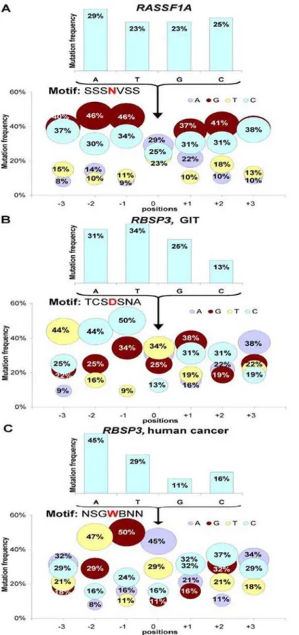
Mutations inRASSF1A,RBSP3AandRBSP3Bare not generated by AID or APOBEC related mechanisms
It has been recently shown that the activation-induced cytidine deaminase (AID) is responsible for somatic hypermutations in activated B cells. Moreover hypermutations generated by this enzyme in oncogenes can cause malignancies in haematopoietic cells [24]. Although much remains to be learned concerning AID, several target sequence motifs for the mutations have been identified, namely WRC, RGYW and DGYW causing C/G mutations. The large family of APOBEC genes, also shown recently to mutate genes on DNA level [21,22], mostly targeted the RCW motifs causing mutations in C/G. Therefore, we checked whether these motifs were targeted or more frequent in
RASSF1 and RBSP3 sequences when compared to the stable
insulin gene. The frequency of the WRC motif per 100 bp varies from 12.3 to 14.3 forRASSF1andRBSP3genes, and the insulin gene contains 16.5 such motifs per 100 bp. Other motifs showed the same distribution (also higher in the insulin gene), arguing against the involvement of these enzymes in hypermutating the
RASSF1andRBSP3genes described here. Indeed, the APOBEC
and AID enzymes cause mutations almost exclusively in C/G nucleotides, while we observed mutations of all 4 nucleotides (Figure 3). The results actually showed that mutations in A/T were even more frequent than in C/G. We tried to find a recognition motif. We studied all mutations (Figure 3A) or a subset of mutations (Figures 3B and 3C), but no obvious motifs have been yet identified.
More studies are needed to resolve this question as this pattern can be different in normal and cancer cells and could be dependent on nucleotide composition of a gene. These small differences in patterns could mask the recognition motif.
RASSF1AandRBSP3mutants from RCC biopsy and lung cancer cell line have significantly reduced growth-inhibiting activity
We tested one RASSF1A gene, isolated from an RCC biopsy that contained two mutations (Cys65Arg and Val211Ala), for growth inhibition under cell culture conditions following transfec-tion into the KRC/Y and prostate cancer LNCaP cells. In KRC/ Y cells the mutated gene had a significantly reduced growth suppression activity (Figure 4A) while in LNCaP it had almost no suppressing activity (same as the empty vector, see [16,17]).
In another experiment, we used RBSP3clones isolated from N417 SCLC cell line with a His139Tyr mutation. Again significant decrease in growth suppressor activity was observed (Figure 4B).Clearly, not all mutations found in this study would inactivate the RBSP3 and RASSF1 genes, and this may be especially true for mutants isolated from normal cells some of which could be polymorphisms. Indeed, different mutants of
RBSP3 had significantly different growth suppression activity
(Figure 4B).
Conclusions
By sequencing 327 RASSF1A and RBSP3 clones, we detected 364 mutations with frequencies reaching 0.70 per 100 bp. Interestingly many clones contained more than 1 mutations (see Table S3A, B, C). Only one SNP was detected inRASSF1Aten clones (exon1a– AAGRCAG, K21Q) and it was excluded from the list of mutations [http://www.ncbi.nlm.nih.gov/SNP/snp_ref. cgi?locusId=11186]. No SNP were found in RBSP3 sequences.
The frequency of mutations was similar to other reported cases of somatic hypermutations found in Rho/TTF, MYC and BCL6 in large-cell lymphomas (MF was from 0.12 for MYC to 0.69 for Figure 2. Flow chart showing accumulation of mutations
(including two founder mutations) in RASSF1A exons 3–5 in
the single cell clone #9. Synonymous mutation Pro122Pro was
caused by nucleotide change ATCRAAC. Mutation GTCRGTA also didn’t result in any amino acid change (Val174Val).
doi:10.1371/journal.pone.0005231.g002
Table 5.Founder mutations in single cell KRC/Y clones.
Cell clone Mutation
Number of sequenced plasmid clones with founder mutation
All 15 clones nt26.735(ARG) 46
1 T196T 3
2 R240R, K241R 2
3 D157N 4
4 I139T 5
4 V225A 4
5 N155S 3
8 K232R 3
9 I139N 3
9 I146N 2
10 L256W, P274P 2
12 E126G 2
14 L260S 4
15 D262G 3
Total founder mutations 86
Figure 3. Distribution of mutations inRASSF1A(A) andRBSP3(BandC).ForRASSF1Aall mutations were analyzed. ForRBSP3mutations found in GIT (B) and in human cancer (C) were analyzed separately. Bubble graphs depict the proportion of substitutions occurring at each of the four bases in theRASSF1AandRBSP3, depending on the distance from the mutated nucleotide (No. 0). N, any nucleotide;B = C, G or T; D = A, G or T; S = G or C; V = A,C, or G; W = A or T.
BCL6). However it was significantly lower than for immunoglob-ulin genes (12.7 mutations per 100 bp, see [25]. However, for the first time we found high frequency of somatic mutations in different tissues including non-haematopoietic and in tumor
suppressor genes contrary to the previous reports where oncogenes were studied.
As AccuPrimeTMPfx DNA polymerase creates maximally one error in 36106bp, our results proved that the observed
Figure 4. Reduced growth inhibiting activity ofRASSF1A(A) andRBSP3A(B) mutants.A. Growth of stably transformed KRC/Y RCC cells with
wild type and mutantRASSF1A(Cys65Arg and Val211Ala) without doxycycline (the gene is on) is presented in A. On day 6, the number of cells with wtRASSF1Awas 36105and the number of cells with mutantRASSF1Awas 56105(1.7 times more than wt). On day 10, the number of cells with wt RASSF1Awas 66105and the number of cells with mutantRASSF1Awas 1.86106(3 times more than wt). The effect of expression of wild type and
hypermutation frequencies in the experiments could not be explained by erroneous performance of polymerases. In our experiment with SCID mice when AccuPrimeTMPfx DNA polymerase and 25 cycles were used, 85% of RBSP3 clones contained mutations.
During the growth of the same cell linesin vitro, 30% ofRBSP3 clones (also 25 cycles and AccuPrimeTMPfx DNA polymerase) were mutant.
In experiments withRASSF1A (391 bp of the first and second exons), 65% of clones contained mutations (experiments with normal cells are not included). Moreover, in our experiments, we used different polymerases with different error rate (see M/M) and no significant differences in mutation frequency were observed, arguing against the generation of the mutations during PCR amplification. Different mutation frequencies betweenin vivoand
in vitroexperiments and in ESTs isolated from normal and cancer
cells is an additional argument against the artificial nature of the hypermutation rate observed inRBSP3andRASSF1genes.
Mutations were detected with similar frequency both in cDNA and genomic DNA for RBSP3 and RASSF1, however, no high mutability either on genomic or cDNA level were found for albumin, insulin, GPR14, TSG p16/INK4a or transcription elongation factor A (SII) TCEA1. Moreover, no mutations were found in experiments with SCID mice for 5 genes:BLU,101F6, PL6, mutFUS1and mutP53. Expression ofRBSP3[10] in six tested samples differed almost 50-fold and on genomic levelRBSP3was present usually in 3–8 copies [26]. Our previous experiments using marker NL3-001 located 90 kb apart from the RASSF1A demonstrated that in tumor cells this region in most cases is present in 1–5 copies [4,5]. Still the frequency of mutations was almost the same. Thus number of template molecules didn’t influence significantly mutability level. Moreover, repeated sequencing of the same plasmid and isolated by different persons and at different time gave identical results (6 RASSF1A and 6
RBSP3 plasmid clones were sequenced) excluding frequent
sequencing errors.
Both genes are CG rich however it seems that although high CG content can induce additional mutations it cannot explain the fact that two genes with significantly different CG content
(RASSF1A, exons 1–2, 72.3%; RASSF1A, exons 1–6, 59.8% and
RBSP3, exons 1–8, 54.3%) both possess high mutability while
other genes with similar CG content (e.g.GPR14, 72.5%; p16/
INK4a, 71.6%; insulin 61.6%) didn’t show any high frequency of
mutations.
Using the same PCR conditions plasmids containingRBSP3and
RASSF1A were amplified from E.coli and no mutations were
discovered arguing against generation of mutations during PCR amplification.
Experiments to find founder mutations with single-cell clones additionally confirm that mutations originate in the cell. Interestingly that from the single cell clone No. 9 we isolated plasmids with one, two or three mutations. This fact clearly showed how these mutations originate from one parental cell clone (Figure 2). Importantly after sequencing exons 3–5 of RASSF1A gene from KRC/Y we discovered founder mutation (destroying splice acceptor site) that was present in approximately 50% of 98 sequenced plasmid clones. This founder mutation appeared in all single cell clones and thus most likely it originated before we started this experiment.
For identification of tumor suppressor genes, we use the gene inactivation test, GIT [26,27]. This test is based on the functional inactivation of the analyzed genes during tumor growth in SCID mice. Our hypothesis was that under selective pressurein vivothe introduced TSG must be inactivated in growing experimental
(xenografted) tumors (by deletion, mutation, promoter methyla-tion) as in the naturally growing tumors. The expression of the tested gene in the GIT was regulated by tetracycline and the level of expression was under physiological conditions. In our published papers [10,23] wild type RBSP3 and wild type and mutated
RASSF1A genes were tested in GIT. The genes were PCR
amplified from tumors and sequenced. In contrast to the wild type
RBSP3andRASSF1genes, that were inactivated (i.e. deleted,
non-expressed, mutated) in all 32 grown tumors, the mutantRASSF1A was not additionally mutated in any of four analyzed tumors. Importantly, in these GIT experiments we used direct sequencing of PCR products. These experiments showed that ‘‘founding mutations’’ really do exist.
Analysis of public EST databases confirmed our experimental data. It should be noted that the frequency of mutations in
RASSF1A and RBSP3 found in EST databases even using very
stringent criteria was significantly higher than found in our experiments. MF for all mutations for RBSP3was 0.63 and for
RASSF1it was 0.22. Probably, this discrepancy could account for
the differences between the cell types analyzed in our experiments and in the EST database.
Unfortunately only 17 RASSF1A clones could be analysed because other EST sequences were either not sufficiently good or could be other isoforms of theRASSF1gene.
Interestingly, mutations of RASSF1A and RBSP3 changing amino acids were found even in clones isolated from normal cell RNA, however, at a lower frequency than in cancer cells (MF ratios for cancer/normal sequences were 3.3 and 3, respectively). This difference for both genes was statistically significant (P,0.001) This probably reflects the selection for and the advantage of coding mutations during cancer progression. Important to mention that ‘‘normal’’ sequences include also non-annotated sequences so we cannot exclude that some of the ‘‘normal’’ sequences actually represent cancer cells.
In fact, these results correlate with the data from the mousein vivoexperiments that showed a higher frequency of mutations in SCID tumors than in the same cells grownin vitro. Interestingly, the same mutations were observed in cells grownin vitroandin vivo, in SCID mice (see Table S2B).
We have also experimentally tested whetherRASSF1A(genomic DNA, exons1 and 2) harbored mutations in normal tissues and found one mutated clone out of 14 in normal kidney (normal control to T356, see section ‘‘Frequent mutations inRASSF1Ain human carcinomas’’). Important to note that so called ‘‘normal’’ kidney could be already partially transformed despite of normal phenotype because it was obtained from tissues adjacent to the tumor. We also sequenced completeRASSF1AcDNA from normal heart and detected six mutated clones out of 15 tested. All six heart mutated clones contained the same two mutations: L214L with codon changed from CTA to CTG and V236V with codon changed from GTA to GTG. Mutations in heartRASSF1cDNA were most likely SNP as they could be also found in otherRASSF1 clones in public databases (e.g. AC002481, NM_170713.2, NM_170714.1). In any case it is clear that mutations inRASSF1 in normal cells are more rare than in cancer cells.
As we found mutations in all 5 coding exons ofRASSF1A(the last 6thexon contains only 48 amino acids = 144 bp). It means that other six known isoforms ofRASSF1are also frequently mutated. Exceptionally high level of germ line SNP mutations in
RASSF1A found in several studies [8] support our data that the
two genes we studied have rather frequent mutations even in normal cells.
by polymerase errors. This is the first report of high mutation frequencies of RASSF1 and RBSP3 genes in different epithelial malignancies. In our preliminary paper [28] we analyzed mutations in RASSF1A gene in NPC samples and the results supported the present observations. In the NPC experiments 35 mutations were detected in 23 patients and mutations were considered real if at least two clones from the same patient contained the same mutation. Ten clones for each sample were sequenced in these experiments. Both DNA strands were sequenced.
At present, we don’t know the nature of the mechanism responsible for this hypermutability, and only speculations could be done for its physiological function(s) in normal cells. There are several DNA polymerases in vertebrate cells that inaccurately copy templates and could be involved in generating hypermutations [29]. One of them, POLH (error rate 361022), has a mutation target
motif WA and may contribute to hypermutagenesis of immuno-globulin genes at A-T bases [30].POLHis expressed in all tissues and, in principle, could cause hypermutations in non-haematopoi-etic cells. We found that 50% of all observed mutations inRBSP3 happened in A or T surrounded by G or C. That means that the mutation target motif for 50% of mutations inRBSP3is SWS and is different from the POLH motif. It is reasonable to suggest that other(s) yet unknown DNA polymerase(s) may be responsible for the high mutability rates we report here and more than one polymerase contributes to hypermutations [29,31].
Our results also argue that mutations are not completely random. They are not correlated with predicted numbers (Tables 1, 2, 3). For example according to statistical calculations our sequences of
RASSF1Aexons 1 and 2 should contain 0.026 nonsense mutations
but in reality we detected 3 nonsense mutations, P,0.001 (see Table S1A). ForRASSF1Aexons 3–5 the predicted number of nonsense mutations is 0.037 and we found 3 such mutations, P,0.001 (see Table S1B). This fact may reflect the nature of cancers and normal tissues studied here. We cannot also exclude that these mutations still have some preferable motif(s).
We mentioned in the text that clonal selection for more aggressive growth of cancer cells could add to changing proportion of different mutations. In our previous paper [28] we also observed an unusual distribution of mutations. Among 35 detected mutations we found 30 transitions, 3 transversions, 2 deletions (frameshift), 3 nonsense (stop), 26 missense and only 4 were synonymous.
High frequency of mutations in different cancers and normal cells was reported earlier for P53 [32]. However, at present it is difficult to compare these results with our study as different methodologies were used and most likely different mechanisms of mutagenesis were involved.
When this manuscript was completed two new publications appeared in PNAS that support our observations and concept [33,34].
Interestingly, in the paper of Yang et al. [35] hypermutability was demonstrated in damaged single-strand DNA formed at double–strand breaks in yeastS. cerevisia. Although yeast data may not apply to human cells, it is worthwhile to note that AP20 and LUCA regions whereRASSF1andRBSP3are located were found extensively damaged (deletions, amplifications) in 90% of studied major epithelial cancers [2, 4, 5, see Introduction].
Materials and Methods
Ethics Statement
All work with mice was performed in special ‘‘Animal House’’ in MTC according to the standard rules. The study was done in
accordance to the guidelines (incl. husbandry) issued by the STOCKHOLMS Norra Djurforsoksetiska Namnd (Animal Ethic Committee of North Stockholm).
Paired tumor/normal samples were obtained from the Blokhin Cancer Research Center, Russian Academy of Medical Sciences after surgical resection of primary tumors and stored in liquid nitrogen.. Top and bottom sections (3–5mm thick) cut from frozen tumor tissues were examined histologically and only samples containing 70% or more tumor cells were used in the study. The samples were collected in accordance to the guidelines issued by the Ethic Committee of the Blokhin Cancer Research Center, Russian Academy of Medical Sciences (Moscow). All patients gave written informed consent that is available upon request. The study was done in accordance with the principles outlined in the Declaration of Helsinki.
Cell lines and experiments with SCID mice
Cell lines were obtained from the MTC-KI (Stockholm, Sweden) cell lines collection. Cell and tumor growth assays were done as described previously [13,16,23,26]. GIT was performed as described previous [16,26,27].
In brief, plasmid DNAs were purified using R.E.A.L. Prep kit (Qiagen, Valencia, CA). Transfections were performed using LipofectAMINE PLUS Reagent (Life Technologies, Rockville, MD) according to the manufacturer’s protocol. After transfection, cells were selected with 5mg/ml Blasticidin and 200 ng/ml doxycycline for two-four weeks. For colony formation assay cells were selected for 2 weeks, fixed, stained with Giemza and counted for transfection efficiency. For isolation of stably transfected cell clones, selection was done for four weeks. PCR positive clones from each recombinant were tested for expression using Northern hybridization and selected clones, 56106 cells/mouse, were
inoculated subcutaneously with or without Matrigel (BD, Franklin Lakes, NJ) into six-week-old female SCID mice. Each mouse received only 1 injection. SCID mice were observed for tumor formation twice a week for up to seven weeks, if tumor formation was observed, tumors were measured using calipers. The tumors were explanted for DNA preparations.
General methods
All molecular biology and microbiology procedures were performed as described previously [10,13,36]. DNA and RNA were isolated from total tumor samples containing less than 30% of non-tumor cells according to histopathology examination.
Construction of pETE vector and KRC/Y and LNCaP cell lines producing tetracycline trans-activator tTA were described in ref. [26].
Polymerases used for PCR
In experiments with cell lines and biopsies we used natural Taq polymerase (New Englands Biolabs, Ipswich, MA, USA) and JumpStartTMAccuTaqLA DNA Polymerase (Sigma-Aldrich, St. Louis, MO, USA). In some experiments (for comparison) we used AccuPrimeTMPfx DNA polymerase (Invitrogen, Carlsbad, CA, USA). No significant difference was observed between these three polymerases. Usually 30 cycles were used.
In experiments with single cell clones and SCID mice AccuPrimeTMPfxDNA polymerase and 25 cycles were employed. Natural Taq polymerase has an error rate 4.5–561025 (i.e.
available (15–26 fold better fidelity than ordinary Taq polymerase) AccuPrimeTMPfxDNA polymerase and only 25 cycles.
The size of theRBSP3Bis 1003 bp and the accuracy of ordinary Taq polymerase is approximately one error in 26105bp. This means that after 30 cycles 15% of clones would be expected to contain mutation(s) in theRBSP3and after 25 cycles 12.5%. In the case of AccuPrimeTMPfxDNA polymerase, after 30 cycles 1% of clones would be mutant and after 25 cycles only 0.84%. In our experiment with SCID mice, 85% of RBSP3 clones contained mutations (AccuPrimeTMPfxDNA polymerase, 25 cycles).
During the growth of the same cell linesin vitro, 30% ofRBSP3 clones (also 25 cycles and AccuPrimeTMPfx DNA polymerase) were mutant.
PCR and Sequencing
PCR primers were purchased from Invitrogen (Carlsbad, CA, USA). PCR was done as described earlier [2]. Initial denaturation was done for 2 min at 95uC following 25–30 cycles: 95uC for 15 sec, 64uC for 30 sec and extension at 68uC for 1 min per 1 kb.
RBSP3A and RBSP3B: gene fragments (ORF) have been
obtained by PCR from cDNA isolated from different cell lines using the following primer sets, according to manufacturer’s manual.
RBSP3B. 120C: 59
-GCGGCCGCCGCGCCGCGCACC-CATGGACGGCCCGGCCATC-39 (nucleotides 1-40) and HYA22C: 59 -AAAACAAAACAGGTAGGCATGGCCA-CATTC-39 (nucleotides 1003-973). See GenBank Accession No. AJ575645
RASSF1A: genomic fragments (GenBank Accession
No. AC002481).
Ex1–Ex2. F2A: 59-GCCCAAAGCCAGCGAAGCAC-39 (nu-cleotides 18051-18070) and EX2F2: 59 -ACCCAGG-CAGCCCTCGAGAA-39(nucleotides 21066- 21047).
Ex3–Ex5. RassF1-2intrF: 59-TGT CCA TGC TGG CCC ATC TTG C-39(nucleotides 26713-26734) and RassF1-5exR: 59-CAC CTC CCC AGA GTC ATT TTC CTT C-39(nucleotides 27530-27554).
RASSF1A: cDNA fragment cDNA (ORF):
F2A: 59-GCCCAAAGCCAGCGAAGCAC-39(nucleotides 97-116) and
F2B: 59-AGCCATACCT GGCTACACCCACAGG-39 (nucle-otides 1343- 1319),
see GenBank Accession No. NM_007182 GPR14: genomic fragment (ORF).
GPR14F: 59 - CCCATCTCAGGGAGTGTCCA - 39 (nucle-otides -52 - 33),
and GPR14R: 59 - GTAGTTCCTGGTGAGCAGCGTG-TAG - 39 (nucleotides 966 - 942), see GenBank Accession No. NM_018949
TCEA1P2: genomic fragment (ORF).
TCEA1F: 59- TTTGTGAGGAAGGGGGCCTA - 39 (nucle-otides 705 - 724),
and TCEA1R: 59 - ATATTTTGCCAATTCTTCCAACT-CAACA - 39 (nucleotides 1775 - 1748), see GenBank Accession No. X73534
pETE primers [26]:
LiTetF: 59- GCCTATATAAGCAGAGCTCGTTTAG - 39 AtetR: 59- CCAAACTCATCAATGTATCTTATCA - 39 Insulin: genomic fragment (ex1–ex3).
InsF: 59-CTGTCACCCAGATCACTGTCCTTC-39 (nucleo-tides 546-569) and InsR: 59
-GGGCTGCGTCTAGTTGCAG-TAGTT-39 (nucleotides 1702-1679), see GenBank Accession No. AY138590.1.
Albumin: cDNA fragment (ex12–ex15). AlbF: 59 -GAAC-CAGTTATGTGTGTTGCATGAGAA-39 (nucleotides 1482 -1508), and AlbR: 59 -CCCACAGAAACTAGAAATCCTC-TACCG-39 (nucleotides 2181 -2155), see GenBank Accession No. NM_000477.3.
All experiments were performed using Gene Amp PCR System 9700 (Perkin Elmer, Foster City, CA, USA).
PCR products were cloned, using the TOPO TA cloning kit for sequencing (Invitrogen). Plasmid DNA was purified using the R.E.A.L.- Prep kit (Qiagen, Valencia, CA). Sequencing was done using an ABI 310 Sequencer (Applied Biosystems, Foster City, CA), according to the manufacturer’s protocol.
Bioinformatics
ForRASSF1Aonly exons 1 and 2 with a total length of 357 bp
(Acc.No. NM_007182) were analyzed. For RBSP3 the longest isoform B (Acc.No. AJ575645; the total length is 831 bp) was analyzed. The gene sequences were searched against GenBank EST division, a collection of expressed sequence tags, or short, single-pass sequence reads from mRNA (cDNA). The statistically significant thresholds for the alignment (score) that provided elimination of alien mRNA sequences was set forRASSF1Aat 462 and for RBSP3 at 404. These thresholds were obtained from expertise estimation to cut off clusters of short and non-significant homologies to the query sequences. An additional manual refinement against low quality sequences was performed. Nucle-otide similarity searches were performed with BLAST 2.2.
In all experiments we always compared a given sequence with the annotated sequences as shown in previous paragraph.
Probabilities of mutation frequency differences were calculated using Poisson distribution.
Supporting Information
Table S1
Found at: doi:10.1371/journal.pone.0005231.s001 (0.39 MB DOC)
Table S2
Found at: doi:10.1371/journal.pone.0005231.s002 (0.16 MB DOC)
Table S3
Found at: doi:10.1371/journal.pone.0005231.s003 (0.15 MB DOC)
Acknowledgments
Authors are grateful to Drs. A. Malyukova and Y. Seryogin for assistance in some experiments and to Ms Laura Geil for help in editing the manuscript. We thank Dr. Bert Vogelstein for important suggestions on the early stage of this work.
Author Contributions
References
1. Alimov A, Kost-Alimova M, Liu J, Li C, Bergerheim U, Imreh S, et al. (2000) Combined LOH/CGH analysis proves the existence of interstitial 3p deletions in renal cell carcinoma. Oncogene 19: 1392–1399.
2. Braga E, Senchenko V, Bazov I, Loginov W, Liu J, Ermilova V, et al. (2002) Critical tumor-suppressor gene regions on chromosome 3P in major human epithelial malignancies: allelotyping and quantitative real-time PCR. Int J Cancer 100: 534–541.
3. Li J, Protopopov A, Wang F, Senchenko V, Petushkov V, Vorontsova O, et al. (2002) NotI subtraction and NotI-specific microarrays to detect copy number and methylation changes in whole genomes. Proc Natl Acad Sci U S A 99: 10724–10729.
4. Senchenko V, Liu J, Braga E, Mazurenko N, Loginov W, Seryogin Y, et al. (2003) Deletion mapping using quantitative real-time PCR identifies two distinct 3p21.3 regions affected in most cervical carcinomas. Oncogene 22: 2984–2992. 5. Senchenko VN, Liu J, Loginov W, Bazov I, Angeloni D, Seryogin Y, et al. (2004) Discovery of frequent homozygous deletions in chromosome 3p21.3 LUCA and AP20 regions in renal, lung and breast carcinomas. Oncogene 23: 5719–5728. 6. Zabarovsky ER, Lerman MI, Minna JD (2002) Tumor suppressor genes on chromosome 3p involved in the pathogenesis of lung and other cancers. Oncogene 21: 6915–6935.
7. Imreh S, Klein G, Zabarovsky ER (2003) Search for unknown tumor-antagonizing genes. Genes Chromosomes Cancer 38: 307–321.
8. Dammann R, Schagdarsurengin U, Strunnikova M, Rastetter M, Seidel C, Liu L, et al. (2003) Epigenetic inactivation of the Ras-association domain family 1 (RASSF1A) gene and its function in human carcinogenesis. Histol Histopathol 18: 665–677.
9. Shivakumar L, Minna J, Sakamaki T, Pestell R, White MA (2002) The RASSF1A tumor suppressor blocks cell cycle progression and inhibits cyclin D1 accumulation. Mol Cell Biol 22: 4309–4318.
10. Kashuba VI, Li J, Wang F, Senchenko VN, Protopopov A, Malyukova A, et al. (2004) RBSP3 (HYA22) is a tumor suppressor gene implicated in major epithelial malignancies. Proc Natl Acad Sci U S A 101: 4906–4911. 11. Buermeyer AB, Deschenes SM, Baker SM, Liskay RM (1999) Mammalian DNA
mismatch repair. Annu Rev Genet 33: 533–564.
12. Protopopov A, Kashuba V, Zabarovska VI, Muravenko OV, Lerman MI, Klein G, et al. (2003) An integrated physical and gene map of the 3.5-Mb chromosome 3p21.3 (AP20) region implicated in major human epithelial malignancies. Cancer Res 63: 404–412.
13. Li J, Wang F, Haraldson K, Protopopov A, Duh FM, Geil L, et al. (2004) Functional characterization of the candidate tumor suppressor gene NPRL2/ G21 located in 3p21.3C. Cancer Res 64: 6438–6443.
14. Spandidos DA (1986) A unified theory for the development of cancer. Biosci Rep 6(8): 691–708.
15. Bishop JM (1987) The molecular genetics of cancer. Science 235(4786): 305–311.
16. Dreijerink K, Braga E, Kuzmin I, Geil L, Duh FM, Angeloni D, et al. (2001) The candidate tumor suppressor gene, RASSF1A, from human chromosome 3p21.3 is involved in kidney tumorigenesis. Proc Natl Acad Sci U S A 98: 7504–7509.
17. Kuzmin I, Gillespie JW, Protopopov A, Geil L, Dreijerink K, Yang Y, et al. (2002) The RASSF1A tumor suppressor gene is inactivated in prostate tumors and suppresses growth of prostate carcinoma cells. Cancer Res 62: 3498–3502. 18. Dammann R, Li C, Yoon JH, Chin PL, Bates S, Pfeifer GP (2000) Epigenetic inactivation of a RAS association domain family protein from the lung tumour suppressor locus 3p21.3. Nat Genet 25: 315–319.
19. Lerman MI, Minna JD (2000) The 630-kb lung cancer homozygous deletion region on human chromosome 3p21.3: identification and evaluation of the
resident candidate tumor suppressor genes. The International Lung Cancer Chromosome 3p21.3 Tumor Suppressor Gene Consortium. Cancer Res 60: 6116–6133.
20. Muto T, Muramatsu M, Taniwaki M, Kinoshita K, Honjo T (2000) Isolation, tissue distribution, and chromosomal localization of the human activation-induced cytidine deaminase (AID) gene. Genomics 68: 85–88.
21. Beale RC, Petersen-Mahrt SK, Watt IN, Harris RS, Rada C, Neuberger MS (2004) Comparison of the differential context-dependence of DNA deamination by APOBEC enzymes: correlation with mutation spectra in vivo. J Mol Biol 337: 585–596.
22. Conticello SG, Thomas CJ, Petersen-Mahrt S, Neuberger MS (2005) Evolution of the AID/APOBEC family of polynucleotide (deoxy)cytidine deaminases. Mol Biol Evol 22: 367–77.
23. Li J, Wang F, Protopopov A, Malyukova A, Kashuba V, Minna JD, et al. (2004b) Inactivation of RASSF1C during in vivo tumor growth identifies it as a tumor suppressor gene. Oncogene 23: 5941–5949.
24. Okazaki IM, Hiai H, Kakazu N, Yamada S, Muramatsu M, Kinoshita K, et al. (2003) Constitutive expression of AID leads to tumorigenesis. J Exp Med 197: 1173–1181.
25. Pasqualucci L, Neumeister P, Goossens T, Nanjangud G, Chaganti RS, Kuppers R, et al. (2001) Hypermutation of multiple proto-oncogenes in B-cell diffuse large-cell lymphomas. Nature 412: 341–346.
26. Protopopov AI, Li J, Winberg G, Gizatullin RZ, Kashuba VI, Klein G, et al. (2002) Human cell lines engineered for tetracycline-regulated expression of tumor suppressor candidate genes from a frequently affected chromosomal region, 3p21. J Gene Med 4: 397–406.
27. Li J, Protopopov AI, Gizatullin RZ, Kiss C, Kashuba VI, Winberg G, et al. (1999) Identification of new tumor suppressor genes based on in vivo functional inactivation of a candidate gene. FEBS Lett 451: 289–294.
28. Pan ZG, Kashuba VI, Liu XQ, Shao JY, Zhang RH, Jiang JH, et al. (2005) High frequency somatic mutations in RASSF1A in nasopharyngeal carcinoma. Cancer Biol Ther 4: 1116–1122.
29. Gearhart PJ, Wood RD (2001) Emerging links between hypermutation of antibody genes and DNA polymerases. Nat Rev Immunol 1: 187–192. 30. Rogozin IB, Pavlov YI, Bebenek K, Matsuda T, Kunkel TA (2001) Somatic
mutation hotspots correlate with DNA polymerase eta error spectrum. Nat Immunol 2: 530–536.
31. Zeng X, Winter DB, Kasmer C, Kraemer KH, Lehmann AR, Gearhart PJ (2001) DNA polymerase eta is an A-T mutator in somatic hypermutation of immunoglobulin variable genes. Nat Immunol 2: 537–541.
32. Strauss BS (2000) Role in tumorigenesis of silent mutations in the TP53 gene. Mutation Research 457: 93–104.
33. Brulliard M, Lorphelin D, Collignon O, Lorphelin W, Thouvenot B, Gothie E, et al. (2007) Nonrandom variations in human cancer ESTs indicate that mRNA heterogeneity increases during carcinogenesis. Proc Natl Acad Sci U S A 104: 7522–7527.
34. Wang J, Gonzalez KD, Scaringe WA, Tsai K, Liu N, Gu D, et al. (2007) Evidence for mutation showers. Proc Natl Acad Sci U S A 104: 8403–8408. 35. Yang Y, Sterling J, Storici F, Resnick MA, Gordenin DA (2008)
Hypermuta-bility of damaged single-strand DNA formed at double-strand breaks and uncapped telomeres in Yeast Saccharomyces cerevisiae. PLoS Genet 4(11): e1000264.