online | memorias.ioc.fiocruz.br
Tissue and serum immune response in chronic hepatitis C
with mild histological lesions
AT R-Viso1/+, MIS Duarte2, C Pagliari2, ER Fernandes2, RA Brasil2, G Benard3, CC Romano3, S Ogusuku3, NP Cavalheiro1, CE Melo1, AA Barone1
1Laboratório de Hepatite 2Laboratório de Patologia e Doenças Transmissíveis 3Laboratório de Dermatologia e Imunodeficiência, Departamento de Doenças Infecciosas e Parasitárias, Faculdade de Medicina, Universidade de São Paulo,
Av. Dr. Enéas de Carvalho Aguiar 470 , Prédio II, sala 12, 0540-3000 São Paulo, SP, Brasil
The immunopathogenesis of chronic hepatitis C virus (HCV) infection is a matter of great controversy and has been suggested to involve a complex balance between cytokines with pro and anti-inflammatory activity. We inves-tigated the expression of inflammatory cells and cytokines in the liver and serum of 51 chronically HCV infected patients and compared them to data from two sets of normal controls: 51 healthy blood donors and 33 liver biopsies of healthy liver donors. We also assessed the relationship between selected cytokines and cell populations in hepatic compartments and the disease stage. Compared with controls, hepatitis C patients had a greater expression of portal
TNF-α, TGF-β and CD4+ and acinar IFN-γ, TNF-α, IL-1β and IL-4, as well as a higher serum concentration of IL-2, IL-10 and TGF-β. Significant positive correlations were found between portal CD4+ and TNF-α, portal CD8+ and TGF-β, portal CD45+RO and TNF-α, acinar CD45+RO and IFN-γ and acinar CD57+ and TGF-β. In conclusion, we
have shown that (i) in this sample of predominantly mild disease, the immune response was associated with a
pro-inflammatory response pattern, (ii) CD4+ T-lymphocytes played a major role in orchestrating the immune response
and (iii) these events primarily took place in the portal space.
Key words: chronic hepatitis C - cytokines - immunohistochemistry - physiopathology of chronic hepatitis C - T-lymphocytes
Hepatitis C virus (HCV) is a major cause of chronic liver disease, with around 130 million people infected worldwide, accounting for an estimated 27% of cirrhosis cases and 25% of primary haepatocellular carcinoma cas-es (Alter 2007). Both viral and host immune mechanisms are involved in chronic infections, with the spontaneous resolution of HCV linked to vigorous and multi-specific T-cell responses and to attenuated CD4+ and CD8+ cell
responses associated with viral persistence (Chang et al. 2001, Thimme et al. 2001, Day et al. 2002).
Several studies have suggested that T-cell immuno-regulatory cytokines play a key role in both HCV viral persistence and in the extent of liver damage. Therefore, while some cytokines may exert a pro-inflammatory
ac-tivity, such as IL-1 (α and β), IFN-α, IL-8, TNF-α and
IL-2, which can prime T-cells towards a Th-1 type im-munity, others have a predominantly anti-inflammatory activity, as is the case for IL-4, IL-6 and IL-10, which are involved in Th-2 immunity. Additionally, some of these
cytokines may have a fibrogenic (e.g., TGF-β) or an anti-fibrogenic (e.g., IFN-α) role (Tilg et al. 2006). Currently,
it is unclear whether several inflammatory processes are the result of pure Th-1 or Th-2 type responses, or, as in hepatitis C, both arms (Th1 + Th2) are involved (Brown
& Neuman 2001). Nevertheless, the expression of some
Financial support: FAPESP (00/14345-0)
+ Corresponding author: [email protected] Received 28 July 2009
Accepted 21 December 2009
cytokines is more closely related to the severity of the
disease. IFN-γ is clearly augmented in the serum of
chronic hepatitis C patients (Tilg et al. 1992, Cacciarelli et al. 1996, Kamal et al. 2004) and its increase has been correlated with an increase in the severity of disease
(Gonzalez-Peralta et al. 1994). Moreover, an increased
expression of IL-1, IL-2, IL-6, IL-10 and IL-18 has been documented in either the blood or liver of chronically
HCV infected patients (Makris et al. 1994, Schvoerer et al. 2003, Hassan et al. 1997, Oyanagi et al. 1999, Polyak et al. 2001). Nonetheless, most studies have been limited
by a reduced sample size, resulting in a lack of statistical power. Additionally, the great variability in the type of patients that were included in the studies, the techniques employed for cytokine assessment and the source of the specimens (peripheral blood or liver) has made it dif-ficult to compare their results.
In this study, we aimed to investigate the immune re-sponse to HCV through expression of inflammatory cells and cytokines in the liver and serum of 51 chronically HCV-infected patients compared to normal controls. We further assessed the relationship between the selected cytokines and cell populations in both the peripheral and hepatic compartments with the stage of disease.
PATIENTS AND METHODS
Patients and controls - Patients were consecutively recruited from the Infectious Diseases Division at the
University Hospital from March 2001-October 2002. Most of the patients were referred from the blood bank
fol-lows: (i) an age between 18-60 years, (ii) a confirmed
HCV infection (by a second or third-generation ELISA or immunoblot technique), (iii) a positive qualitative PCR for HCV-RNA, (iv) a 1.54-4.00 fold increase in serum
alanine aminotransferase (ALT) and (v) a liver biopsy confirming chronic hepatitis, graded and staged
accord-ing to Ishak et al. (1995). Patients with any other hepatic
diseases, previous immunosuppressive treatment, HIV or human T cell lymphotropic virus infection, or concurrent malignant disease were excluded. Two sets of controls were selected for this study; healthy blood donors were selected for cytokine pattern comparisons and liver biop-sy specimens from the organ donors were used as controls for immunohistochemistry features. The institutional re-view board approved the study and all of the patients and blood donors provided written informed consent.
Virological markers - Quantification of HCV-RNA
was performed with an Amplicor HCV Monitor (Roche Diagnostic Systems, Mannheim, Germany) at a lower de
-tection limit of 600 IU/mL. Genotyping was carried out by using the purified product of the nested-PCR, as de
-limited by NCR4 primers. The samples were submitted to sequencing (ABI Prism DNA Sequencer) using the ABI Prism Big Dye Terminator Cycle Sequencing Ready Re -action version 2.0 (Applied Biosystems). The sequences were then analysed and compared with compatible geno-types within the HCV Sequence Data Base with BLAST (Basic Local Alignment Search Tool, 2003).
Tissue sample processing - The biopsy specimens, obtained during a routine diagnosis of the patients at the University Hospital, were fixed in 10% formalin and em-bedded in paraffin for light microscopy. Only the biopsy specimens measuring at least 1 cm or bearing at least 10 portal spaces were analyzed. The specimens were then submitted to haematoxylin-eosin staining, as well
as Masson’s trichromate, reticulin and Perls’ techniques.
Two experienced histopathologists independently per-formed all of the analyses. The case specimens were then graded and staged according to the semi-quantita-tive Ishak score (Ishak et al. 1995).
Immunohistochemistry - Expression of cytokines in the biopsy specimens of HCV-cases and liver donor controls was semi-quantitatively assessed through im-munomarking by immunohistochemistry, following the streptavidin-biotin-peroxidase immunohistochemical method of detection. The following specific monoclonal
antibodies were employed: IFN-γ (MAB285), TNF-α (AF-210-NA), IL-1α (AF-200-NA), IL-1 β (AF-201-NA), IL-2 (AF-202-NA), IL-4 (AF-204-NA), IL-6 [AF-206-NA and IL-10 (MAB217) (all from R&D Systems)] and transforming growth factor (TGF)-β (MCA 797; SEROTEC). Cell subpopulations were assessed with
the following specific monoclonal antibodies: mouse anti-CD45+RO (M742), mouse anti-CD4 (M834), mouse
anti-CD8 (M7103), mouse anti-CD68 (M876), rabbit
anti-S100 (Z311) (all from DAKO) and mouse anti-CD57
(1166 CH; IMMUNOTECH).
The sections were dewaxed in xylene and hydrated through a graded ethanol series. After this, 3% hydrogen peroxide was used to block endogenous peroxidase. The
sections were incubated overnight with primary mouse antibodies and the streptavidin-biotin-peroxidase im-munohistochemical method of detection was used. The Catalysed Signal Amplification-CSA, based on the per-oxidase catalysed deposition of a biotinylated phenolic compound, was used to better visualise the positive cells. Biotinylated anti-mouse antibody was applied to the sample for 15 min at 37ºC, following application of
the primary antibodies. Peroxidase-labelled streptavi -din-biotin complex was then applied for 15 min at 37ºC, followed by the biotinyl tyramide reagent for 15 min and a secondaryreaction with streptavidin peroxidase. The slides were counterstained with Harris haematoxylin
and 3.3 diamino-benzidine tetrahydrochloride (SIGMA
Chemical Company) was used as chromogen.
Counting of the cytokines and cell populations was performed with an optical microscope and a square op-tical grid (1 cm2). Screening began at the first portal
space on the left upper extremity and then moved to the right and downwards, with the grid sequentially centred on each portal space and the surrounding area. Within each screening square, positive cells were counted in the portal and the periportal spaces at a 400X magnifi-cation. At least 5-10 portal spaces were counted on each slide. The number of positive cells was divided by the number of counted spaces and this result was divided by 0.0625 (Weibel 1979).
Cytokine assay - A quantitative measurement of se-rum cytokines was performed for the cases and blood donor controls using a commercially available enzyme-linked immunosorbent assay (R&D Systems), according
to the manufacturer’s instructions. The following cyto-kines were assessed: IL-1α, IL-2, IL-4, IL-6, IL8, IL-10, TNF-α, TNF-β, IFN-γ and TGF-β. The minimum detec
-tion limit was 10 pg/mL; thus, any measures below this
threshold were considered to be zero.
Statistical analysis - For descriptive analyses, the means and standard deviations, or the medians and in-terquartile ranges, are reported. Comparisons of propor-tions between the cases and controls were assessed by
Chi-square test or Fisher’s exact test, when appropriate. Continuous and ordinal data were compared by Mann-Whitney rank sum test. The Spearman’s rank correlation
coefficient was used to assess correlations between the continuous variables. A significance level of 0.05 was used and all of the p values are two-sided. All of the
analyses were conducted in SPSS version 12 (2002).
RESULTS
Characteristics of the cases and controls - Fifty-one HCV-patients were included in the study and compared with 51 healthy blood donors and 33 liver donors. The demographic and epidemiological characteristics of the cases and controls are shown in Table I. Thirty (58.8%) cases were male, with a median age of 38 years (range 20-59). The cases and controls did not differ significantly
with regard to gender, age or race. For 41 cases, the mean
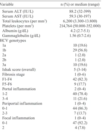
The detailed laboratory and histological findings of the cases are listed in Table II. In summary, the median
aspartate aminotransferase and ALT levels were 45 IU/L (range 30-197) and 69 IU/L (range 32-399), respectively;
11 patients (21.5%) had hypoalbuminaemia and 16 pa-tients (31.4%) had hypergammaglobulinaemia. The ma-jority of patients (76.5%) were infected with HCV
geno-type 1 and 59.2% had an HCV RNA viral load greater than 850.000 IU/mL.
The median Ishak score for histology was 5 (range 3-14) and a minimal and mild disease accounted for 78.4% of the pathologic findings (31.4% and 47.1%, re-spectively). Confluent hepatocyte necrosis was not seen.
Grade I fibrosis (F1) was seen in 30 cases (58.8%) and
portal inflammation was found in 29 (56.8%) cases. All of the cases had Councilman bodies in the hepatic pa-renchyma and the hepatic sinusoids presented with mild hyperplasia and hypertrophy in Kupffer cells, with a predominance of lymphocyte infiltrates.
The majority (78%) of cases showed some evidence of steatosis, which was either mild, moderate or severe, either focused or generalized and either macro or micro-vacuolar. Biopsies from the liver donor biopsies showed no evidence of pathologic findings. A mild haemosid-erin deposition and glycogenic degeneration was seen in one and two samples, respectively.
Cytokine patterns in the cases and blood donor con-trols - Compared with the blood donor controls (Table III), the cases had significantly higher serum levels of
IL-2 (426 ± 362 vs. 91 ± 124 pg/mL; p < 0.01), IL-10 (294 ± 289 vs. 38 ± 141 pg/mL; p < 0.01), TNF-β (17 ± 35 vs. 7 ± 11 pg/mL; p < 0.01) and TGF-β (898 ± 348 vs. 1 ± 904 pg/mL; p < 0.01). On the other hand, the cases showed
significant lower levels of the following cytokines: IL-4
(27 ± 21 vs. 62 ± 93 pg/mL; p = 0.02), IL-6 (11 ± 30 vs. 48 ± 69 pg/mL; p < 0.01), IL-8 (58 ± 84 vs. 263 ± 337 pg/mL; p < 0.01) and IFN-γ (4 ± 9 vs. 26 ± 73 pg/mL; p < 0.01).
Cell populations and cytokine patterns in liver bi-opsies of the cases and liver donor controls - In immu-nohistochemistry reactions, the positive cytokines and
cell populations were stained brown, as shown in Figs
1, 2, respectively. The cell populations from the cases
TABLE I
Demographic characteristics of chronically hepatitis C virus (HCV) infected cases and controls
Cases Blood donors Liver donors
Variable n = 51 n = 51 n = 33
Male gender - n (%) 30 (58.8) 28 (54.9) 15 (45.5)
Age years - median (range) 38 (20-59) 33 (18-58) 36 (18-60)
White race - n (%) 34 (66.7) 36 (70.6) 21 (63.6)
Risk factors
History of injection drug abuse 3 (5.9) NA NA
Previous transfusion 21 (41.2) NA NA
Previous surgery 27 (52.9) NA NA
Duration of HCV infectiona 18.2 (3-31) – –
a: available for 41 patients and defined as time in years from suspected exposure to biopsy; NA: not available.
TABLE II
Biochemical, virological and histological characteristics of 51 cases
Variable n (%) or median (range)
Serum ALT (IU/L) 88.2 (32-399)
Serum AST (IU/L) 59.3 (30-197)
Total leukocytes (per mm3) 6,200 (3.300-13.800) Platelets (per mm3) 214,764 (50.000-352.000)
Albumin (g/dL) 4.2 (2.7-5.1)
Gammaglobulin (g/dL) 1.56 (0.7-2.6)
HCV genotypes
1a 10 (19.6)
1b 29 (56.8)
2a 1 (2.0)
2b 1 (2.0)
3a 10 (19.6)
Ishak score (overall) 5 (3-14)
Fibrosis stage 1 (0-6)
F1-F4 42 (82.3)
F5-F6 9 (17.7)
Portal inflammation 2 (0-4)
1-2 40 (78.4)
3-4 11 (21.6)
Periportal inflammation 1 (0-4)
0-1 44 (86.3)
2-3 7 (13.7)
Focal inflammation 1 (0-4)
0-1 47 (92.2)
2 4 (7.8)
ALT: alanine aminotransferase; AST: aspartate aminotrans-ferase; HCV: hepatitis C virus.
had a higher expression of portal CD4+ (706.4 ± 388.0
vs. 294.5 ± 129.2 cells/mm2; p < 0.01) and CD8+ (196.8 ±
113.8 vs. 66.3 ± 57.8 cells/mm2; p < 0.01) than the con
-trols (Table IV).
The cytokine patterns of the cases had a higher
ex-pression of acinar IL-4 (2.5 ± 2.0 vs. 1.1 ± 1.2 cells/mm2;
p < 0.01), TNF-α (3.8 ± 2.8 vs. 0.9 ± 1.6 cells/mm2; p <
and TGF-β (5.1 ± 3.9 vs. 1.7 ± 1.4 cells/mm2; p < 0.01),
compared to the controls. The cases also expressed
signif-icantly higher portal TNF-α (3.6 ± 3.0 vs. 1.6 ± 2.5 cells/
mm2; p < 0.01) and TGF-β (3.6 ± 3.8 vs. 0.8 ± 1.4 cells/
mm2; p < 0.01) than the controls. The cases showed a sig
-nificantly lower expression of portal S100 dendritic cells
(DC) (22.5 ± 20.2 vs. 64.8 ± 42.3 cells/mm2; p < 0.01),
ac-inar S100 (2.3 ± 4.7 vs. 8.5 ± 7.6 cells/mm2; p < 0.01),
ac-inar IL-1β (0.2 ± 0.5 vs. 0.8 ± 1.1 cells/mm2; p < 0.01) and
acinar IL-10 (1.3 ± 1.6 vs. 2.7 ± 2.7 cells/mm2; p < 0.01).
Correlation patterns of cell population and cytokines among cases - The correlations between the cell markers and cytokines was analysed at either the portal spaces or the acinar areas (Table V). Significant positive correla-tions were seen between the following measures at the portal spaces: CD45+RO and TNF-α, CD4+ and TNF-α,
CD8+ and TGF-β, CD68+ and TNF-α, CD57+ and IL-10,
S-100 and IL-10. At the acinar level, CD45+ and IFN-γ,
TABLE III
Cytokine profiles in peripheral blood of chronically hepatitis C virus (HCV) infected cases and healthy blood donors
Cases Blood donors
n = 51 n = 51
Cytokine mean (SD) mean (SD) p value
IL-2 426.373 (361.594) 90.636 (123.531) < 0.01 IL-10 294.444 (289.346) 37.744 (140.680) < 0.01 TNF-β 16.804 (34.935) 6.866 (10.858) < 0.01 TGF-β 897.553 (347.819) 1.334 (904) < 0.01 IL-1α 1.280 (3.325) 5.640 (21.594) < 0.01
IL-4 27.178 (20.612) 61.583 (92.659) 0.02
IL-6 10.742 (30.466) 47.593 (69.181) < 0.01 IL-8 57.817 (84.190) 262.941 (336.897) < 0.01 IFN-γ 4.187 (9.217) 25.569 (73.028) < 0.01 TNF-α 64.437 (149.211) 24.473 (41.016) 0.07
SD: standard deviations.
Fig. 1: typical staining of immunohistochemistry detection of cytok
-ines: A: IFN-γ in acinar area (400X); B: IFNγ in portal space; C: IL2
in acinar area; D: IL6 in acinar area. Arrows indicate cells staining positive for the marker.
Fig. 2: typical staining of immunohistochemistry detection of cell
populations. A: CD4 T-lymphocytes in acinar area (400X); B: CD4 T-lymphocytes in portal areas (200X); C: S100 cells in portal space (400X); D: CD45+ cells in acinar area (400X). Arrows indicate cells staining positive for the marker.
TABLE IV
Cell populations and cytokine patterns in liver biopsy of chronically hepatitis C virus (HCV) infected cases
and liver donors
Cases Liver donors
n = 51 n = 33
Variable mean (SD) mean (SD) p value
Portal CD45 412.3 (290.9) 304.5 (111.8) 0.20
Acinar CD45 33.1 (35.7) 38.2 (30.8) 0.07
Portal CD4 706.4 (388.0) 294.5 (129.2) < 0.01
Acinar CD4 46.4 (33.0) 36.7 (23.2) 0.15
Portal CD8 196.8 (113.8) 66.3 (57.8) < 0.01
Acinar CD8 12.5 (13.2) 13.8 (17.2) 0.55
Portal CD68 107.4 (106.9) 64.5 (42.7) 0.12
Acinar CD68 104.6 (65.6) 92.4 (38.6) 0.70
Portal S100 22.5 (20.2) 64.8 (42.3) < 0.01 Acinar S100 2.3 (4.7) 8.5 (7.6) < 0.01 Portal CD57 24.3 (33.9) 12.5 (10.3) 0.39
Acinar CD57 11.1 (10.4) 6.4 (3.8) 0.07
Portal TNF-α 3.6 (3.0) 1.6 (2.5) < 0.01 Acinar TNF-α 3.8 (2.8) 0.9 (1.6) < 0.01 Portal TGF-β 3.6 (3.8) 0.8 (1.4) < 0.01 Acinar TGF-β 5.1 (3.9) 1.7 (1.4) < 0.01
Portal INF-γ 3.1 (5.2) 1.5 (2.3) 0.14
Acinar INF-γ 2.8 (2.8) 0.5 (1.2) < 0.01
Portal IL-1α 1.1 (1.9) 0.6 (1.4) 0.28
Acinar IL-1α 0.3 (0.7) 0.6 (2.0) 0.67
Portal IL-1β 1.3 (2.6) 0.7 (1.2) 0.74
Acinar IL-1β 0.2 (0.5) 0.8 (1.1) < 0.01
Portal IL-2 1.0 (2.2) 0.9 (2.2) 0.78
Acinar IL-2 1.7 (1.8) 1.5 (2.0) 0.45
Portal IL-4 1.7 (2.8) 0.8 (1.3) 0.06
Acinar IL-4 2.5 (2.0) 1.1 (1.2) < 0.01
Portal IL-6 0.2 (0.6) 0.0 (0.3) 0.36
Acinar IL-6 0.7 (1.5) 0.8 (1.0) 0.17
Portal IL-10 0.5 (1.1) 0.5 (1.2) 0.57
Acinar IL-10 1.3 (1.6) 2.7 (2.7) < 0.01
CD45+ and IL-1α, CD57+ and TGF-β and CD57+ and IL-2
were positively correlated, while CD68+ and IL-6 and
CD68+ and IL-10 were negatively correlated.
The correlations between the degree of liver dam-age and the cell populations and cytokines were also as-sessed. In immunohistochemistry analyses of the cases, periportal activity was positively correlated with portal CD4+ expression (r = 0.4; p < 0.01), while focal inflam
-mation was positively correlated with acinar CD4+ (0.33;
p = 0.02). A significant positive correlation for cytokine
density was found between periportal hepatitis and
por-tal IL-1β (r = 0.36; p < 0.01) and between acinar activity and acinar TGF-β (r = 0.34; p = 0.02). Following a corre -lation analysis of the serum cytokines and the degree of liver lesions, IL-8 was positively correlated with fibrosis
(r = 0.29; p = 0.04) and IL1-α was negatively correlated with focal inflammation (r = -0.29; p = 0.04); the other
serum cytokines did not show any correlation with liver compromised liver (data not shown).
DISCUSSION
In this study, we systematically assessed the role of a comprehensive array of cytokines and cell populations in chronically HCV infected patients and compared them with normal controls. Among the cases, we found that
there was a greater expression of portal TNF-α, TGF-β,
CD4+ and CD8+ and acinar IFN-γ, TNF-α, TGF-β and
IL-4, as well as a higher serum concentration of IL-2,
IL-10 and TGF-β compared to the controls.
Cytokines are key mediators of inflammation, apop-tosis, necrosis and fibrosis and they are actively involved in the regeneration process of liver tissue after injury. It has been hypothesised that successful treatment of hepa-titis C depends on a complex balance between pro and anti-inflammatory responses (Bertoletti et al. 1997, Tsai et al. 2003, Wan et al. 2009). In our study, we found both pro and anti-inflammatory responses, with a predomi-nant pro-inflammatory response pattern in the liver
mediated by IFN-γ and TNF-α and modulated by CD4+,
CD8+ and CD45RO+ T lymphocytes. This complex
bal-ance is further supported by the significant correlations found between: (i) portal CD4+ and TNF-α, (ii) portal
CD8+ and TGF-β, (ii) portal CD45+RO and TNF-α and
(iv) acinar CD45+RO and IFN-γ. The latter correlations
that involve CD45+RO suggest that there is an ongoing
activation of the cellular immune response, which is in
agreement with the study by Murata et al. (2002). In line with other reports (Minutello et al. 1993, Sakai et al. 1999, Exley et al. 2002), our results demonstrate
the compartmentalised nature of the immune response to HCV. The correlation between periportal activity and portal CD4+, as seen in a preliminary study (Viso et al.
TABLE V
Correlations between cell populations and cytokine patterns by immuhohistoquemistry in liver biopsy of chronically hepatitis C virus (HCV) infected cases
Cell Citokines
populations Portal TNF-α Acinar TNF-α Portal TGF-β Acinar TGF-β Portal IFN-γ Acinar IFN-γ
Portal CD45 rs 0.32 NA 0.06 NA 0.08 NA
p 0.023a NA 0.692 NA 0.592 NA
Acinar CD45 rs NA 0.00 NA -0.08 NA 0.29
p NA 0.992 NA 0.562 NA 0.042a
Portal CD4 rs 0.40 NA 0.10 NA 0.12 NA
p 0.004a NA 0.496 NA 0.407 NA
Acinar CD4 rs NA -0.09 NA 0.23 NA -0.17
p NA 0.514 NA 0.107 NA 0.220
Portal CD8 rs 0.00 NA 0.30 NA 0.13 NA
p 0.994 NA 0.030a NA 0.354 NA
Acinar CD8 rs NA -0.06 NA 0.04 NA -0.01
p NA 0.697 NA 0.796 NA 0.932
PortalCD68 rs 0.29 NA 0.04 NA 0.07 NA
p 0.042a NA 0.797 NA 0.635 NA
Acinar CD68 rs NA 0.17 NA 0.17 NA 0.20
p NA 0.241 NA 0.227 NA 0.166
Portal S100 rs 0.09 NA -0.06 NA 0.04 NA
p 0.540 NA 0.689 NA 0.766 NA
Acinar S100 rs NA 0.03 NA -0.10 NA 0.04
p NA 0.854 NA 0.478 NA 0.768
Portal CD57 rs -0.28 NA 0.17 NA 0.06 NA
p 0.049a NA 0.224 NA 0.688 NA
Acinar CD57 rs NA -0.13 NA 0.29 NA -0.26
p NA 0.358 NA 0.040a NA 0.064
2007) and the increased expression of portal TNF-α and acinar IFN-γ, TNF-α and IL-4, which was not replicated
in serum, suggests that there was an intra-hepatic re-sponse. Apparently, most of the immune response oc-curred at the portal space level, supported by the lack of correlation between the hepatic and serum cytokines among the cases.
TNF-α production is an early event in the pathogen -esis of liver damage, triggering the synth-esis of other cytokines. It is required for the proliferation of normal hepatocytes in liver regeneration, but it also mediates hepatocyte death (Zhu et al. 1998). The increased
ex-pression of portal and acinar TNF-α and acinar IFN-γ
in the cases compared to the controls reinforces the ma-jor role of these pro-inflammatory cytokines in the liver disease caused by HCV.
IL-2 is considered to be a Th1 type cytokine and is involved in enhancing the proliferation and activation of
most T-lymphocytes, NK cells and B-lymphocytes. Liver
sinusoidal and inflammatory cells have been reported to be sources of IL-2 and no consensus exists on the pre-dictive value of this cytokine. Apparently, expression of IL-2 is associated with a more advanced stage of
dis-ease, as previously reported (Makris et al. 1994, Napoli
et al. 1996, Bozkaya et al. 2000, Gramenzi et al. 2005). In our sample, although a higher concentration of IL-2 was found in the serum, no significant expression was found in the liver from the cases compared to the normal controls. This could possibly be due to the relatively mild disease that was observed in most of our cases, agree-ing with reports from other authors (Woitas et al. 1997,
Falasca et al. 2006). Indeed, it has been hypothesised that failure to secrete IL-2 might lead to a disruption of IFN-γ
and proliferative capacity, contributing to the develop-ment of a persistent infection (Semmo et al. 2005). Our findings agree with those of Gramenzi et al. (2005), who reported that there was an inverse correlation between ALT levels and IL-2 expression when cytokine profiles of patients with chronic disease were compared with pa-tients who had persistently normal serum ALT levels.
IL-6 is a major mediator of the inflammatory process in the acute phase response, acting as a blocker of cell apoptosis (Baumann & Gauldie 1990) and promoting the differentiation of naïve T-cells into Th1 cells. IL-6 has been associated with disease progression and has been reported to be elevated in both hepatic and peripheral
compartments (Malaguarnera et al. 1997, Oyanagi et al. 1999, Lapinski et al. 2001, Falaska et al. 2006). Interest -ingly, in this study, IL-6 was not associated with either chronic HCV infection or with the stage of disease. Our results are in contrast to those of other researches, but agree with those of Cribier et al. (1998), who found simi-lar patterns in serum IL-6 between HCV-infected and non-infected patients. Although, it should be noted that IL-6 was above the cut-off value in only 11 patients, pos-sibly due to the overall lower severity of disease sample. However, these findings deserve further investigation with appropriately powered studies.
TGF-β has been implicated as a mediator of hepatic
fibrogenesis and is known to have negative regulatory
effects on the immune system (Nakatsukasa et al. 1990,
Nelson et al. 1997, Roulot et al. 1999, Sakaguchi et al. 2002). In this study, expression of TGF-β was signifi -cantly higher in both the liver (portal space and lobule) and peripheral compartments of the cases compared to the controls. These findings confirm those of other
au-thors (Flisiak et al. 2000, Kinnman et al. 2000, Ray et al. 2003) and provide additional evidence that TGF-β acts
as an immune mediator in chronic HCV infection. The negative correlation of S100 DC with peri-portal activity among the cases and its lower expression com-pared to the controls suggests that there was a lack of HCV recognition by these cells, which, in turn, might have facilitated viral persistence. The mechanisms in-volved in this down regulation are still unclear and a few hypotheses have been raised. It has been suggested that HCV proteins might modulate T-cell responses by decreasing the stimulatory ability of DC (Sarobe et al. 2003) and that there would be an allostimulatory defect of monocyte-derived DC because these cells would con-stitute an extra-hepatic reservoir for the virus (Bain et al. 2001). Jinushi et al. (2004) also suggested that the
aberrant expression of natural killer (NK) receptors as -sociated with chronic infection might have an impact on the magnitude and direction of DC activation of T-cells. The positive correlation between portal S100 cells and IL-10 in our study reinforces this hypothesis. IL-10 has a modulatory effect on hepatic fibrogenesis by down-regulating the pro-inflammatory response, including the antigen-presenting cells and in a controlled study, IL-10
has also been shown to reduce liver damage (Nelson et
al. 2000, 2003). Studies have reported that disease pro-gression is associated with an increased expression of circulating IL-10 (Gramenzi et al. 2005), a correlation
with low liver alterations (Piazzolla et al. 2000) and a decreased expression at intrahepatic compartments (Na -poli et al. 1996). In our study, the cases had higher serum concentrations of IL-10, but lower intrahepatic expres-sion, than did the respective controls, thus reinforcing the predominance of pro-inflammatory factors.
The possible correlations between the duration of infection (n = 41 patients, whose duration of infection
could be estimated) or the age of the patient (n = 51) with
the immunological parameters was also analysed but did not reveal any biologically meaningful correlations (data not shown). The only possibly relevant finding was the observation that age was positively correlated with por-tal CD45+RO (p = 0.012, r = 0.35) (as expected), as well
as with acinar CD8+ (p = 0.020, r = 0.33), but was nega
-tively correlated with acinar IL-10 (p = 0.024, r = -0.32).
Therefore, the duration of the infection or the age of the patient is unlikely to explain our findings.
Our study has some limitations. Firstly, it is possible
that the observed balance towards a pro-inflammatory type of response was biased by the inclusion of cases with a milder disease; only ~20% presented a more
se-vere classification (F5-F6). For our study, we split the
patients into two groups: one group containing the nine
patients with severe histology (F5-F6) and the other
group containing the 42 patients with mild histology
(F1-F4). We then reassessed the correlation analyses
and blood (cytokines) and we did not find any statisti-cal differences between the two groups in any of the comparisons (data not shown). Also, the data from the 42 patients remained similar to the data from all of the patients (n= 51) (data not shown). Secondly, the cross-sectional nature of the study precludes causal inferences and a prospective assessment of these cases is warrant-ed. However, our results devise several hypotheses for future research. Thirdly, genetic polymorphisms associ-ated with cytokine expression have been shown to play a role in the ability to respond to therapy (Yee et al. 2001,
Milton et al. 2005) and were not assessed in our study. Fourth, the function of T lymphocytes was not analysed in this study as it has been in other studies (Moonka et al. 2008). Finally, some comparisons might have been
affected by a lack of statistical power, particularly those with a considerable floor effect that was caused by the lower limit of detection for the cytokine assays.
In conclusion, we simultaneously assessed pro and anti-inflammatory related cytokines and cell populations in the liver and sera and have shown that (i) the immune response in the samples of predominantly mild disease was associated with several pro-inflammatory cytok-ines, (ii) CD4+ T-lymphocytes played a major role in
or-chestrating the immune response and (iii) these events took place primarily at the portal space. Our results sug-gest that this balance is highly dynamic, particularly in less severe disease and further research is needed to ex-plore the longitudinal aspects of this relationship.
REFERENCES
Alter MJ 2007. Epidemiology of hepatitis C virus infection. World J
Gastroenterol13: 2436-2441.
Bain C, Fatmi A, Zoulim F, Zarski JP, Trepo C, Inchauspe G 2001.
Impaired allostimulatory function of dendritic cells in chronic hepatitis C infection. Gastroenterology120: 512-524.
Baumann H, Gauldie J 1990. Regulation of hepatic acute phase plas-ma protein genes by hepatocyte stimulating factors and other me-diators of inflammation. Mol Biol Med7: 147-159.
Bertoletti A, D’Elios MM, Boni C, De Carli M, Zignego AL, Durazzo M, Missale G, Penna A, Fiaccadori F, Del Prete G, Ferrari C 1997.
Different cytokine profiles of intraphepatic T cells in chronic hepatitis B and hepatitis C virus infections. Gastroenterology
112: 193-199.
Bozkaya H, Bozdayi AM, Aslan N, Türkay C, Sarioglu M, Cetinkaya H, Akdogan M, Cinar K, Erden E, Köse K, Sentür H, Akkiz H,
Sarayalcin S, Yurdaydin C, Uzunalimoglu Ö 2000. Circulating IL-2 and IL-10 in chronic active hepatitis C with respect to the
response to IFN treatment. Infection28: 309-313.
Brown PMJ, Neuman MG 2001. Immunopathogenseis of hepatitis C viral infection: Th1/Th2 responses and the role of cytokines. Clin Biochemistry34: 167-171.
Cacciarelli TV, Martinez OM, Gish RG, Villanueva JC, Krams SM
1996. Immunoregulatory cytokines in chronic hepatitis C virus infection: pre- and post-treatment with interferon alfa. Hepatol-ogy24: 6-9.
Chang KM, Thimme R, Melpolder JJ, Oldach D, Pemberton J, Moor
-head-Loudis J, McHutchison JG, Alter HJ, Chisari FV 2001. Dif-ferential CD4(+) and CD8(+) T-cell responsiveness in hepatitis C virus infection. Hepatology33: 267-276.
Cribier B, Schmitt C, Rey D, Lang JM, Kirn A, Stoll-Keller F 1998. Production of cytokines in patients infected by hepatitis C virus. J Med Virol55: 89-91.
Day CL, Lauer GM, Robbins GK, McGovern B, Wurcel AG, Gandhi
RT, Chung RT, Walker BD2002. Broad specificity of virus-spe-cific CD4+ T-helper-cell responses in resolved hepatitis C virus infection. J Virol76: 12584-12595.
Exley MA, He Q, Cheng O, Wang RJ, Cheney CP, Balk SP, Koziel MJ 2002. Cutting edge: compartmentalization of Th1-like nonin-variant CD1d-reactive T cells in hepatitis C virus-infected liver.
J Immunol168: 1519-1523.
Falasca K, Ucciferri C, Dalessandro M, Zingariello P, Mancino P, Petrarca C, Pizzigallo E, Conti P, Vecchiet J 2006. Cytokine pat-terns correlate with liver damage in patients with chronic hepati-tis B and C. Ann Clin Lab Sci36: 144-150.
Flisiak R, Pytel-Krolczuk B, Prokopowicz D 2000. Circulating trans -forming growth factor beta(1) as an indicator of hepatic function impairment in liver cirrhosis. Cytokine12: 677-681.
Gonzalez-Peralta RP, Fang JW, Davis GL, Gish R, Tsukiyama-Koha
-ra K, Koha-ra M, Mondelli MU, Lesniewski R, Phillips MI, Mizo
-kami M 1994. Optimization for the detection of hepatitis C virus
antigens in the liver. J Hepatol20: 143-147.
Gramenzi A, Andreone P, Loggi E, Foschi FG, Cursaro C, Margotti M, Biselli M, Bernardi M 2005. Cytokine profile of peripheral blood mononuclear cells from patients with different outcomes of hepatitis C virus infection. J Viral Hepat12: 525-530.
Hassan G, Moreno S, Massimi M, Di Biagio P, Stefanini S 1997. Inter -leukin-1-producing plasma cells in close contact with hepatocytes in patients with chronic active hepatitis. J Hepatol27: 6-17.
Ishak K, Baptista A, Bianchi L, Callea F, De Groote J, Grudat F, Denk H, Desmet V, Korb G, MacSween RNM, Phillips MJ, Portmann BG, Poulsen H, Scheuer PJ, Schmid M, Thaler H 1995. Histological grading and staging of chronic hepatitis. J Hepatol22: 696-699.
Jinushi M, Takehara T, Tatsumi T, Kanto T, Miyagi T, Suzuki T, Ka
-nazawa Y, Hiramatsu N, Hayashi N 2004. Negative regulation of NK cell activities by inhibitory receptor CD94/NKG2A leads to altered NK cell-induced modulation of dendritic cell functions in
chronic hepatitis C virus infection. J Immunol173: 6072-6081.
Kamal SM, Graham CS, He Q, Bianchi L, Tawil AA, Rasenarch JW, Khalifa KA, Massoud MM, Koziel MJ 2004. Kinetics of intrahe -patic hepatitis C virus (HCV)-specific CD4+ T cell responser in HCV and Schistosoma mansoni coinfection: relation to progres-sion of liver fibrosis. J Infect Dis189: 1140-1150.
Kinnman N, Andersson U, Hultcrantz R 2000. In situ expression of transforming growth factor-beta1-3, latent transforming growth factor-beta binding protein and tumor necrosis factor-alpha in liver tissue from patients with chronic hepatitis C. Scand J Gas -troenterol35: 1294-1300.
Lapinski TW 2001. The levels of IL-1beta, IL-4 and IL-6 in the serum and the liver tissue of chronic HCV-infected patients. Arch Im-munol Ther Exp (Warsz)49: 311-316.
Makris M, Preston FE, Ralph S 1994. Increased soluble IL-2 recep -tor levels in HCV-infected haemophiliacs: a possible indica-tor of liver disease severity. Br J Haematol 87: 419-421.
Malaguarnera M, Di Fazio I, Romeo MA, Restuccia S, Laurino A, Trovato BA 1997. Elevation of interleukin 6 levels in patients
with chronic hepatitis due to hepatitis C virus. J Gastroenterol
32: 211-215.
Minton EJ, Smillie D, Smith P, Shipley S, McKendrick MW, Gleeson
associ-ated with single nucleotide polymorphisms in the IL-1, -6 or -10 genes. Hum Immunol66: 127-132.
Minutello MA, Pileri P, Unutmaz D, Censini S, Kuo G, Houghton M, Brunetto MR, Bonino F, Abrignani S 1993. Compartmentaliza-tion of T lymphocytes to the site of disease: intrahepatic CD4+ T
cells specific for the protein NS4 of hepatitis C virus in patients
with chronic hepatitis C. J Exp Med178: 17-25.
Moonka D, Milkovich KA, Rodriguez B, Abouljoud M, Lederman MM, Anthony DD 2008. Hepatitis C virus-specific T-cell gamma
interferon and proliferative responses are more common in peri-hepatic lymph nodes than in peripheral blood or liver. J Virol82: 11742-11748.
Murata M, Nabeshima S, Maeda N, Nakashima H, Kashiwagi S, Hayashi J 2002. Increased frequency of IFN-gamma-producing
peripheral CD8+ T cells with memory-phenotype in patients with chronic hepatitis C. J Med Virol67: 162-170.
Nakatsukasa H, Evarts RP, Hsia CC, Thorgeirsson SS 1990. Transform -ing growth factor-beta 1 and type I procollagen transcripts dur-ing regeneration and early fibrosis of rat liver. Lab Invest 63: 171-180.
Napoli J, Bishop GA, McGuinness PH, Painter DM, McCaughan GW 1996. Progressive liver injury in chronic hepatitis C infection cor -relates with increased intrahepatic expression of Th1-associated cytokines. Hepatology24: 759-765.
Nelson DR, Gonzalez-Peralta RP, Qian K, Xu Y, Marousis CG, Davis
GL, Lau JY 1997. Transforming growth factor-beta 1 in chronic hepatitis C. J Viral Hepat4: 29-35.
Nelson DR, Lauwers GY, Lau JY, Davis GL 2000. Interleukin 10 treat -ment reduces fibrosis in patients with chronic hepatitis C: a pilot trial of interferon nonresponders. Gastroenterology118: 655-660.
Nelson DR, Tu Z, Soldevila-Pico C, Abdelmalek M, Zhu H, Xu YL,
Cabrera R, Liu C, Davis GL 2003. Long-term interleukin 10 therapy in chronic hepatitis C patients has a proviral and anti-inflammatory effect. Hepatology 38: 859-868.
Oyanagi Y, Takahashi T, Matsui S, Takahashi S, Boku S, Takahashi K, Furukawa K, Arai F, Asakura H 1999. Enhanced expression of
interleukin-6 in chronic hepatitis C. Liver19: 464-472.
Piazzolla G, Tortorella C, Schiraldi O, Antonaci S 2000. Relationship
between interferon-gamma, interleukin-10 and interleukin-12 production in chronic hepatitis C and in vitro effects of interfer-on-alpha. J Clin Immunol20: 54-61.
Polyak SJ, Khabar KS, Rezeiq M, Gretch DR 2001. Elevated levels of
interleukin-8 in serum is associated with hepatitis C virus infec-tion and resistance to interferon therapy. J Virol75: 6209-6211.
Ray S, Broor SL, Vaishnav Y, Sarkar C, Girish R, Dar L, Seth P, Broor S
2003. Transforming growth factor beta in hepatitis C virus infection:
in vivo and in vitro findings. J Gastroenterol Hepatol18: 393-403.
Roulot D, Sevcsik AM, Coste T, Strosberg AD, Marullo S 1999. Role
of transforming growth factor beta type II receptor in hepatic fi-brosis: studies of human chronic hepatitis C and experimental fibrosis in rats. Hepatology29: 1730-1738.
Sakaguchi E, Kayano K, Segawa M, Okamoto M, Sakaida I, Okita
K 2002. Th1 down-regulation at the single-lymphocyte level in
HCV-related liver cirrhosis and the effect of TGF-beta on Th1
response: possible implications for the development of hepatoma.
Hepatol Res24: 282.
Sakai A, Kaneko S, Honda M, Matsushita E, Kobayashi K 1999.
Quasispecies of hepatitis C virus in serum and in three different parts of the liver of patients with chronic hepatitis. Hepatology
30: 556-561.
Sarobe P, Lasarte JJ, Zabaleta A, Arribillaga L, Arina A, Melero I, Borras-Cuesta F, Prieto J 2003. Hepatitis C virus structural pro-teins impair dendritic cell maturation and inhibit in vivo induc-tion of cellular immune responses. J Virol77: 10862-10871.
Schvoerer E, Navas MC, Thumann C, Fuchs A, Meyer N, Habersetzer F, Stoll-Keller F 2003. Production of interleukin-18 and interleu -kin-12 in patients suffering from chronic hepatitis C virus infec-tion before antiviral therapy. J Med Virol70: 588-593.
Semmo N, Day CL, Ward SM,Lucas M, Harcourt G, Loughry A, Kle
-nerman P 2005. Preferential loss of IL-2-secreting CD4+ T helper cells in chronic HCV infection. Hepatology 41: 1019-1028.
SPSS 2002. SPSS for Windows. Statistical Package for the Social Sci -ences, Release 12.0 ed, Chicago.
Thimme R, Oldach D, Chang KM, Steiger C, Ray SC, Chisari FV
2001. Determinants of viral clearance and persistence during acute hepatitis C virus infection. J Exp Med194: 1395-1406.
Tilg H, Kaser A, Moschen AR 2006. How to modulate inflammatory
cytokines in liver diseases. Liver Int26: 1029-1039.
Tilg H, Wilmer A, Vogel W, Herold M, Nolchen B, Judmaier G, Huber
C 1992. Serun levels of cytokynes in chronic liver diseases. Gas -troenterology103: 264-274.
Tsai SL, Sheen IS, Chien RN, Chu CM, Huang HC, Chuang YL, Lee TH, Liao SK, Lin CL, Kuo GC, Liaw YF 2003. Activation of Th1 immunity is a common immune mechanism for the successful treatment of hepatitis B and C: tetramer assay and therapeutic implications. J Biomed Sci10: 120-135.
Viso AT, Barbosa Tde C, Yamamoto L, Pagliari C, Fernandes ER, Brasil RA, Andrade Jr HF Jr, Duarte MI, Barone AA 2007. Portal CD4+ and CD8+ T lymphocyte correlate to intensity of interface hepatitis in chronic hepatitis C. Rev Inst Med Trop Sao
Paulo49: 371-378.
Wan L, Kung YJ, Lin YJ, Liao CC, Sheu JJ, Tsai Y, Lai HC, Peng CY, Tsai FJ 2009. Th1 and Th2 cytokines are elevated in
HCV-infected SVR(-) patients treated with interferon-alpha. Biochem Biophys Res Commun379: 855-860.
Weibel ER 1979. Stereological methods. Practical methods for bio -logical morphometry, vol. 1, Academic Press, London, 415 pp.
Woitas RP, Lechmann M, Jung G, Kaiser R, Sauerbruch T, Spengler
U 1997. CD30 induction and cytokine profiles in hepatitis C virus core-specific peripheral blood T lymphocytes. J Immunol
159: 1012-1018.
Yee LJ, Tang J, Gibson AW, Kimberly R, Van Leeuwen DJ, Kaslow RA 2001. Interleukin 10 polymorphisms as predictors of sus-tained response in antiviral therapy for chronic hepatitis C infec-tion. Hepatology33: 708-712.
Zhu N, Khoshnan A, Schneider R, Matsumoto M, Dennert G, Ware C, Lai MM 1998. Hepatitis C virus core protein binds to the