Lack of Interleukin 1-beta Expression Following
Orthodontic Induced Root Resorption
Massoud Seifi, D.D.S., M.S.D.1*, Bahram Kazemi, Ph.D.2, Elahe Vahid-Dastjerdi, D.D.S., M.S.D.3, Mojgan Bandehpour, Ph.D.2, Milad Karamlou, D.D.S.3
1. Dentofacial Deformities Research Center and Orthodontic Department, School of Dentistry, Shahid Beheshti University of Medical Sciences, Tehran, Iran
2. Cellular and Molecular Biology Research Center, Shahid Beheshti University of Medical Sciences, Tehran, Iran 3. Orthodontic Department, School of Dentistry, Shahid Beheshti University of Medical Sciences, Tehran, Iran
* Corresponding Address: Dentofacial Deformities Research Center and Orthodontic Department, School of Dentistry, Shahid Beheshti University of Medical Sciences, Tehran, Iran
Email: mseifi@dent.sbmu.ac.ir
Received: 9/Dec/2009, Accepted: 16/Mar/2010
Abstract
Objective: To determine the effect of orthodontic tooth movement on the expression of interleukin-1β mRNA in rats using RT-PCR.
Materials and Methods: Sample consisted of eighteen 8-week-old male Wistar rats. The right maxillary first molar of each animal was protracted using an orthodontic protraction ap-pliance. The left maxillary first molar received no treatment and was assigned as the control group. On day 21, all rats were sacrifice and divided in two equal groups. The first group, group (A), was histologically evaluated for the presence and size of potential resorptive la-cunae. The second group, group (B), was investigated using RT-PCR in order to determine IL-1β mRNA expression.
Results: Measurements revealed that the mean tooth movement was 0.23 mm in group A and 0.24 mm in group B. The mean depth of the resorptive lacunae was 0.17×10⁻¹¹ mm2 in the control group and 4.9×10⁻¹¹ mm2 in the intervention group (control group: left maxillary first molars; right maxillary first molars were divided to group A & B, histologic study of group A assures the existence of resorptive lacunae and its extent relative to control group). The difference between the two groups was statistically significant (p<0.05). The RT-PCR evalu-ation showed no significant differences in IL-1β mRNA expressions of resorptive lacunae between the treated and untreated groups.
Conclusion: Although interleukin1-beta is the most potent stimulator of bone resorption and mediator of inflammatory response, the present study showed that the IL-1beta mRNA was not expressed more significantly in root resorption lacunae of the treated molars rela-tive to the control group.
Keywords: Interleukin-1 beta, Gene Expression, Root Resorption, Orthodontics
Cell Journal(Yakhteh), Vol 12, No 4, Winter 2011, Pages: 463-468
Introduction
Orthodonticaly induced root resorption is the most frequent unwanted side effect of orthodontic tooth movement that is unavoidable (1). The sterile in-flammatory process initiated by force application is the major cause of root resorption and bone re-modeling (1, 2). Many experiments have dealt with problems associated with root resorption. Root resorption is a multicausal problem; for instance, systemic (3-5), iatrogenic, and age (6, 7) factors have been considered.
One of the iatrogenic causes is orthodontic treat-ment. The duration of orthodontic force, whether constant or intermittent, and its direction play im-portant roles (8, 9).
Interleukin-1 is a multifunctional cytokine with a wide variety of activities. IL-1α and IL-1β are two active peptides encoded by two separate gene
products. They have identical activities and po-tentials. Gowen et al. showed that IL-1 is the first polypeptide mediator of immune cell func-tion that regulates bone resorpfunc-tion (10). Canalis showed that IL-1 regulates bone formation (11) and demonstrated that this cytokine, as an osteo-clast activating factor (OAF), is a major contrib-utor to bone resorption. Lorenzo et al. reported that IL-1 is the most potent stimulator of bone resorption and promoter of prostaglandin synthe-sis in bone (12).
increase in prostaglandin synthesis is a stimulator of bone resorption activity (12).
Proinflammatory cytokines, such as interleukin-1, TNF-alpha and IL-6 are believed to play a role in biological processes involved in the course of or-thodontic tooth movement and especially root re-sorption (14, 15).
The interleukin-1 family (IL-1 alpha, IL-1 beta, and IL-1 RA) has been shown to be a potent stimulus for bone resorption. According to pre-vious studies, the external apical root resorption (EARR) phenomena associated with IL-1 in par-ticular, is caused by peptide hormones which act as signaling substances in the immune system and amplify osteoclast differentiation which in turn affects resorption (15, 16). Previous studies have mentioned that the presence of elevated levels of IL-1 beta in the periodontal tissue and gingival crevicular fluid of patients after orthodontic tooth movement also plays a role in orthodontic tooth movement (17-20). Inflammatory process occurs in an aseptic environment during application of mechanical force to teeth following palatal expan-sion. IL-1beta has been shown to be present in the process of bone resorption in patients undergoing orthodontic tooth movement (21, 22) and it also can be a contributing factor to root resorption. Al-Qawasmi et al. showed that absence of IL-1 beta cytokine activity did not affect baseline root resorption (1). Gulden et al. showed that there is no genetic predisposition for EARR caused by the IL-1 beta allele (23); however, they did note a correlation between this IL-1 variant and EARR. Furthermore, in a previous clinical finding report-ed that IL-1 beta cytokine is a significant factor in root resorption associated with orthodontic tooth movement (24).
The aim of this study was to determine the rela-tion of orthodontic tooth movement induced root resorption to IL-1 beta gene expression in resorp-tive lacunae.
Materials and Methods
Eighteen -week-old Wistar male rats weighing 300g have been used in this experimental split-mouth study. During the study, rats were fed a soft food diet ad libitum. Our animal selection and or-thodontic protocol were approved by Shahid Be-heshti University of Medical Sciences’ School of Dentistry Institutional Ethics Committee. In this experiment, right first upper molars of the rats were protracted with orthodontic appliance and taken as the case group, while the left maxillary molars which had no treatment were considered as the control group.
Anesthesia and orthodontic appliance installation On the first day, all the rats underwent operation under sterile conditions. The animals were sedated by an IP injection of ketamin 44 mg/kg (Proke-Davis) and xylazine hydrochloride 2% (0.1 mg/ kg). A slot was prepared with the aid of a hand-piece bur on the distobuccal portion of the max-illary incisors. Orthodontic appliance including a 9 mm long NiTi closed coil spring (Dentaurum, 0.008 × 0.02 inch) was tightened to the first molar posteriorly and to the upper incisor anteriorly us-ing a steel ligature wire. No-mix composite was utilized for wire fixation on the teeth. Orthodontic force (60 g) was applied to the teeth for 21 days to produce the protracting movement of the maxil-lary first molar.
Specimen preparation
After being treated for twenty one days, the ani-mals were euthanized with an overdose of pento-barbital. They were divided into two equal groups (A & B) and their resulting orthodontic tooth movements (OTMs) between the right maxillary first and second molars were measured by us-ing calibrated gauges with an accuracy of 0.05 mm. Furthermore, the molars from experimental (OTM) side of group A (9 teeth) were histological-ly examined, and those of group B (9 teeth) were investigated for RT-PCR.
The left maxillary first molars of both groups served as the control group and did not receive protraction force. This group was also divided into two equal parts and examined for the histology and RT-PCR of the root surfaces.
Histologic evaluation
The maxillae were dissected out, divided into two halves which were fixed in neutral buffered for-malin and decalcified with formic acid 10% for 7 days. They were then dehydrated in an ascend-ing series of alcohol rinses. In order to increase their translucency, they were also placed in 50% and 100% methyl salicylate solutions for 2 and 5 hours respectively. These prepared blocks were then embedded in paraffin.
Serial sections with the thickness of 5μm in a
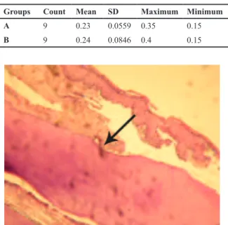
Images were captured from the mesial portion of each first molar under a microscope with a Fujix Hc-300Zi digital camera (Nikon, Japan). Midme-sial sections of the roots were chosen and the mean surface areas of the resorptive lacunae in the mesial portion of the first molar teeth were histomorpho-metrically measured.
The RT-PCR evaluation
The mesial root of each first maxillary molar with the alveolar bone was extracted by means of rongeur forceps. The root and bone were detached and placed separately in normal saline in -20°C. The tooth and bone were placed in EDTA 5% for twenty four hours, and were sonicated to achieve complete lyses. One milliliter of RNX plus buff-er was added to each specimen. Aftbuff-er staying in room temperature for 10 minutes, 200 μL of chlo-roform was added to the mixture. It was then cen-trifuged for 15 minutes in 4°C at 12000 rpm. The upper phase was moved to another microtube and pure ethylic alcohol, in equal volume to the upper phase, was added to the specimen. The RNA
sedi-ment was rinsed with 100μL alcohol (70%) and
was centrifuged for 2 minutes at 12000 rpm. The
RNA sediment was solved in 25 μL of
diethyl-pyrocarbonate (DEPC) treated water. The speci-mens were then heated to 70°C in order to open the probable RNA loops before reaction reverse transcription.
cDNA synthesis
The transcriptase reaction was prepared with the
fol-lowing: 1 μg of RNA, 1×RT buffer, 0.3 mM dNTP,
100 units RNasine, 200 units of reverse transcriptase enzyme, DEPC treated water up to 20 μl. The reac-tion was incubated at 42°C for one hour.
PCR amplification
The PCR reaction included the following: total synthesized cDNA, 0.2 mM dNTP, 1.5 mM Mg-Cl2, 40 pmol each of forward and reverse primers, 1× PCR buffer, 1.25 units of Taq DNA polymerase (CinnaGen, Iran), and ddH2O to give a final vol-ume of 50 μL. PCR cycling parameters included an initial 5 min preincubation at 94°C for 5 minutes, with incubation cycles consisting of denaturation at 94°C for 30 seconds, annealing temperature of 65°C for 60 seconds and extension at 72°C for 30 seconds. 30 cycles were repeated followed by a fi-nal incubation at 72°C for 5 minutes.
Nested PCR
The nested PCR was prepared as the PCR. In the
pre-sented study, PCR product was used instead of cDNA. The PCR product was electrophoresed in 3% agar-ose gel, stained with ethidium bromide and observed under a UV transilluminator devise. The PCR prod-uct for IL1-β was considered as 297 nucleotides.
Results
Orthodontic tooth movement observation
After comparing the degree of orthodontic tooth movements (OTMs) between groups A and B, it was concluded that a significant difference did not exist. Acccording to normal distribution of data, statistical test was student t-test and p=0.872, sta-tistical package of SPSS 11.5. It was hence de-duced that both groups would experience the same phenomenon from the root resorption perspective. Consequently, group A was assigned for histologic evaluation and group B was assigned for RT-PCR evaluation. Furthermore, the tooth movements between control and experimental groups were significantly different because the control group did not receive any protraction force and was only used for histologic and RT-PCR examinations. The results of the OTM experiment have been shown in table 1 and the examination of root re-sorption in table 2. According to the Mann-Whit-ney U test the magnitude of root resorption in group A and control groups were significantly dif-ferent (p<0.001). It shows that a significant ortho-dontic tooth movement is capable of creating root resorptive craters and lacunae (Fig 1).
Table 1: Orthodontic tooth movement in the histologically examined group A and the RT-PCR evaluated group B in millimeters
Minimum Maximum
SD Mean Count Groups
0.15 0.35
0.0559 0.23
9
A
0.15 0.4
0.0846 0.24
9
B
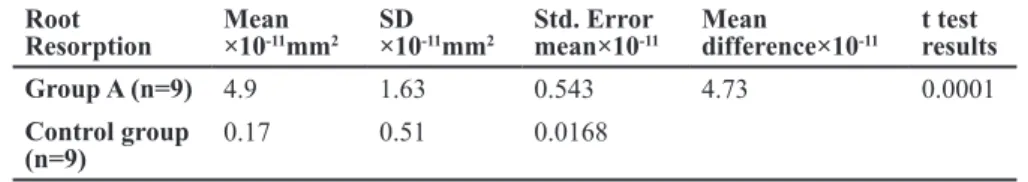
Fig 2: PCR product electrophoresis in 3% agarose gel. Lane 1= DNA ladder marker, lane 2= A 297 bp fragment as the IL-1 beta PCR products.
RT-PCR Evaluation
According to the RT-PCR for the IL-1β mRNA
gene expression in the root resorptive lacunae of group B and root craters or erosions in the control group, the authors conclude that there is no signifi-cant difference between the groups in relation to IL-1β gene expression (Fig 2).
Discussion
The investigation data regarding the IL-1 beta mRNA expression did not correlate with resorptive lacunae production, however further cellular and molecular analysis of larger animal groups is need-ed to confirm the irrelevance of IL-1beta genetic expression and root resorption during orthodontic tooth movement.
In the presented experiment, histological changes were considerably pronounced after force appli-cation to allow for the evaluation of the root for amount of resorption and IL-1β expression. This study was focused on the correlation between IL-1
beta expression and root resorption following orthodontic force application, and showed that there was no fundamental connection between specific IL-1β alleles and sporadic external root resorption.
The present investigation data fail to confirm the association reported by Al-Qawasmi et al. between external apical root resorption and IL-1β expres-sion (1). The aforementioned authors divided the control rats into two groups and IL1-beta in two groups of treated and untreated. They supported findings that the IL-1 cytokine was a significant factor in root resorption associated with orthodon-tic tooth movement. Knock out mice have enabled scientists to understand the function of a gene in its absence. At the end of an inbred line, the knocked-out gene is expressed in the complete absence of its viable copy of the gene.
Gene expression hypothetically may take place on the root surface or precisely in the resorptive la-cunae. By knocking out the gene, its expression would be inhibited in a complete manner. Conse-quently, it is an intervening factor that is eliminat-ed during research.
In the presented study, the resorptive phenomenon was surveyed locally and gene expression was investigated in the resorptive lacunae in an effort to eliminate the role of IL-1 in the bone. In their study, Al-Qawasmi et al. express the inequality of root resorption between wild and knockout ex-perimental groups without mentioning the degree of orthodontic tooth movement (1). The present study, the molars in the control and experimental groups were matched because a decreased rate of alveolar bone catabolic bone remodelling may re-sult in the prolonged compression of root against the alveolar bone.
Gulden et al. demonstrated a correlation between
IL-1α polymorphism and EARR (23); however,
Table 2: Measurement of root resorption in group A and control group in square millimeters
t test results Mean
difference×10-11 Std. Error
mean×10-11 SD
×10-11mm2 Mean
×10-11mm2 Root
Resorption
0.0001 4.73
0.543 1.63
4.9 Group A (n=9)
0.0168 0.51
0.17 Control group (n=9)
Table 3: IL-1β index comparison between the experimental and control groups using Wilcoxon signed rank test
P-value z
Sum of ranks Mean
rank Negative
amount Positive
amounts Count
Groups
0.157 -1.414 0.00
0.00 3
2 5 Experiment
3.00 1.5
1 4
they did not find IL-1β as a predisposing factor in EARR which shows consistency with our ex-periment. It is worthy to mention that osteopontin, osteonectin, cytokeratin 8 and granulocyte colony-stimulation factor (G-CSF) are other predisposing factors for root resorption (23).
Alhashimi et al. performed an in situ hybridiza-tion in order to measure the messenger RNA ex-pressions of IL-1β, IL-6, and TNF-α at 3, 7, and 10 days after application of orthodontic force on the maxillary first molars of 12 rats. They showed that the mRNA for IL-1 and IL-6 had reached their maximal expression on day three and finally they concluded that these proinflam-matory cytokines may play important roles in bone resorption after application of orthodontic force (15).
Ogasawara et al. evaluated the in situ expression of receptor activator of nuclear factor -κB ligand (RANKL), receptor activator of nuclear factor
-κB (RANK), osteoprotegerin, interleukin-1 β
(IL-1β) and tumour necrosis factor α (TNFα) in osteoclasts of rat periodontal tissue (25). They dis-covered that an autocrine mechanism of RANKL-RANK exists in the osteoclast, and is increased in pathological conditions. They also observed that in osteoclasts, the autocrine mechanism of IL-1 beta and TNF alpha is evident under pathologi-cal conditions. Hence, they concluded that the au-tocrine mechanisms seem to both pathologically and physiologically regulate the osteoclast func-tion in both physiological and pathological condi-tions (25).
Similarity between resorptive processes of bone and root, from a genetic point of view, is sup-ported by RANKL mRNA expression during root resorption (26) and osteopontin expression dur-ing OTM (27). Given the genetic proximity be-tween these two hard tissues i.e. bone and tooth, it should be emphasized that they are different tissues and some genetic diversity exists among them leading to different genetic expression in a similar situation.
Conclusion
Although interleukin-1 beta is the most potent stimulator of bone resorption and mediator of in-flammatory response, the presented study shows that the IL-1beta mRNA is not expressed signifi-cantly higher in root resorption lacunae of the ex-perimental group relative to the control group. In quantitative measurements, the IL-1beta expres-sion in root resorption currently reveals no indica-tion of its possible predisposing role in resorptive lacunae production.
Acknowledgments
The authors wish to thank the director of research at Shahid Beheshti University of Medical Scienc-es’ school of dentistry for his support and Dr. Dar-wish for her assistance in preparation of the sam-ples. There is no conflict of interest in this study and its related article.
References
Al-Qawasmi RA, Hartsfield JK, Hartsfield JK Jr, Everett 1.
ET, Weaver MR, Foroud TM, et al. Root resorption associ-ated with orthodontic force in IL-1Beta knockout mouse. J Musculoskelet Neuronal Interact. 2004; 4(4): 383-385. Davidovitch Z. Tooth movement. Crit Rev Oral Biol Med. 2.
1991; 2(4): 411-450.
Goldie RS, King GJ. Root resorption and tooth movement 3.
in orthodontically treated, calcium deficient and lactating rats. Am J Orthod. 1984; 85: 424-430.
Newman WG. Possible etiologic factors in external root 4.
resorption. Am J Orthod 1975; 67: 522-539.
Nishioka M, Loi H, Nakata S, Nakasima A, Counts A. Root 5.
resorption and immune system factors in Japanese. An-gle Orthod. 2006; 76: 103-108.
Oyama K, Motoyoshi M, Hirabayashi M, Hosoi K, Shimizu 6.
N. Effects of root morphology on stress distribution at the root apex. Eur J Orthod. 2007; 29: 113-117.
Hartsfield JK Jr, Everett ET, Al-Qawasmi RA. Genetic 7.
factors in external apical root resorption and orthodontic treatment. Crit Rev Oral Biol Med. 2004; 15(2): 115-122. Harry MR, Sims MR. Root resorption in bicuspid intrusion: 8.
a scanning electron microscopic study. Angle Orthod. 1982; 52: 235-258.
Reitan K. Effect of force magnitude and direction of tooth 9.
movement on different alveolar bone types. Angle Orthod. 1964; 44: 244-255.
Gowen M, Wood DD, Ihrie EJ, McGuire MK, Russell RG. 10.
An interleukin 1 like factor stimulates bone resorption in vitro. Nature. 1983; 306(5941): 378-380.
Canalis E. Interleukin-1 has independent effects on deox-11.
yribonucleic acid and collagen synthesis in cultures of rat calvariae. Endocrinology. 1986; 118(1): 74-81.
Lorenzo JA, Sousa SL, Alander C, Raisz LG, Dinarello 12.
CA. Comparison of the bone-resorbing activity in the su-pernatants from phytohemagglutinin-stimulated human peripheral blood mononuclear cells with that of cytokines through the use of an antiserum to interleukin 1. Endo-crinology. 1987; 121(3): 1164-1170.
Seifi M, Eslami B, Saffar AS. The effect of prostaglandin 13.
E2 and calcium gluconate on orthodontic tooth movement and root resorption in rats. Eur J Orthod. 2003; 25(2): 199-204.
Grieve WG III, Johnson GK, Moore RN, Reinhardt RA, 14.
DuBois LM. Prostaglandin E (PGE) and interleukin-1 beta (IL-1 beta) levels in gingival crevicular fluid during human orthodontic tooth movement. Am J Orthod Dentofacial Or-thop. 1994; 105: 369-374.
Alhashimi N, Frithiof L, Brudvik P, Bakhiet M. Orthodontic 15.
tooth movement and de novo synthesis of proinflamma-tory cytokines. Am J Orthod Dentofacial Orthop. 2001; 119: 307-312 .
Wei S, Kituara H, Zhou P , Ross FP, Teitelbaum SL. IL-1 16.
Mediates TNF induced osteoclastogenesis. J Clin Invest. 2005; 115: 282-290.
Tzannetou S, Efstratiadis S, Nicolay O, Grbic J, Lamster 17.
Jäger A, Zhang D, Kawarizadeh A, Tolba R, Braumann B, 18.
Lossdörfer S, et al. Soluble cytokine receptor treatment in experimental orthodontic tooth movement in the rat. Eur J Orthod. 2005; 27: 1-11.
Sari E, Ucar C. Interleukin 1beta levels around microscrew 19.
implants during orthodontic tooth movement. Angle Or-thod. 2007; 77(6): 1073-1078.
Maeda A, Soejima K, Bandow K, Kureoe K, Kakinoto K, 20.
Miyawaki S, et al. Force-induced IL-8 from periodontal ligament cells requires IL-1beta. J Dent Res. 2007; 86(7): 629-634.
Meikle MC. The tissue, cellular, and molecular regulation 21.
of orthodontic tooth movement: 100 years after Carl Sand-stedt. Eur J Orthod. 2006; 28: 221-240.
Tzannetou S, Efstratiadis S, Nicolay O, Grbic J, Lamster I. 22.
Interleukin-1beta and beta-glucuronidase in gingival crev-icular fluid from molars during rapid palatal expansion. Am J Orthod Dentofacial Orthop. 1999; 115: 686-696. Gulden N, Eggermann T, Zerres K, Beer M, Meinelt A, Die-23.
drich P. Interleukin-1 polymorphism in relation to external
apical root resorption (EARR). J Orofac Orthop. 2009; 70: 20-38.
Al-Qawasmi RA, Hartsfield JK Jr, Everett ET, Flury L, Liu 24.
L, Foroud TM, et al. Genetic predisposition to external apical root resorption in orthodontic patients: linkage of chromosome-18 marker. J Dent Res. 2003; 82(5): 356-360.
Ogasawara T, Yoshimine Y, Kiyoshima T, Kobayashi I, 25.
Matsuo K, Akamine A, et al. In situ expression of RANKL, RANK, osteoprotegerin and cytokines in osteoclasts of rat periodontal tissue. J Periodontal Res. 2004; 39(1): 42-49.
Seifi M, Jessri M. Expression of RANKL mRNA during root 26.
resorption induced by orthodontic tooth movement in rats. Yakhteh Medical Journal. 2009; 11(3): 293-298.
Seifi M, Jessri M, Vahid-Dastjerdi E. Identification of a 27.