Mice
Ravisankar A. Ramadas1,2*, Susan L. Ewart3, Yoichiro Iwakura4, Benjamin D. Medoff1,2, Ann Marie LeVine5*
1Center for Immunology and Inflammatory Diseases, Harvard Medical School, Massachusetts General Hospital, Boston, Massachusetts, United States of America,
2Pulmonary and Critical Care Unit, Massachusetts General Hospital, Boston, Massachusetts, United States of America,3Department of Large Animal Clinical Sciences, Michigan State University, East Lansing, Michigan, United States of America,4Center for Experimental Medicine and Systems Biology, University of Tokyo, Tokyo, Japan,
5Department of Pediatrics and Communicable Diseases, University of Michigan, Ann Arbor, Michigan, United States of America
Abstract
Interleukin (IL-) 36 cytokines (previously designated as novel IL-1 family member cytokines; IL-1F5– IL-1F10) constitute a novel cluster of cytokines structurally and functionally similar to members of the IL-1 cytokine cluster. The effects of IL-36 cytokines in inflammatory lung disorders remains poorly understood. The current study sought to investigate the effects of IL-36a (IL-1F6) and test the hypothesis that IL-36a acts as a pro-inflammatory cytokine in the lungin vivo. Intratracheal instillation of recombinant mouse IL-36ainduced neutrophil influx in the lungs of wild-type C57BL/6 mice and IL-1ab2/2 micein vivo. IL-36ainduced neutrophil influx was also associated with increased mRNA expression of neutrophil-specific chemokines CXCL1 and CXCL2 in the lungs of C57BL/6 and IL-1ab2/2micein vivo. In addition, intratracheal instillation of IL-36a enhanced mRNA expression of its receptor IL-36R in the lungs of C57BL/6 as well as IL-1ab2/2 mice in vivo. Furthermore, in vitro incubation of CD11c+cells with IL-36aresulted in the generation of neutrophil-specific chemokines
CXCL1, CXCL2 as well as TNFa. IL-36aincreased the expression of the co-stimulatory molecule CD40 and enhanced the ability of CD11c+cells to induce CD4+T cell proliferationin vitro. Furthermore, stimulation with IL-36aactivated NF-kB in
a mouse macrophage cell line. These results demonstrate that IL-36a acts as a pro-inflammatory cytokine in the lung without the contribution of IL-1aand IL-1b. The current study describes the pro-inflammatory effects of IL-36ain the lung, demonstrates the functional redundancy of IL-36awith other agonist cytokines in the IL-1 and IL-36 cytokine cluster, and suggests that therapeutic targeting of IL-36 cytokines could be beneficial in inflammatory lung diseases.
Citation:Ramadas RA, Ewart SL, Iwakura Y, Medoff BD, LeVine AM (2012) IL-36aExerts Pro-Inflammatory Effects in the Lungs of Mice. PLoS ONE 7(9): e45784. doi:10.1371/journal.pone.0045784
Editor:Bernhard Ryffel, French National Centre for Scientific Research, France
ReceivedMay 17, 2012;AcceptedAugust 22, 2012;PublishedSeptember 20, 2012
Copyright:ß2012 Ramadas et al. This is an open-access article distributed under the terms of the Creative Commons Attribution License, which permits unrestricted use, distribution, and reproduction in any medium, provided the original author and source are credited.
Funding:This research work was supported by Grants from the National Institute of Allergy and Infectious Diseases 1R03AI101366 (R.A.R) and the National Heart, Lung & Blood Institute 5T32HL007874 (B.D.M). The funders had no role in study design, data collection and analysis, decision to publish, or preparation of the manuscript.
Competing Interests:The authors have declared that no competing interests exist.
* E-mail: [email protected] (RAR); [email protected] (AML)
Introduction
The Interleukin 1 (IL-1) cytokine cluster consists of a network of agonist, antagonist and receptor molecules that are expressed in a variety of immune and structural cells [1]. Since its initial identification as the human leukocytic pyrogen that causes fever [2], IL-1 has emerged as highly organized facilitator of early immune events in a plethora of inflammatory diseases [3]. The role of IL-1 cytokines in the development of acute and chronic inflammatory disorders of the lung has been extensively docu-mented and mechanistically characterized in human patients and in animal models of human lung disorders. For example, increased levels of IL-1 protein or mRNA, reflective of increased IL-1 cytokine activity, has consistently been observed in tissues from human patients with inflammatory lung disorders such as asthma [4–7], acute lung injury [8–10] and pulmonary fibrosis [11]. Similarly, studies from animal models have demonstrated that IL-1 cytokines such as IL-IL-1a and IL-1b play critical roles in the pathogenesis of asthma [12–14], chronic obstructive pulmonary disease [15,16], acute lung injury [17] and pulmonary fibrosis [18– 20]. In pulmonary infection models, IL-1 promoted or dampened
bacterial clearance from the lungs depending on the bacterial pathogen [21–23]. While IL-1 induced the recruitment of neutrophils and pulmonary inflammation during the early stages of influenza infection in mice [24], it was critical in conferring protective immunity and enhancing survival of mice during the later stages of the disease [24–26].
which consists of agonists (IL-1a, IL-1b) and an antagonist (IL-1Ra) which binds to a functional heterodimeric receptor complex containing IL-1 receptor I (IL-1R1) and IL-1RAcP [35]. Both the classical and novel IL-1 cytokine cluster agonists induce the activation of nuclear factor kappa B (NF-kB) and mitogen associated protein kinase (MAPK) to initiate pro-inflammatory pathways [31,35]. While the role of classical IL-1 cytokines have been extensively characterized in the pathogenesis of inflamma-tory disorders [35,36], the disease-relevant functions of the novel IL-1 like cytokines (IL-36 cytokines) remain poorly characterized. We have previously demonstrated that IL-36cis constitutively expressed and inducibly upregulated in the lungs of mice following allergic inflammation [37,38]. Airway epithelial cells express IL-36aand IL-36c[38,39], and the mRNA expression of IL-36aand IL-36cin airway epithelial cells is increased in response to several inflammatory stimuli [39,40]. IL-36a and IL-36cdirectly act on IL-36R expressing lung fibroblasts to activate NF-kB and MAPK pathways [39]. We have demonstrated that intratracheal in-stillation of recombinant IL-36c increased airway hyperrespon-siveness, induced neutrophil influx and neutrophil-specific che-mokine production the lungsin vivo[38]. We also demonstrated that IL-36c activated NF-kB in whole lung tissue and alveolar macrophage cell lines [38]. Reports from other groups have demonstrated that the mRNA expression of IL-36a and IL-36c
were also increased in human bronchial epithelial cells exposed to
Pseudomonas aeruginosa[40] and in recurrent respiratory papillomas [41]. Recent reports have also demonstrated the critical role of IL-36a, IL-36Ra and IL-36R in the development of psoriasis [42– 45], and that a single amino acid mutation in IL-36Ra can lead to unchecked cytokine production and pustular psoriasis [46]. Accumulating evidence thus points to the existence of a novel, underexplored signaling pathway in inflammatory diseases, driven by the IL-36 cytokine cluster.
Despite the recent progress in elucidating the role of IL-36 cytokines in inflammatory diseases, thein vivo role of IL-36a has not been extensively investigated in inflammatory lung disorders. Since multiple cytokines contribute to shaping the overall immune response in inflammatory disorders, it is necessary to characterize the proportional contribution and functional redundancy of individual cytokines to comprehensively understand disease pathogenesis and devise therapeutic strategies. Since it has been demonstrated that the mRNA expression of IL-36ain the airway epithelial cells are increased in response to multiple pro-inflammatory stimuli [39], we hypothesized that the presence of IL-36ain the lungs may induce pulmonary inflammationin vivo. Here we demonstrate that intratracheal instillation of recombinant mouse IL-36a induces neutrophil influx and the production of neutrophil-specific chemokines in wild-type mice as well as mice that lack IL-1aand IL-1b. Furthermore, we also demonstrate that IL-36aacts directly on CD11c+
cells to induce the expression of neutrophil-specific chemokines, T cell costimulatory molecules and the enhancement of CD4+T cell proliferation. Finally, we also demonstrate that IL-36aactivates NF-kB in mouse macrophages. These results suggest that IL-36a acts as a pro-inflammatory cytokine in the lung, and that IL-36acould increase inflammatory responses in disease conditions which involve release of IL-36a
into the lungs.
Materials and Methods
Mice and Procedures
Six to eight week old mice (C57BL/6, C3H/HeJ and OTII TCR transgenic strains) were purchased from Jackson Laboratory (Bar Harbor, ME). Mice deficient in IL-1aand IL-1b(IL-1a/b2/
2) were obtained from Dr. Yoichiro Iwakura [47] and backcrossed
to C57BL/6 background. All mice were housed or bred in specific pathogen free facility at Massachusetts General Hospital. For intratracheal instillations, mice were anesthetized with a mixture of xylazine (8 mg/kg) and ketamine (45 mg/kg). Recombinant protein preparation or phosphate buffered saline (PBS) controls in a 50ml volume were intratracheally (i.t.) instilled using a non-invasive method. Mice were euthanized with an overdose of pentobarbital, and tissues were collected at indicated time points following the procedure. For cell isolation protocols that do not require isolation of lung tissue, mice were euthanized by carbon-dioxide asphyxiation.
Ethic Statement
Mice were housed in a temperature controlled specific-pathogen free facility at Massachusetts General Hospital, and provided unrestricted access to food and water. All protocols involving the use of mice were approved by Subcommittee on Research Animal Care at Massachusetts General Hospital.
Generation of Recombinant IL-36a
A cDNA clone containing full length mouseIl36a(Il1f6, Clone
ID: MMM1013-99829107) was obtained (Open Biosystems). A PCR product encoding a fusion protein comprised of an N-terminal FLAG tag followed by the full length coding sequence of
Il36awas generated from this clone using the following
restriction-site engineered primers: Forward 59 -CGGAATTCCgattacaaggat-gacgatgacaagAATAAGGAGAAAGAACTAAGAG-39 and re-verse 59 -AAGGAAAAAAGCGGCCGCTTAATGTACCA-CAATCATCTC-39. The PCR product and pETDuet-1 vector (Clontech) were digested with EcoRI and NotI. The digested PCR product was cloned in-frame with the Histidine purification tags into the pETDuet-1 vector. The ligation reactions were trans-formed intoE. coliand positive clones were identified. Expression of His_FLAG_IL-F6 fusion protein was induced in positive clones by addition of 1 mM Isopropyl b-D-1-thiogalactopyranoside (IPTG) to log-phase bacterial cultures for 4 h. Following in-duction, bacterial pellets were frozen at 220uC until protein isolation. IL-36awas purified from bacterial lysate by Ni2+
column chromatography (B-PER 6xHis Fusion Protein Kit, Pierce), and dialyzed against PBS. Purified, dialyzed IL-36awas treated with enterokinase (EK-Max, Invitrogen) to cleave His_FLAG tags from IL-36a protein. Enterokinase was removed from the tag-cleaved IL-36a preparation by treating with EK-Away (Invitrogen), according to the manufacturer’s protocols. The purified, tag-cleaved, enterokinase-removed recombinant IL-36a preparation was dialyzed and the dialyzed preparation was passed through a Ni2+ chromatography column. Cleaved tags bound to the column and the tag-free IL-36apreparations recovered from the flow-through were dialyzed against PBS, treated with polymyxin-B agarose beads (Detoxi-Gel, Pierce) to remove LPS contamination. LPS concentration in the purified IL-36apreparation was,0.01 EU/mg protein, as measured by Limulus Amebocyte Lysate assay (Lonza). Tag-removed, polymyxin-treated recombinant mouse IL-36awas used forin vitroandin vivostudies.
Coomassie Staining
and microwaved multiple times and washed in water to destain the gel.
Western Immunoblotting
Increasing amounts of IL-36a(5, 10 and 20 ng) were separated on a SDS-polyacrylamide gel under reducing and denaturing conditions. Proteins were transferred to polyvinylidene fluoride membranes, probed with rat anti-mouse IL-36aantibody (R&D Systems, Catalog No. MAB2297) and detected with a horseradish peroxidase conjugated goat anti-rat secondary antibody (Abcam) and ECL Plus chemiluminescent detection system (Amersham).
Tissue Collection and Analysis
Mice were euthanized with an overdose of pentobarbital, and the treacheas were catheterized. Lungs were lavaged three times with 1 mL of ice cold-PBS. Bronchoalveolar lavage (BAL) fluid was centrifuged at 1,500 rpm for 15 minutes and the supernatants were stored at280uC. Cells in the pellet were used to calculate total cell numbers (Cellometer, Nexelcom), as well as differential cell counts on cytospun slides stained with Hema-3 White Cell Differential Staining Kit (Fisher). More than 100 cells were counted per sample by light microscopy to calculate differential cell counts. Upper right lobe of the lung was collected for RNA analysis, and the left lung was inflation fixed with 10% neutral buffered formalin for histological analysis.
RNA Isolation and Quantitative Real-time PCR
Total RNA was isolated from lung tissue using Trizol (Invitrogen) and cell cultures using RNeasy (Qiagen) and treated with Amplification grade DNaseI (Invitrogen) according to manufacturer’s protocols. Equal concentrations of DNaseI treated RNA was reverse transcribed into cDNA using Taqman Reverse Transcription reagents (Applied Biosystems) and used as input for quantitative real-time PCR using Power SYBR Green kit (Applied Biosystems). Primers used in real-time PCR analyses were designed using the mouse qPrimerdepot (http:// mouseprimerdepot.nci.nih.gov/). Cycle threshold (CT) values
obtained from the assays were analyzed and reported as copies of target genes per copy of GAPDH, a housekeeping gene.
Measurement of Protein Levels in the BAL Fluid
Cytometric bead array Flex-kits (BD Biosciences) for IL-1a, IL-1b, TNFa and CXCL1 were used according to manufacturer’s protocols to evaluate the protein levels in the BAL fluid recovered from the mice.
Measurement of Lung Function in Mice
Six to eight week old mice were intratracheally administered 10mg IL-36aor 50ml PBS, and lung function measurements were performed 24 h later using the Flexivent system (Scireq, Montreal, Quebec, Canada), as described before [38]. Briefly, mice were anesthetized with an intraperitoneal injection of xylazine (12 mg/ kg) and pentobarbital (70 mg/kg). The trachea of anesthetized mice was cannulated and the mice were ventilated with 6 ml/kg tidal volume at 150 breaths per minute. To suppress spontaneous breathing during measurement of lung function, mice were intraperitoneally injected with pancuronium bromide (2 mg/kg). Incremental doses of neublized methacholine were used to determine total lung resistance and compliance according to the Snapshot-150 perturbation provided by the Flexivent equipment. Thirteen data points were collected for each methacholine dose, and only data with a coefficient of determination greater than 0.95 were included in analyses. The survival of mice during the
procedure was simultaneously monitored using electrocardio-grams.
Isolation and Culture of CD11c+Cells
Splenic CD11c+ cells were isolated from wildtype C57BL/6 using magnetic separation according to manufacturer’s (Miltenyi Biotec) protocol. Briefly, single cell suspensions prepared from spleens of wildtype C57BL/6 mice were incubated with red blood cell lysis solution (Invitrogen) to lyse RBCs. The resulting cell suspensions were incubated with mouse anti-CD11c microbeads (Miltenyi Biotec), and CD11c+cells were isolated by two rounds of positive selection to enhance purity. The purity of isolated CD11c+ cells were routinely .90%. The isolated CD11c+
cells were resuspended in RPMI-1640 media containing 10% heat inacti-vated fetal bovine serum (FBS), L-glutamine and 1% penicillin-streptomycin. According to the experiment, aliquots of resus-pended cells were mixed with various concentrations of IL-36a
(0.1mg/mL, 1.0mg/mL or 10.0mg/mL) or an equivalent volume of PBS. For RNA isolation, cells mixed with IL-36aor PBS were plated at a concentration of 2.56105 cells per well in a 48-well plate in a volume of 250ml. The cells were incubated for 2 hours to adhere. The media was gently removed from the plates by pipetting, and the cells were washed once with sterile, cold PBS. RNA was isolated from the adherent cells using RNeasy RNA isolation kit (Qiagen) according to manufacturer’s protocols. In some experiments, the media containing IL-36a was removed from the wells 2 hours after incubation, replaced with RPMI-1640 growth media and allowed to incubate for an additional 22 hours before evaluating cell surface expression of co-stimulatory molecules by flow cytometry.
Isolation and Culture of CD4+T Cells
Spleens and pooled lymph nodes from wildtype C57BL/6 mice were used to isolate CD4+
cells using magnetic separation according to manufacturer’s (Miltenyi Biotec) protocol to a routine purity of .95%. Isolated CD4+ cells were stained with CFSE (Celltrace CFSE staining kit, Invitrogen) and resuspended in culture media. For CD4+
and CD11c+
co-culture studies, CD11c+ cells were isolated using magnetic separation protocols, mixed with the indicated concentrations of IL-36a, plated at a concentration of 16105cells per well in a 96-well plate and allowed to adhere for 2 hours. Following incubation, media from the wells was removed by pipetting, cells were washed once with cold PBS, and 46105 CFSE-stained CD4+
T cells were incubated with the adherent CD11c+cells for 96 hours. Following incubation for 96 h, cells were removed and analyzed by flow cytometry. To evaluate antigen-specific responses, isolated splenic CD11c+
cells were plated at a concentration of 16105 cells per well with indicated concentrations of IL-36aand 100 ng/mL OVA323-339for 2 hours.
Following incubation, media from the wells was removed by pipetting, cells were washed once with cold PBS, and 46105 CFSE-stained CD4+ T cells from OTII TCR transgenic mice were incubated with the adherent CD11c+
cells for 96 hours. Following incubation for 96 h, cells were removed and analyzed by flow cytometry.
Flow Cytometry
by gating on live CD4+
cells based on forward- and side-scatter properties. Flow cytometry data were acquired by an Accuri C6 flow cytometer (Accuri) and analyzed using CFlow software (Accuri) or Flowjo (Treestar).
Isolation of Alveolar Macrophages from Naı¨ve Mice Naı¨ve C57BL/6 mice were euthanized with an overdose of pentobarbital. Lungs of mice were lavaged three times with 1 mL of ice cold-PBS. Pooled BAL fluids from three mice were centrifuged at 1,500 rpm for 15 minutes. An aliquot of cells were used for cytospin analysis. RNA was isolated from the remaining cells using RNeasy RNA isolation kit (Qiagen). Isolated RNA was treated with DNaseI, reverse transcribed and the resulting cDNA was used as the input for PCR.
NF-kB Activation Assays in Mouse Macrophage Cell Lines A mouse macrophage cell line (RAW-ELAM) stably transfected with the NF-kB responsive E-Selectin promoter driving the expression of enhanced green fluorescent protein [48] was used to investigate if IL-36a induced NF-kB activity in mouse macrophages. Cells were cultured in RPMI-1640 with 10% fetal bovine serum and 1% penicillin and streptomycin. One day before the experiments, cells were plated at a density of 56105cells per well. Media was removed the day of the experiment, and IL-36a
mixed with media was added to the cells and incubated for 6 h. After incubation, media was removed and cells were washed twice with ice-cold PBS. Cells were collected, and eGFP fluorescence reflective of NF-kB activity was measured using an Accuri C6 flow cytometer.
Statistical Analysis
Data from BAL cell enumeration experiments and real-time PCR quantification on lung tissue were analyzed by two-tailed student’s t-test. Real-time PCR data from cell culture experiments were analyzed by two tailed student’s t-test. Lung resistance and compliance measurements were analyzed by two-way repeated measures ANOVA. Statistical significance was accepted at
P,0.05.
Results
IL-36aInduces Neutrophil Influx in the Lungs of Mice We and others have previously demonstrated that IL-36c is predominantly expressed in airway epithelial cells and that i.t. instillation of IL-36cinduces neutrophil influx in the lungs of mice [38,39]. IL-36aand IL-36cmRNA expression levels are increased in airway epithelial cells in response to various stimuli [39]. To investigate if IL-36a induces proinflammatory responses in the lung in vivo, we generated pure recombinant murine IL-36a
protein (Fig. 1A & 1B). A single i.t. instillation of 10mg IL-36a
induced neutrophil influx (Fig. 1C) in the lungs of wild-type C57BL/6 mice 24 h following instillation. As expected, the majority of cells in the lungs of PBS instilled mice were
macrophages (Fig. 1C). The total number of cells (Fig. 1D), percentage of neutrophils (Fig. 1E) and the total number of neutrophils (Fig. 1F) were significantly increased in the lungs of mice that received i.t. IL-36acompared to mice that received PBS. Flow cytometric evaluation of BAL cells also revealed that majority of cells recovered from BAL fluid of IL-36a instilled mice were CD11c2CD11b+
Ly6G+
neutrophils, while a majority of cells recovered from the BAL fluid of PBS instilled mice were CD11c+
CD11b2Ly6G2cells, reflective of macrophages (Fig. 1G). Examination of Hematoxylin & Eosin stained lung sections revealed significant cellular infiltration in the lungs of IL-36a
instilled mice (Fig. 1H) compared to the lungs of mice that received PBS. While we used polymyxin-treated, highly pure IL-36a, to ensure that the results we obtained are not due to trace amounts of contaminating LPS from the bacteria-derived IL-36a, we performed the same set of experiments in C3H/HeJ mice, which are unresponsive to LPS due to a spontaneous loss-of-function mutation in Toll like receptor 4 [49]. It has been previously demonstrated that LPS does not induce neutrophil influx or the production of neutrophil-specific chemokines CXCL1 and CXCL2 in the lungs of C3H/HeJ mice [50]. In the current study, a single i.t. instillation of 10mg IL-36ainduced neutrophil influx into the lungs (Fig. S1A), increased the total number of cells (Fig. S1B), percentage of neutrophils (Fig. S1C) and total number of neutrophils (Fig. S1D) in the lungs as assessed from BAL fluid recovered from C3H/HeJ mice 24 h following instillation. Macrophages were the main cell types present in the BAL fluid of PBS instilled C3H/HeJ mice. These results strongly suggest that the neutrophil influx observed in the lungs of mice is due to the agonist activity of IL-36a and not due to contaminants in the protein preparation. Taken together, these data demonstrate that neutrophil influx is the predominant response to overproduction of IL-36ain the lungs.
IL-36aInduces the Expression of Early Response Cytokines and Neutrophil Specific Chemokines
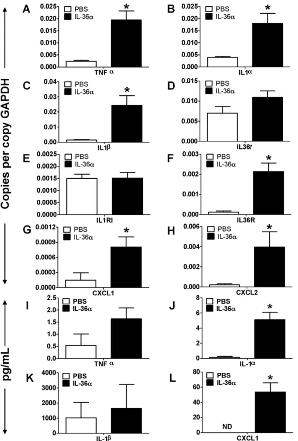
To determine IL-36adriven molecular mechanisms that induce neutrophil influx into the lungs, we collected lung tissue from wildtype C5BL/6 mice 24 h following a single i.t. instillation of IL-36a and profiled the mRNA expression of selected cytokines, chemokines as well as agonists and receptors in the IL-1 cytokine network. The mRNA expression of the early response cytokine TNFa was significantly increased in the lungs of mice which received i.t. instillation of IL-36a, compared to saline controls (Fig. 2A). Similarly, the mRNA expression of IL-1a(Fig. 2B) and IL-b(Fig. 2C) were also significantly increased in the lungs of IL-36a instilled mice, while the expression of IL-36cwas unaltered (Fig. 2D). We did not detect the mRNA expression of other agonist IL-36 cytokines in the lungs under the conditions used (data not shown). While the mRNA expression of the classical IL-1 receptor (1RI) was unchanged (Fig. 2E), the mRNA expression of the IL-36R, the receptor for IL-36a and IL-36c, was significantly increased in the lungs of IL-36a instilled mice (Fig. 2F). In Figure 1. Intratracheal instillation of IL-36ainduced neutrophil influx in the lungs of wild-type C57BL/6 mice.A) Coomassie blue stained gel demonstrating the purity of IL-36apreparation. Tenmg IL-36awas loaded on the lane. B) Western immunoblotting of IL-36aprotein preparation detects a band around 18 KDa, the predicted molecular weight of mouse IL-36a. C) Cytospin preparations demonstrating neutrophil influx in the BAL fluid recovered from mice 24 h following a single i.t. instillation of PBS or 10mg IL-36a. D) Total cell counts from BAL fluid recovered from mice 24 h following a single i.t. instillation of PBS or 10mg IL-36a. E) Differential cell count percentages and F) Differential cell count numbers in the BAL fluid recovered from mice 24 h following a single i.t. instillation of PBS or 10mg IL-36a. *Indicates significant differences (P,0.05) compared
to PBS treated mice. Data represent mean6SEM from 4–5 mice/group. G) Flow cytometry on cells recovered from BAL fluid from mice 24 h following a single i.t. instillation of PBS or 10mg IL-36a. A majority of cells from the IL-36ainstilled lungs were CD11c2CD11b+Ly6G+neutrophils. Depicted flow
cytometry plots are representative of 4–5 mice/group. H) Hematoxylin & Eosin stained lung sections isolated from mice 24 h following a single i.t instillation of PBS or 10mg IL-36a. Depicted sections are representative of 4–5 mice/group.
Figure 2. Intratracheal instillation of IL-36aincreased the mRNA expression of proinflammatory mediators in the lungs of wild-type C57BL/6 mice.A–H) Transcript expression of early response cytokines (TNFa, IL-1a, IL-1b, IL-36c), the classical IL-1 receptor IL-1R1, the novel IL-1 cytokine cluster receptor IL-36R and the neutrophil specific chemokines CXCL1 and CXCL2 in the lungs of mice 24 h following a single i.t instillation of PBS or 10mg IL-36a. Transcript expression was evaluated by SYBR-Green based quantitative real-time PCR. I–K) Protein expression of TNFa, IL-1a, IL-1band CXCL1 in the BAL fluid recovered from mice 24 h following a single i.t instillation of PBS or 10mg IL-36a. Protein expression was quantified by multiplexed cytometric bead arrays. *Indicates significant differences (P,0.05) compared to PBS treated mice. Data represent mean6SEM from 4–
5 mice/group.
doi:10.1371/journal.pone.0045784.g002
addition, the mRNA expression of the neutrophil recruiting chemokines CXCL1 (Fig. 2G) and CXCL2 (Fig. 2H) were also significantly increased in the lungs of IL-36a instilled mice compared to PBS controls. The protein levels of IL-1a (Fig. 2J) and CXCL1 (Fig. 2L) were significantly increased in the BAL fluid recovered from IL-36ainstilled mice, however the protein levels of TNFa and IL-1b were not significantly increased. These results suggest that by inducing the expression of its receptor (IL-36R), IL-36amay potentially contribute to the initiation of a feedback signaling loop to enhance proinflammatory responses. Further-more, these results also suggest that IL-36aacts on structural or immune cells in the lung to induce the production of CXCL1 and CXCL2, leading to neutrophil influx in the lungs.
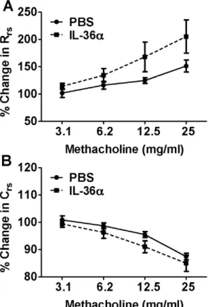
A Single i.t. Instillation of IL-36adoes not Induce Airway Hyperresponsiveness in Mice
To determine if IL-36ainduced proinflammatory effects in the lungs results in differences in physiological responses, wildtype C57BL/6 mice were intratracheally instilled with a single dose of IL-36a. Pulmonary function parameters evaluated 24 h later using invasive plethysmography revealed that the total lung resistance (Fig. 3A) and lung compliance (Fig. 3B) were not significantly different between IL-36aand PBS instilled mice.
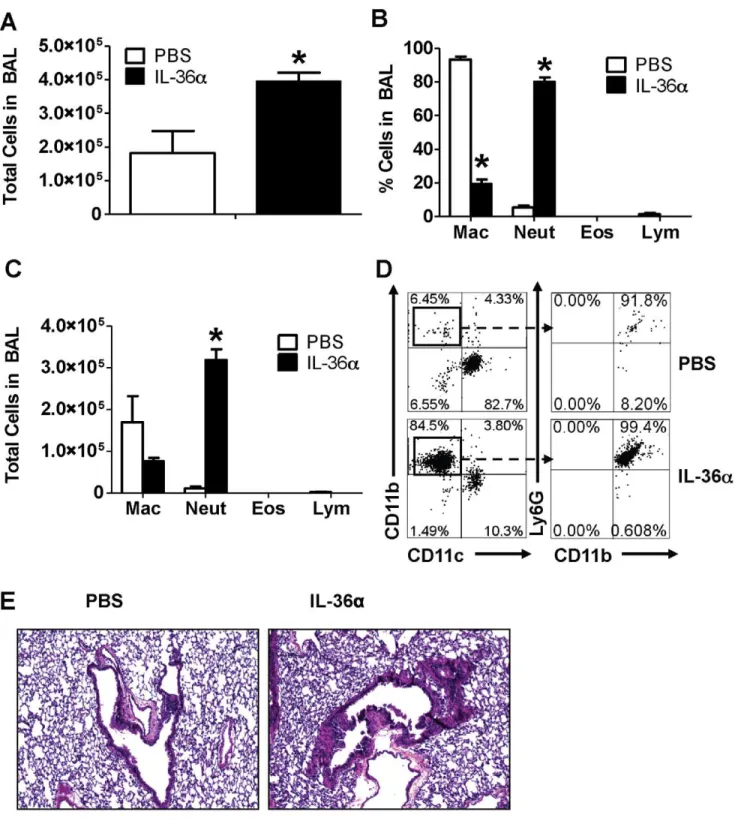
IL-36aInduces Neutrophil Influx in the Lungs of IL-1a and IL-1b Deficient Mice
Agonist members of the IL-1 cytokine network often induce overlapping and redundant pro-inflammatory responses. For example, IL-1aand IL-1b can induce neutrophil influx into the lung and induce the production of a variety of proinflammatory mediators that are induced by IL-36a and IL-36c. We have demonstrated that IL-36a and IL-36c induce the production of IL-1a and IL-1b ([38] and the current study). Therefore, to determine if IL-1aand IL-1bmay also contribute in part to the IL-36a induced pro-inflammatory responses in the lung, we performed a single i.t. instillation of IL-36ainto the lungs of mice genetically deficient in both IL-1aand IL-1b(IL-1ab2/2mice). A
single i.t. instillation of 10mg IL-36a induced neutrophil influx (Fig. 4A) in the lungs of IL-1ab2/2 mice 24 h following
instillation. The majority of cells in the lungs of PBS instilled mice were macrophages. The total number of cells (Fig. 4A), percentage of neutrophils (Fig. 4B) and the total number of neutrophils (Fig. 4C) were significantly increased in the lungs of IL-1ab2/2mice that received i.t. IL-36acompared to IL-1ab2/2
mice that received PBS. Flow cytometric evaluation of BAL cells also revealed that majority of cells recovered from the BAL fluid of IL-36a instilled mice were CD11c2CD11b+
Ly6G+
neutrophils, while a majority of cells recovered from the BAL fluid of PBS instilled mice were CD11c+
CD11b2Ly6G2 cells (Fig. 4D). Examination of hematoxylin & eosin stained lung sections also revealed significant cellular infiltration in the lungs of IL-36a
instilled mice (Fig. 4E) compared to the lungs of mice that received PBS. These data demonstrate that IL-36acan induce neutrophil influx in the lungs in an IL-1a/b-independent manner.
IL-36aInduces the Expression of Early Response Cytokines and Neutrophil Specific Chemokines in the Lungs of IL-1aand IL-1bDeficient Mice
To determine if IL-36a induced cytokine and chemokine expression is altered in the absence of IL-1a and IL-1b, we collected lung tissue from IL-1ab2/2 mice 24 h following
a single i.t. instillation of IL-36a and profiled the mRNA expression of selected cytokines, chemokines as well as agonists and receptors in the IL-1 cytokine network. The mRNA expression of the early response cytokine TNFa(Fig. 5A) as well as IL-36c Fig. (5B) was significantly increased in the lungs of IL-1ab2/2 mice i.t. instilled with IL-36a, compared to saline
controls. As expected, mRNA expression of neither IL-1a nor IL-1b was detected in the lungs of IL-1ab2/2 mice (data not
shown). While the mRNA expression of the classical IL-1 receptor (IL-1RI) was unchanged (Fig. 5C), the mRNA expression of IL-36R was significantly increased in the lungs of IL-36a instilled IL-1ab2/2 mice (Fig. 5D). In addition, the
mRNA expression of the neutrophil recruiting chemokines CXCL1 (Fig. 5E) and CXCL2 (Fig. 5F) were also increased in the lungs of IL-36a instilled IL-1ab2/2 mice compared to PBS
controls. While TNFa protein levels were increased in the BAL fluid recovered from the lungs of IL-36a instilled IL-1ab2/2
mice, the increase was not statistically significant (Fig. 5G). However, the protein levels of CXCL1 were significantly increased in the BAL fluid recovered from the lungs of IL-36a instilled IL-1ab2/2 mice (Fig. 5H). These results suggest
that IL-36a can induce the expression of its receptor (IL-36R) and the neutrophil chemokines CXCL1 and CXCL2 in the absence of both IL-1a and IL-1b.
Figure 3. Intratracheal instillation of IL-36adoes not induce airway hypresponsiveness. A) Total lung resistance and B) lung compliance were not significantly different in the lungs of IL-36a
challenged mice compared to PBS controls. Airway responses in mice were measured using invasive plethysmography 24 h following i.t instillation of IL-36aor PBS. Data presented are percentage changes from baseline (0mg/mL) measurements. Data represent mean6SEM from 5–7 mice/group.
Figure 4. Intratracheal instillation of IL-36ainduced neutrophil influx in the lungs of IL-1ab2/2mice.A) Total cell counts from BAL fluid recovered from mice 24 h following a single i.t. instillation of PBS or 10mg IL-36a. B) Differential cell count percentages and C) Differential cell count numbers in the BAL fluid recovered from mice 24 h following a single i.t. instillation of PBS or 10mg IL-36a. *Indicates significant differences (P,0.05)
compared to PBS treated mice. Data represent mean6SEM from 3–4 mice/group. D) Flow cytometry on cells recovered from BAL fluid from mice 24 h following a single i.t. instillation of PBS or 10mg IL-36a. A majority of cells from the IL-36ainstilled lungs were CD11c2CD11b+Ly6G+neutrophils.
Depicted flow cytometry plots are representative of 3–4 mice/group. H) Hematoxylin & Eosin stained lung sections isolated from mice 24 h following a single i.t instillation of PBS or 10mg IL-36a. Depicted sections are representative of 3–4 mice/group.
doi:10.1371/journal.pone.0045784.g004
IL-36aInduces the mRNA Expression of Early Response Cytokines and Neutrophil-specific Chemokines in Splenic CD11c+Cells
IL-36a and IL-36c have been reported to be expressed and inducibly upregulated in airway epithelial cells in response to various stimuli [39]. In addition, recent reports demonstrated that bone marrow derived dendritic cells express IL-36R, the receptor for IL-36aand IL-36c[51]. Since more than 95% of immune cells in a naı¨ve mouse lung are CD11c+ alveolar macrophages, we hypothesized that under inflammatory conditions, overproduced IL-36amay activate CD11c+cells, leading to the production of inflammatory mediators that orchestrate the cellular immune responses in the lung. To determine if IL-36ahas direct effects on cytokine production and antigen-presenting abilities of CD11c+ cells, we first isolated splenic CD11c+ cells and incubated them
with increasing concentrations of IL-36a for 2 h. The mRNA expression of TNFa (Fig. 6A), IL-36c(Fig. 6B), IL-1a (Fig. 6C) and IL-1b(Fig. 6D) were increased in a dose-dependent manner upon incubation with IL-36a. However, the expression of neither IL-1RI (Fig. 6E) nor IL-36R (Fig. 6F) was increased in CD11c+ cells upon incubation with IL-36a. It is important to note that the baseline expression of IL-36R in CD11c+
cells was relatively high (,0.3 copies/copy GAPDH, Fig. 6F), suggesting that CD11c+cells
may readily respond to IL-36a and IL-36c. In addition, IL-36a
also induced a dose-dependent increase in the mRNA expression of neutrophil recruiting chemokines such as CXCL1 (Fig. 6G) and CXCL2 (Fig. 6H) in CD11c+
cells. These data also demonstrate that IL-36adirectly acts on CD11c+
cells to induce the production of neutrophil recruiting chemokines, elucidating a molecular mechanism that potentially drives the influx of neutrophils to the lung following i.t. instillation of IL-36a. While the results described Figure 5. Intratracheal instillation of IL-36aincreased the mRNA expression of proinflammatory mediators in the lungs of IL-1ab2/2
mice.A–F) Transcript expression of early response cytokines (TNFaand IL-36c), the classical IL-1 receptor IL-1R1, the novel IL-1 cytokine cluster receptor IL-36R and the neutrophil specific chemokines CXCL1 and CXCL2 in the lungs of IL-1ab2/2mice 24 h following a single i.t instillation of PBS or 10mg IL-36a. Transcript expression was evaluated by SYBR-Green based quantitative real-time PCR. G–H) Protein expression of TNFaand CXCL1 in the BAL fluid recovered from IL-1ab2/2mice 24 h following a single i.t instillation of PBS or 10mg IL-36a. Protein expression was quantified by multiplexed cytometric bead arrays. *Indicates significant differences (P,0.05) compared to PBS treated mice. Data represent mean6SEM from 3–4
mice/group.
Figure 6. IL-36ainduced the expression of proinflammatory cytokines and chemokines in splenic CD11c+cells.
A–H) Transcript expression of early response cytokines (TNFa, IL-1a, IL-1b, IL-36c), the classical IL-1 receptor IL-1R1, the novel IL-1 cytokine cluster receptor IL-36R as well as the neutrophil specific chemokines CXCL1 and CXCL2 in splenic CD11c+
above were obtained using splenic CD11c+
cells, we also demonstrate that alveolar macrophages isolated from the lungs of naı¨ve wild-type mice (Fig. 6I) express 1R1, 36R and 1RAcP (Fig. 6J). In addition to previous reports demonstrating IL-36R expression in BMDCs and T cells, here we show that alveolar macrophages also have the molecular machinery to respond to IL-36 cytokines. Further studies are required to determine if other cells that infiltrate the lung during an immune response also express IL-36R.
IL-36aInduces the Expression of co-stimulatory
Molecules in Splenic CD11c+Cells and Enhances CD4+T
Cell Proliferation
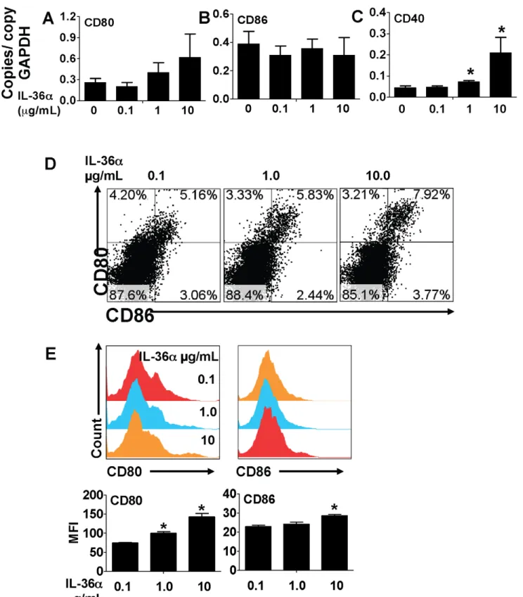
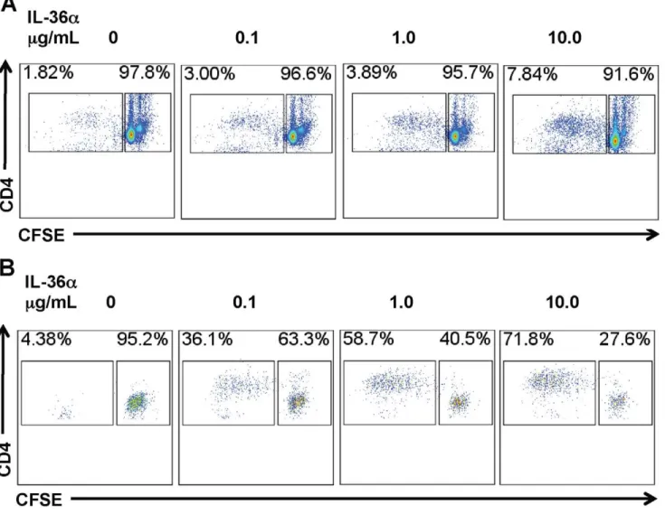
Activation and excessive proliferation of T cells is a critical component in the pathogenesis of a multitude of inflammatory disorders, including lung disorders such as asthma. A recent report demonstrated that CD4+T cells and CD11c+cells express IL-36R, the receptor for IL-36a and the other IL-36 cytokines [51]. To determine if IL-36aregulated T cell proliferation by modulating costimulatory molecule expression in APCs, we incubated splenic CD11c+
cells with increasing concentrations of IL-36a and measured the mRNA expression of costimulatory molecules at earlier timepoints and cell surface expression of the costimulatory molecules at the later timepoints. While incubation of CD11c+ cells with IL-36adid not induce the mRNA expression of CD80 (Fig. 7A) or CD86 (Fig. 7B) in CD11c+ cells 2 h following incubation, the expression of the co-stimulatory molecule CD40 was significantly increased in a dose-dependent manner (Fig. 7C). Incubation of splenic CD11c+
cells with IL-36a for 2 h was sufficient to induce a modest, but dose-dependent increase in the percentage of CD80+
CD86+
cells 24 hours later (Fig. 7D). Furthermore, incubation of splenic CD11c+
cells with IL-36afor 2 h also significantly increased cell surface CD80 expression and a modest, but significant increase in cell surface CD86 expression 24 hours later (Fig. 7E). In addition, the proliferation of CD4+
T cells was proportionally increased when co-cultured with CD11c+ cells pre-incubated with increasing concentrations of IL-36a
(Fig. 8A). Antigen-specific CD4+
T cell proliferation was also proportionally increased when CD4+T cells from OVA-specific OTII TCR transgenic mice were co-cultured with CD11c+
cells pre-incubated with increasing concentrations of IL-36aand a fixed concentration of OVA323–339peptide, the cognate antigen. Since
the IL-36acontaining media was removed from the CD11c+cells before addition of CD4+
cells, these data suggest that IL-36a
induced expression of costimulatory molecules or mediators may indirectly contribute to T cell proliferation responses.
IL-36aInduces the Activation of NF-kB in Mouse Macrophage Cell Lines
Previous studies have reported that agonist IL-36 cytokines such as IL-36a and IL-36c induce NF-kB activation in Jurkat cells transfected with IL-36R [31]. We have previously demonstrated that IL-36cinduces the activation of NF-kB in macrophage cell lines in a dose-dependent manner [38]. Here, we demonstrate that similar to IL-36c, IL-36aalso induces the activation of NF-kB in a mouse macrophage cell line (Fig. 9). These data suggest that IL-36a induced NF-kB activity may be a critical mechanism in
driving the production of proinflammatory mediators by macro-phages and proliferation of T cells by enhancing the stimulatory properties of dendritic cells.
Discussion
The current study demonstrates that IL-36a acts as a pro-inflammatory cytokine in the lungs independent of both IL-1a
and IL-1b. Intratracheal instillation of IL-36a induced neutro-phil influx and increased the expression of pro-inflammatory cytokines and chemokines in the lungs of wild-type C57BL/6 mice as well as mice deficient in both IL-1a and IL-1b. IL-36a
acted directly on CD11c+antigen presenting cells to induce the production of early response cytokines and neutrophil-specific chemokines. In addition, IL-36a increased the expression of T cell co-stimulatory molecules on CD11c+ cells and enhanced their ability to stimulate CD4+
T cell proliferation. Stimulation of a macrophage cells line with IL-36a induced NF-kB activation. Collectively, these data suggest that disease-induced overproduction of IL-36a in the lungs may play an important role in inflammatory lung disease.
Intratracheal instillation of IL-36ainduced neutrophil influx in the lungs of wildtype and IL-1ab2/2mice. Transgenic mice that
conditional overexpress IL-1b in the airway epithelium have neutrophil influx, increased mucus production and the develop-ment of emphysematous and fibrotic changes in the lung [52]. Prior reports also demonstrated that intratracheal administration of IL-1 induced neutrophil influx into the lungs of mice [53], and that IL-1 is necessary for lipopolysaccharide (LPS) mediated neutrophil influx in the murine lung [54,55]. We previously reported that intratracheal administration of IL-36calso induced neutrophil influx into the lungs and the expression of neutrophil-specific chemokines [38]. Here we show that IL-36aalso resulted in neutrophil chemotaxis in the lungs, along with an increase in the mRNA expression of the classical IL-1 agonists IL-1aand IL-1bin wild-type mice. This supports our previous findings [38] and those of others [39] demonstrating that agonist IL-36 cytokines induce the production of classical IL-1 cytokines. Importantly, in the current study intratracheal instillation of IL-36a induced neutrophil influx into the lungs of IL-1ab2/2mice, suggesting that IL-36adriven neutrophil recruitment to the lungs is independent of IL-1aand IL-1b.
The mRNA expression of early response cytokines such as TNFa, IL-1a and IL-1bwere increased in the lungs of wildtype mice following intratracheal instillation of IL-36a. These results are consistent with our previous reports demonstrating that i.t. instillation of IL-36cinto the lungs of mice induced the production of IL-1a in a dose-dependent manner [38]. While the mRNA expression of IL-1R1 was unchanged, the expression of IL-36R was significantly increased in the lungs of IL-36a treated mice, demonstrating that IL-36a can induce the expression of its own receptor in lung tissue either by directly acting on structural or immune cells in the lung, or indirectly by inducing the production of other mediators in the lung that increase IL-36R expression. Since the expression of 36R was also induced in the lungs of IL-36a instilled IL-1ab2/2 mice, it is possible to speculate that
neither IL-1a nor IL-1b contribute to IL-36a driven IL-36R expression. IL-1 induced the mRNA expression of multiple 36a. Transcript expression was evaluated by SYBR-Green based quantitative real-time PCR. *Indicates significant differences (P,0.05) compared to
0mg/mL group. Data represent mean6SD of quadruplicate samples from one of two representative experiments. I) Diff-quik stained cells from cytospun BAL cells from the lungs of naı¨ve mice demonstrates that the majority of lung resident immune cells are alveolar macrophages. J) PCR on cDNA from naı¨ve mouse alveolar macrophages demonstrating the constitutive mRNA expression of an endogenous control (b-actin), IL-1R1, IL-36R and IL-1RAcP. Image of a DNA electrophoresis gel has been color-inverted for clarity. bp – base pairs.
Figure 7. IL-36ainduced the expression of T cell costimulatory molecules in splenic CD11c+cells.A–C) Transcript expression of the
co-stimulatory molecules CD80, CD86 and CD40 in splenic CD11c+
cells 2 h following incubation with increasing concentrations of IL-36a. Transcript expression was evaluated by SYBR-Green based quantitative real-time PCR. *Indicates significant differences (P,0.05) compared to 0mg/mL group.
Data represent mean6SD from quadruplicate samples from one of two representative experiments. D) Flow cytometric evaluation of splenic CD11c+
cells 24 h following incubation with increasing concentrations of IL-36afor 2 h. E) Cell surface expression of co-stimulatory molecules in splenic CD11c+
cells 24 h following incubation with increasing concentrations of IL-36afor 2 h. MFI – mean fluorescence intensity. *Indicates significant differences (P,0.05) compared to 0.1mg/mL group. Data represent mean6SD from triplicate samples from one of two representative experiments.
doi:10.1371/journal.pone.0045784.g007
chemokines in airway epithelial cells in vitro[56–58] and in vivo
[52]. Furthermore, overexpression of IL-1b in the airway epithelium increased the mRNA expression of CXCL1, CXCL2, CCL2 and CCL7 chemokines and the accumulation of neutro-phils in the lungs of mice [59]. Increased mRNA expression of CXCL1 and CXCL2 in the lungs of IL-36ainstilled wildtype as well as IL-1ab2/2mice in the current study suggests that IL-36a
can induce the expression of neutrophil-specific chemokines in the lungs independent of IL-1aand IL-1b.
We have previously demonstrated that a single intratracheal instillation of IL-36c induces airway hyperresponsiveness in response to aerosolized methacholine [38]. However, in the current study, a single i.t. instillation of a similar amount of IL-36a
did not increase airway hyperresponsiveness. This discrepancy
could be due to inherent differences in the ability of IL-36aand IL-36cin inducing AHR. For example, we have shown that unlike IL-36c, IL-36a is not constitutively expressed in lungs [38]. Furthermore, the magnitude of induction of IL-36a mRNA in response to cytokines and other inflammatory stimuli appears to be consistently lower than that of IL-36c mRNA expression in airway epithelial cells [39], bone marrow derived dendritic cells (BMDCs) and T cells [51]. Therefore, it is possible that a higher concentration of IL-36ais required to produce similar magnitude of responses driven by IL-36c at a lower concentration. In addition, the discrepancy could also be due to differences in AHR-specific genetic susceptibilities between A/J mice used in our previous report [38] and the C57BL/6 strain of mice used in this study.
Figure 8. Incubation with IL-36aenhances the ability of splenic CD11c+cell mediated CD4+T cell proliferation.A) Flow cytometric
evaluation of CD4+
T cell proliferation responses induced by IL-36astimulated splenic CD11c+
cells. Splenic CD11c+
cells were incubated with increasing doses of IL-36afor 2 h. Following incubation, media containing IL-36awas removed and CFSE-labeled CD4+
T cells were co-cultured with IL-36astimulated CD11c+
cells. CFSE dilution was used to evaluate T cell proliferation responses 96 h following co-culture. CD4+
T cell proliferation was proportional to the concentration of IL-36aused for stimulating CD11c+
cells used in the co-culture. Flow cytometry plot presented is representative of quadruplicate samples in one out of two independent experiments. B) Flow cytometric evaluation of antigen-specific CD4+T cell
proliferation responses induced by IL-36astimulated splenic CD11c+
cells. Splenic CD11c+
cells were incubated with increasing doses of IL-36aand 100 ng/mL OVA323-339for 2 h. Following incubation, media containing IL-36aand the OVA peptide was removed and CFSE-labeled CD4+T cells from
OTII TCR transgenic mice were co-cultured with IL-36astimulated, OVA peptide pulsed CD11c+
cells. CFSE dilution in the culture was used to evaluate T cell proliferation responses 96 h following co-culture. CD4+T cell proliferation was proportional to the concentration of IL-36aused for stimulating
CD11c+
cells used in the co-culture. Flow cytometry plot presented is representative of quadruplicate samples in one out of two independent experiments.
IL-36a acted directly on splenic CD11c+ cells to induce the production of neutrophil-specific chemokines. A recent report demonstrated that IL-36R was expressed in murine BMDCs and CD4+
T cells and that the stimulation of BMDCs with IL-36a, IL-36band IL-36cinduced the production of multiple cytokines and chemokines [51]. In the current study we demonstrate that IL-36a
acts directly on splenic CD11c+
cells to induce the production of the neutrophil-specific chemokines CXCL1, CXCL2 as well as other early response cytokines such as TNFa, IL-1a and IL-1b. Interestingly, the mRNA expression of neither IL-1R1 nor IL-36R was increased in IL-36astimulated splenic CD11c+cells under the conditions tested. Since we observed increased IL-36R expression in whole lung tissue following a single IL-36a challenge, we speculate that IL-36a either directly increases IL-36R in non-CD11c+cells in the lung, or induces the production of another endogenous mediator from an IL-36a responsive, non-CD11c+ cell type in the lungs to increase IL-36R mRNA expressionin vivo. Furthermore, we have also demonstrated that similar to splenic CD11c+
cells, alveolar macrophages from naı¨ve mice also express IL-36R mRNA at levels higher than IL-1R1, suggesting that alveolar macrophages are poised to respond to IL-36 cytokines released into the lung airspaces.
IL-36a acted directly on CD11c+ cells to induce the expression of co-stimulatory molecules that regulate T cell activation and proliferation. While activation and proliferation of T cells are essential to confer protection against certain pathogens, aberrant activation and proliferation of T cells leads to detrimental responses in lung diseases such as asthma [60]. Apart from T cell receptor activation, engagement of co-stimulatory molecules expressed on T cells (such as CD28 and CD40L) with those expressed on antigen presenting cells (such as CD80, CD86 and CD40) are critical for T cell activation and proliferation [61]. Previous reports have demonstrated that IL-1b induces antigen-specific proliferation of CD4+ T cells during the primary and secondary (memory) immune responses [62], and that CD4+
T cells from IL-1ab2/2mice are defective
in proliferation and the production of T helper 2 (Th2) cytokines [13]. It has also been demonstrated that IL-1a
induced CD40 expression in human dendritic cellsin vitro[63]. Consistent with a recent report [51], we also found that IL-36a
directly induced the mRNA expression of CD40 in CD11c+ cells. Although both CD80 and CD86 mRNA were constitu-tively expressed in CD11c+ cells in our experiments, mRNA expression was not significantly increased 2 h following stimulation with IL-36a. However, we found that the cell surface expression of CD80 and CD86 were increased in splenic CD11c+cells 24 hours following 2 hours of stimulation with IL-36a. We also found that CD4+ T cell proliferation responses were enhanced when co-cultured with IL-36a stimulated CD11c+
cells, suggesting that IL-36a mediated upregulation of costimulatory molecules may be important for T cell pro-liferation. Interaction of CD40 with CD40L is important for Th1 cell differentiation [64]. Vigne et al., recently demonstrated that IL-36b (IL-1F8) increased CD40 mRNA expression and enhanced Th1 responses in vivo [51]. Collectively, these data support an important role of novel IL-1 cytokine agonists in regulating T cell activation and proliferation by modulating antigen presentation and co-stimulatory abilities of APCs. Experiments in the current study were performed using splenic CD11c+
cells, which may have different cytokine secretion and antigen-presenting capabilities than CD11c+
cells that reside in or are recruited to the lungs during pulmonary inflammation [65]. We have shown that alveolar macrophages isolated from naı¨ve wild-type mice express IL-36R and IL-1RAcP, suggesting Figure 9. IL-36a directly induced NF-kB activation in mouse
macrophage cell lines.A) Cells from a mouse macrophage NF-kB reporter cell line (RAW-ELAM cells) were stimulated with increasing concentrations of IL-36a. Green fluorescent protein (GFP) expression, indicative of NF-kB activation, was increased in a dose-dependent manner upon incubation with IL-36a. Flow cytometry plot presented is representative of triplicate samples in one out of two independent experiments.
doi:10.1371/journal.pone.0045784.g009
that alveolar macrophages are poised to respond to IL-36 cytokines in the lung. While we speculate that the responses of lung CD11c+ cells to IL-36a would be largely similar to the responses reported in the current study, further studies are required to determine the functional effects of IL-36a on lung specific CD11c+ cells and subsets.
IL-36aactivated NF-kB in mouse macrophages. Activation of NF-kB in the lungs leads to airway inflammation, mucus cell metaplasia and the production of proinflammatory cytokine and chemokines [66–68]. Agonist members of the novel IL-1 cytokine cluster induced NF-kB and MAPK activation in Jurkat cells transfected with IL-36R [31]. We previously demonstrated that IL-36c induced NF-kB activation in mouse macrophages following in vitro stimulation and in total lung tissue following a single intratracheal challenge with IL-36c [38]. In addition, IL-36c also induced NF-kB activation in lung fibroblasts [39]. Consistent with these findings, IL-36a also induced NF-kB activation in mouse macrophages, which may be a potential mechanism driving the production of neutrophil-specific che-mokines from macrophages and dendritic cells in the lung resulting in recruitment of neutrophils. Furthermore, these results also confirm previous findings that NF-kB is a critical signaling molecule downstream of both IL-1R1 and IL-36R mediated responses.
To date, the role of IL-36ahas not been examined in human inflammatory lung disease. It is interesting that very high concentrations of the IL-36 agonists including IL-36a are required to stimulate in vitrocellular responses [30,31]. A recent report demonstrates that truncation variants of IL-36a, IL-36b
and IL-36c exert enhanced agonist activity in vitro, suggesting that a yet unidentified protease may process these cytokines to more biologically potent forms in vivo [69]. Although the magnitude of the cellular responses was enhanced by the truncation variants, the underlying IL-36 cytokine driven mechanisms such as NF-kB activation remain similar. There-fore, we conclude that although that the results reported in this study using the full-length IL-36a reflect the functional consequences of IL-36a overproduction in the lung, further
studies are required to determine if the truncated versions of IL-36a proteins would induce more severe inflammation in the lung in vivo. Furthermore, since the IL-36 cytokine agonists do not possess a leader peptide that is necessary for cytokine secretion [70], thein vivo mechanisms by which these cytokines are released into the extracellular spaces such as BAL fluid remains to be clarified. While there are several reports demonstrating increased mRNA expression of novel IL-1 cytokine agonists, there is a paucity of reports on the in vivo
detection of the cytokines at the protein level in extracellular spaces. Among the several mechanisms by which IL-1b has been proposed to become available in the extracellular space [71–75], plasma membrane breakdown has been suggested to be the most plausible mechanism by which intracellular IL-36a
could become available in the extracellular space [76]. The current study determined that IL-36a acts as a pro-inflamma-tory cytokine in the lung, and that IL-36a may increase inflammatory responses in disease conditions which involve release of IL-36a into the lungs.
Supporting Information
Figure S1 Intratracheal instillation of IL-36ainduced neutrophil influx in the lungs of endotoxin resistant C3H/HeJ mice. A) Total cell counts from BAL fluid recovered from mice 24 h following a single i.t. instillation of PBS or 10mg IL-36a. C) Differential cell count percentages and D) Differential cell count numbers in the BAL fluid recovered from mice 24 h following a single i.t. instillation of PBS or 10mg IL-36a. *Indicates significant differences (P,0.05) compared to PBS treated mice. Data represent mean6SEM from 4–5 mice/group.
(TIFF)
Author Contributions
Conceived and designed the experiments: RAR BDM AML. Performed the experiments: RAR. Analyzed the data: RAR. Contributed reagents/ materials/analysis tools: SLE YI. Wrote the paper: RAR AML.
References
1. Arend WP, Palmer G, Gabay C (2008) IL-1, IL-18, and IL-33 families of cytokines. Immunol Rev 223: 20–38.
2. Dinarello CA, Renfer L, Wolff SM (1977) Human leukocytic pyrogen: purification and development of a radioimmunoassay. Proc Natl Acad Sci U S A
74: 4624–4627.
3. Dinarello CA (2011) Interleukin-1 in the pathogenesis and treatment of inflammatory diseases. Blood 117: 3720–3732.
4. Borish L, Mascali JJ, Dishuck J, Beam WR, Martin RJ, et al. (1992) Detection of alveolar macrophage-derived IL-1 beta in asthma. Inhibition with corticoster-oids. J Immunol 149: 3078–3082.
5. Brasier AR, Victor S, Boetticher G, Ju H, Lee C, et al. (2008) Molecular phenotyping of severe asthma using pattern recognition of bronchoalveolar lavage-derived cytokines. J Allergy Clin Immunol 121: 30–37 e36.
6. Tonnel AB, Gosset P, Tillie-Leblond I (2001) Characteristics of the In-flammatory response in bronchial lavage fluids from patients with status asthmaticus. Int Arch Allergy Immunol 124: 267–271.
7. Hastie AT, Moore WC, Meyers DA, Vestal PL, Li H, et al. (2010) Analyses of asthma severity phenotypes and inflammatory proteins in subjects stratified by sputum granulocytes. J Allergy Clin Immunol 125: 1028–1036 e1013. 8. Ranieri VM, Suter PM, Tortorella C, De Tullio R, Dayer JM, et al. (1999)
Effect of mechanical ventilation on inflammatory mediators in patients with acute respiratory distress syndrome: a randomized controlled trial. JAMA : the journal of the American Medical Association 282: 54–61.
9. Ranieri VM, Giunta F, Suter PM, Slutsky AS (2000) Mechanical ventilation as a mediator of multisystem organ failure in acute respiratory distress syndrome. JAMA : the journal of the American Medical Association 284: 43–44. 10. Frank JA, Pittet JF, Wray C, Matthay MA (2008) Protection from experimental
ventilator-induced acute lung injury by IL-1 receptor blockade. Thorax 63: 147– 153.
11. Zhang Y, Lee TC, Guillemin B, Yu MC, Rom WN (1993) Enhanced IL-1 beta and tumor necrosis factor-alpha release and messenger RNA expression in
macrophages from idiopathic pulmonary fibrosis or after asbestos exposure. Journal of immunology 150: 4188–4196.
12. Johnson VJ, Yucesoy B, Luster MI (2005) Prevention of IL-1 signaling attenuates airway hyperresponsiveness and inflammation in a murine model of toluene diisocyanate-induced asthma. J Allergy Clin Immunol 116: 851–858. 13. Nakae S, Komiyama Y, Yokoyama H, Nambu A, Umeda M, et al. (2003) IL-1 is
required for allergen-specific Th2 cell activation and the development of airway hypersensitivity response. Int Immunol 15: 483–490.
14. Wang CC, Fu CL, Yang YH, Lo YC, Wang LC, et al. (2006) Adenovirus expressing interleukin-1 receptor antagonist alleviates allergic airway inflamma-tion in a murine model of asthma. Gene Ther 13: 1414–1421.
15. Botelho FM, Bauer CM, Finch D, Nikota JK, Zavitz CC, et al. (2011) IL-1alpha/IL-1R1 expression in chronic obstructive pulmonary disease and mechanistic relevance to smoke-induced neutrophilia in mice. PloS one 6: e28457.
16. Pauwels NS, Bracke KR, Dupont LL, Van Pottelberge GR, Provoost S, et al. (2011) Role of IL-1alpha and the Nlrp3/caspase-1/IL-1beta axis in cigarette smoke-induced pulmonary inflammation and COPD. The European respiratory journal : official journal of the European Society for Clinical Respiratory Physiology 38: 1019–1028.
17. Kolb M, Margetts PJ, Anthony DC, Pitossi F, Gauldie J (2001) Transient expression of IL-1beta induces acute lung injury and chronic repair leading to pulmonary fibrosis. The Journal of clinical investigation 107: 1529–1536. 18. Ortiz LA, Dutreil M, Fattman C, Pandey AC, Torres G, et al. (2007) Interleukin
1 receptor antagonist mediates the antiinflammatory and antifibrotic effect of mesenchymal stem cells during lung injury. Proceedings of the National Academy of Sciences of the United States of America 104: 11002–11007. 19. Piguet PF, Vesin C, Grau GE, Thompson RC (1993) Interleukin 1 receptor
20. Wilson MS, Madala SK, Ramalingam TR, Gochuico BR, Rosas IO, et al. (2010) Bleomycin and IL-1beta-mediated pulmonary fibrosis is IL-17A de-pendent. The Journal of experimental medicine 207: 535–552.
21. Jones MR, Simms BT, Lupa MM, Kogan MS, Mizgerd JP (2005) Lung NF-kappaB activation and neutrophil recruitment require IL-1 and TNF receptor signaling during pneumococcal pneumonia. Journal of immunology 175: 7530– 7535.
22. Schultz MJ, Rijneveld AW, Florquin S, Edwards CK, Dinarello CA, et al. (2002) Role of interleukin-1 in the pulmonary immune response during Pseudomonas aeruginosa pneumonia. American journal of physiology Lung cellular and molecular physiology 282: L285–290.
23. Zwijnenburg PJ, van der Poll T, Florquin S, Roord JJ, Van Furth AM (2003) IL-1 receptor type IL-1 gene-deficient mice demonstrate an impaired host defense against pneumococcal meningitis. Journal of immunology 170: 4724–4730. 24. Schmitz N, Kurrer M, Bachmann MF, Kopf M (2005) Interleukin-1 is
responsible for acute lung immunopathology but increases survival of respiratory influenza virus infection. Journal of virology 79: 6441–6448.
25. Ichinohe T, Lee HK, Ogura Y, Flavell R, Iwasaki A (2009) Inflammasome recognition of influenza virus is essential for adaptive immune responses. The Journal of experimental medicine 206: 79–87.
26. Perrone LA, Szretter KJ, Katz JM, Mizgerd JP, Tumpey TM (2010) Mice lacking both TNF and IL-1 receptors exhibit reduced lung inflammation and delay in onset of death following infection with a highly virulent H5N1 virus. The Journal of infectious diseases 202: 1161–1170.
27. Smith DE, Renshaw BR, Ketchem RR, Kubin M, Garka KE, et al. (2000) Four new members expand the interleukin-1 superfamily. J Biol Chem 275: 1169– 1175.
28. Kumar S, McDonnell PC, Lehr R, Tierney L, Tzimas MN, et al. (2000) Identification and initial characterization of four novel members of the interleukin-1 family. J Biol Chem 275: 10308–10314.
29. Nicklin MJ, Barton JL, Nguyen M, FitzGerald MG, Duff GW, et al. (2002) A sequence-based map of the nine genes of the human interleukin-1 cluster. Genomics 79: 718–725.
30. Debets R, Timans JC, Homey B, Zurawski S, Sana TR, et al. (2001) Two novel IL-1 family members, IL-1 delta and IL-1 epsilon, function as an antagonist and agonist of NF-kappa B activation through the orphan IL-1 receptor-related protein 2. J Immunol 167: 1440–1446.
31. Towne JE, Garka KE, Renshaw BR, Virca GD, Sims JE (2004) Interleukin (IL)-1F6, IL-1F8, and IL-1F9 signal through IL-1Rrp2 and IL-1RAcP to activate the pathway leading to NF-kappaB and MAPKs. J Biol Chem 279: 13677–13688. 32. Dinarello C, Arend W, Sims J, Smith D, Blumberg H, et al. (2010) IL-1 family
nomenclature. Nat Immunol 11: 973.
33. Barksby HE, Lea SR, Preshaw PM, Taylor JJ (2007) The expanding family of interleukin-1 cytokines and their role in destructive inflammatory disorders. Clin Exp Immunol 149: 217–225.
34. van de Veerdonk FL, Stoeckman AK, Wu G, Boeckermann AN, Azam T, et al. (2012) IL-38 binds to the IL-36 receptor and has biological effects on immune cells similar to IL-36 receptor antagonist. Proceedings of the National Academy of Sciences of the United States of America 109: 3001–3005.
35. Dinarello CA (2009) Immunological and inflammatory functions of the interleukin-1 family. Annu Rev Immunol 27: 519–550.
36. Dinarello CA (1996) Biologic basis for interleukin-1 in disease. Blood 87: 2095– 2147.
37. Ramadas RA, Li X, Shubitowski DM, Samineni S, Wills-Karp M, et al. (2006) IL-1 Receptor antagonist as a positional candidate gene in a murine model of allergic asthma. Immunogenetics 58: 851–855.
38. Ramadas RA, Ewart SL, Medoff BD, LeVine AM (2011) Interleukin-1 family member 9 stimulates chemokine production and neutrophil influx in mouse lungs. Am J Respir Cell Mol Biol 44: 134–145.
39. Chustz RT, Nagarkar DR, Poposki JA, Favoreto S, Jr., Avila PC, et al. (2011) Regulation and Function of the IL-1 Family Cytokine IL-1F9 in Human Bronchial Epithelial Cells. Am J Respir Cell Mol Biol 45: 145–153. 40. Vos JB, van Sterkenburg MA, Rabe KF, Schalkwijk J, Hiemstra PS, et al. (2005)
Transcriptional response of bronchial epithelial cells to Pseudomonas aerugi-nosa: identification of early mediators of host defense. Physiol Genomics 21: 324–336.
41. DeVoti JA, Rosenthal DW, Wu R, Abramson AL, Steinberg BM, et al. (2008) Immune dysregulation and tumor-associated gene changes in recurrent respiratory papillomatosis: a paired microarray analysis. Mol Med 14: 608–617. 42. Blumberg H, Dinh H, Dean C, Jr., Trueblood ES, Bailey K, et al. (2010) IL-1RL2 and its ligands contribute to the cytokine network in psoriasis. J Immunol 185: 4354–4362.
43. Blumberg H, Dinh H, Trueblood ES, Pretorius J, Kugler D, et al. (2007) Opposing activities of two novel members of the IL-1 ligand family regulate skin inflammation. J Exp Med 204: 2603–2614.
44. Johnston A, Xing X, Guzman AM, Riblett M, Loyd CM, et al. (2010) IL1F5, -F6, -F8, and -F9: a novel IL-1 family signaling system that is active in psoriasis and promotes keratinocyte antimicrobial peptide expression. J Immunol 186: 2613–2622.
45. Carrier Y, Ma HL, Ramon HE, Napierata L, Small C, et al. (2011) Inter-regulation of Th17 cytokines and the IL-36 cytokines in vitro and in vivo: implications in psoriasis pathogenesis. The Journal of investigative dermatology 131: 2428–2437.
46. Marrakchi S, Guigue P, Renshaw BR, Puel A, Pei XY, et al. (2011) Interleukin-36-receptor antagonist deficiency and generalized pustular psoriasis. The New England journal of medicine 365: 620–628.
47. Horai R, Asano M, Sudo K, Kanuka H, Suzuki M, et al. (1998) Production of mice deficient in genes for interleukin (IL)-1alpha, IL-1beta, IL-1alpha/beta, and IL-1 receptor antagonist shows that IL-1beta is crucial in turpentine-induced fever development and glucocorticoid secretion. J Exp Med 187: 1463– 1475.
48. Stacey KJ, Young GR, Clark F, Sester DP, Roberts TL, et al. (2003) The molecular basis for the lack of immunostimulatory activity of vertebrate DNA. Journal of immunology 170: 3614–3620.
49. Poltorak A, He X, Smirnova I, Liu MY, Van Huffel C, et al. (1998) Defective LPS signaling in C3H/HeJ and C57BL/10ScCr mice: mutations in Tlr4 gene. Science 282: 2085–2088.
50. Jeyaseelan S, Chu HW, Young SK, Freeman MW, Worthen GS (2005) Distinct roles of pattern recognition receptors CD14 and Toll-like receptor 4 in acute lung injury. Infection and immunity 73: 1754–1763.
51. Vigne S, Palmer G, Lamacchia C, Martin P, Talabot-Ayer D, et al. (2011) IL-36R ligands are potent regulators of dendritic and T cells. Blood.
52. Lappalainen U, Whitsett JA, Wert SE, Tichelaar JW, Bry K (2005) Interleukin-1beta causes pulmonary inflammation, emphysema, and airway remodeling in the adult murine lung. Am J Respir Cell Mol Biol 32: 311–318.
53. Leff JA, Baer JW, Bodman ME, Kirkman JM, Shanley PF, et al. (1994) Interleukin-1-induced lung neutrophil accumulation and oxygen metabolite-mediated lung leak in rats. Am J Physiol 266: L2–8.
54. Calkins CM, Bensard DD, Shames BD, Pulido EJ, Abraham E, et al. (2002) IL-1 regulates in vivo C-X-C chemokine induction and neutrophil sequestration following endotoxemia. Journal of endotoxin research 8: 59–67.
55. Ulich TR, Yin SM, Guo KZ, del Castillo J, Eisenberg SP, et al. (1991) The intratracheal administration of endotoxin and cytokines. III. The interleukin-1 (IL-1) receptor antagonist inhibits endotox and IL-1-induced acute in-flammation. The American journal of pathology 138: 521–524.
56. Heiman AS, Abonyo BO, Darling-Reed SF, Alexander MS (2005) Cytokine-stimulated human lung alveolar epithelial cells release eotaxin-2 (CCL24) and eotaxin-3 (CCL26). J Interferon Cytokine Res 25: 82–91.
57. Jedrzkiewicz S, Nakamura H, Silverman ES, Luster AD, Mansharamani N, et al. (2000) IL-1beta induces eotaxin gene transcription in A549 airway epithelial cells through NF-kappaB. Am J Physiol Lung Cell Mol Physiol 279: L1058– 1065.
58. Manzer R, Wang J, Nishina K, McConville G, Mason RJ (2006) Alveolar epithelial cells secrete chemokines in response to IL-1beta and lipopolysaccha-ride but not to ozone. Am J Respir Cell Mol Biol 34: 158–166.
59. Bry K, Whitsett JA, Lappalainen U (2007) IL-1beta disrupts postnatal lung morphogenesis in the mouse. Am J Respir Cell Mol Biol 36: 32–42. 60. Afshar R, Medoff BD, Luster AD (2008) Allergic asthma: a tale of many T cells.
Clin Exp Allergy 38: 1847–1857.
61. Lenschow DJ, Walunas TL, Bluestone JA (1996) CD28/B7 system of T cell costimulation. Annual review of immunology 14: 233–258.
62. Ben-Sasson SZ, Hu-Li J, Quiel J, Cauchetaux S, Ratner M, et al. (2009) IL-1 acts directly on CD4 T cells to enhance their antigen-driven expansion and differentiation. Proceedings of the National Academy of Sciences of the United States of America 106: 7119–7124.
63. McLellan AD, Sorg RV, Williams LA, Hart DN (1996) Human dendritic cells activate T lymphocytes via a CD40: CD40 ligand-dependent pathway. European journal of immunology 26: 1204–1210.
64. Howland KC, Ausubel LJ, London CA, Abbas AK (2000) The roles of CD28 and CD40 ligand in T cell activation and tolerance. Journal of immunology 164: 4465–4470.
65. Kugathasan K, Roediger EK, Small CL, McCormick S, Yang P, et al. (2008) CD11c+antigen presenting cells from the alveolar space, lung parenchyma and spleen differ in their phenotype and capabilities to activate naive and antigen-primed T cells. BMC immunology 9: 48.
66. Choi IW, Kim DK, Ko HM, Lee HK (2004) Administration of antisense phosphorothioate oligonucleotide to the p65 subunit of NF-kappaB inhibits established asthmatic reaction in mice. Int Immunopharmacol 4: 1817–1828. 67. Desmet C, Gosset P, Pajak B, Cataldo D, Bentires-Alj M, et al. (2004) Selective
blockade of NF-kappa B activity in airway immune cells inhibits the effector phase of experimental asthma. J Immunol 173: 5766–5775.
68. Poynter ME, Cloots R, van Woerkom T, Butnor KJ, Vacek P, et al. (2004) NF-kappa B activation in airways modulates allergic inflammation but not hyperresponsiveness. J Immunol 173: 7003–7009.
69. Towne JE, Renshaw BR, Douangpanya J, Lipsky BP, Shen M, et al. (2011) Interleukin-36 (36) ligands require processing for full agonist (36alpha, IL-36beta, and IL-36gamma) or antagonist (IL-36Ra) activity. The Journal of biological chemistry 286: 42594–42602.
70. Sims JE, Smith DE (2010) The IL-1 family: regulators of immunity. Nature reviews Immunology 10: 89–102.
71. MacKenzie A, Wilson HL, Kiss-Toth E, Dower SK, North RA, et al. (2001) Rapid secretion of interleukin-1beta by microvesicle shedding. Immunity 15: 825–835.
72. Hamon Y, Luciani MF, Becq F, Verrier B, Rubartelli A, et al. (1997) Interleukin-1beta secretion is impaired by inhibitors of the Atp binding cassette transporter, ABC1. Blood 90: 2911–2915.
73. Andrei C, Dazzi C, Lotti L, Torrisi MR, Chimini G, et al. (1999) The secretory route of the leaderless protein interleukin 1beta involves exocytosis of endolysosome-related vesicles. Molecular biology of the cell 10: 1463–1475. 74. Andrei C, Margiocco P, Poggi A, Lotti LV, Torrisi MR, et al. (2004)
Phospholipases C and A2 control lysosome-mediated IL-1 beta secretion: Implications for inflammatory processes. Proceedings of the National Academy of Sciences of the United States of America 101: 9745–9750.
75. Qu Y, Franchi L, Nunez G, Dubyak GR (2007) Nonclassical IL-1 beta secretion stimulated by P2X7 receptors is dependent on inflammasome activation and correlated with exosome release in murine macrophages. Journal of immunology 179: 1913–1925.