Multiple Mouse Models Efficiently Elicit Tumors in
Immune-Competent Hosts
Natasza A. Kurpios1¤a, Adele Girgis-Gabardo1, Robin M. Hallett, Stephen Rogers1, David W. Gludish1, Lisa Kockeritz2, James Woodgett2¤b, Robert Cardiff3, John A. Hassell1*
1Centre for Functional Genomics, Department of Biochemistry and Biomedical Sciences, McMaster University, Hamilton, Ontario, Canada,2Department of Medical Biophysics, University of Toronto, Toronto, Ontario, Canada,3Department of Pathology, University of California Davis, Davis, California, United States of America
Abstract
The tumor-initiating cell (TIC) frequency of bulk tumor cell populations is one of the criteria used to distinguish malignancies that follow the cancer stem cell model from those that do not. However, tumor-initiating cell frequencies may be influenced by experimental conditions and the extent to which tumors have progressed, parameters that are not always addressed in studies of these cells. We employed limiting dilution cell transplantation of minimally manipulated tumor cells from mammary tumors of several transgenic mouse models to determine their tumor-initiating cell frequency. We determined whether the tumors that formed following tumor cell transplantation phenocopied the primary tumors from which they were isolated and whether they could be serially transplanted. Finally we investigated whether propagating primary tumor cells in different tissue culture conditions affected their resident tumor-initiating cell frequency. We found that tumor-initiating cells comprised between 15% and 50% of the bulk tumor cell population in multiple independent mammary tumors from three different transgenic mouse models of breast cancer. Culture of primary mammary tumor cells in chemically-defined, serum-free medium as non-adherent tumorspheres preserved TIC frequency to levels similar to that of the primary tumors from which they were established. By contrast, propagating the primary tumor cells in serum-containing medium as adherent populations resulted in a several thousand-fold reduction in their tumor-initiating cell fraction. Our findings suggest that experimental conditions, including the sensitivity of the transplantation assay, can dramatically affect estimates of tumor initiating cell frequency. Moreover, conditional on cell culture conditions, the tumor-initiating cell fraction of bulk mouse mammary tumor cell preparations can either be maintained at high or low frequencyin vitrothus permitting comparative studies of tumorigenic and non-tumorigenic cancer cells.
Citation:Kurpios NA, Girgis-Gabardo A, Hallett RM, Rogers S, Gludish DW, et al. (2013) Single Unpurified Breast Tumor-Initiating Cells from Multiple Mouse Models Efficiently Elicit Tumors in Immune-Competent Hosts. PLoS ONE 8(3): e58151. doi:10.1371/journal.pone.0058151
Editor:Alana L. Welm, Huntsman Cancer Institute, University of Utah, United States of America
ReceivedOctober 18, 2012;AcceptedJanuary 31, 2013;PublishedMarch 26, 2013
Copyright:ß2013 Kurpios et al. This is an open-access article distributed under the terms of the Creative Commons Attribution License, which permits unrestricted use, distribution, and reproduction in any medium, provided the original author and source are credited.
Funding:The authors’ research was supported by grants from the Canadian Breast Cancer Research Alliance, the Ontario Institute for Cancer Research, the Canadian Breast Cancer Foundation (Ontario) and the Canadian Stem Cell Network. The funders had no role in study design, data collection and analysis, decision to publish, or preparation of the manuscript.
Competing Interests:The authors have declared that no competing interests exist.
* E-mail: [email protected]
¤a Current address: Department of Molecular Medicine, College of Veterinary Medicine, Cornell University, Ithaca, New York, United States of America ¤b Current address: Samuel Lunenfeld Research Institute, Mount Sinai Hospital, Toronto, Ontario, Canada
Introduction
Tumor-initiating cells (TICs), often termed cancer stem cells, are functionally defined by their capacity to re-grow a new tumor after transplant into experimental animals that recapitulates the phenotype of the primary tumor from which the cells were isolated, and which can be serially transplanted thus demonstrat-ing their capacity to differentiate and self-renew [1]. TICs were first identified in acute myelogenous leukemia [2], and thereafter in many solid tumors [3–7] including those of the breast [8]. TICs and tissue-specific adult stem cells share phenotypic and functional properties leading to the suggestion that they originate from adult stem cells or from progenitor cells that acquire stem cell traits [9– 11]. TICs are infrequent in most human tumors, rarely exceeding 0.01% of the bulk tumor cell population [3–6,8,12,13]. However, recent findings in mouse cancer models [14–20] and human melanomas [21] demonstrate that TIC frequencies can approach
25% of the bulk tumor cell population calling into question the generality of the cancer stem cell model. However, various parameters influence TIC frequency in bulk tumor cell prepara-tions including the methods used to isolate and process tumor samples, the site of tumor cell injection, the extent of the immune-deficiency of the recipient host, the duration of the observational period following tumor cell transplant, and whether agents that facilitate tumor cell engraftment such as Matrigel or stromal cells are co-injected with the tumor cells [21]. Hence the frequency of TICs in tumors is insufficient to distinguish malignancies that follow the cancer stem cell model from those that do not.
cell population thus compromising their specific study [8,22]. To overcome these limitations we investigated whether mammary tumors of transgenic mice might afford a more plentiful and renewable source of TICs for investigation. Whereas the available mouse models of breast cancer do not wholly reproduce the diversity of human breast tumor subtypes, in part because most mouse mammary tumors rarely express the estrogen receptor, morphological analyses [23,24], biomarker studies [25] and global transcript profiling [26] suggests that they provide approximate replicas of their human subtype counterparts. For example, mammary tumors occurring in the Neu and polyomavirus middle tumor antigen (mT) models are morphologically similar to certain human breast tumor histological subtypes [24,25], and share a gene expression signature characteristic of the luminal molecular subclass of human breast tumors [26]. Similarly mouse mammary tumors resulting from overexpression of Wnt/beta (b) – catenin pathway components mimic those of the basal-like molecular subtype of human breast tumors [26].
Mouse mammary TICs have been identified in p53-null mice and in transgenic mice genetically engineered to overexpress Wnt-1 or Neu (Erbb2 or Her-2) in their mammary epithelium [Wnt-17–20]. The TIC frequency among tumors from each of the models varies between 0.01%–1% of the bulk tumor cell population. Whereas the frequency of TICs in different tumors arising in the same model as determined by an individual research group is roughly equivalent, different laboratories report up to 50-fold differences in TIC frequencies in tumors arising in the same model [17–20,27]. For example, the TIC frequency reported by Vaillant et al in tumors of a Wnt-1 transgenic strain (0.56%) supersedes that reported by Cho et al (0.013%) by over 40 fold [17,19].
We employed several mouse models of human breast cancer, where the mouse mammary tumor virus (MMTV) promoter directs transcription of the rat neu proto-oncogene [28] or oncogenes encoding either mT [29] or a stable mutant form of b-catenin in mammary epithelial cells to determine their tumor-resident TIC frequency and to learn whether this parameter differed among the models. The tumors arising in the transgenic strains display characteristic histopathologies, vary in their cellular composition and have been inferred to originate from the oncogenic transformation of mammary epithelial stem cells or particular progenitor cells [30,31]. Neu-induced tumors comprise a relatively homogenous tumor cell population that primarily expresses luminal-lineage markers [24], whereas tumors induced by expression of mT or by activation of the Wnt/b-catenin pathway are heterogeneous comprising both luminal- and myoepithelial-lineage-biased cells [8,24,30,32]. The extent of the cellular heterogeneity of Wnt-1 induced tumors varies among individual tumors [24] and that of the mT-generated tumors increases during their progression [25].
We determined the TIC fraction in multiple mammary tumors arising in each of the models and found that, independent of the model, all the tumors comprised between 15% and 50% TICs. Culture of primary tumor cells from tumors of the MMTV-Neu model in serum-free medium, conducive for stem cell self-renewal [33], led to the formation of non-adherent spheres, termed tumorspheres [34], that comprised a TIC frequency similar to that of the tumor from which the cells were isolated. By contrast, when the primary tumor cells were placed in serum-containing medium, conditions that initiate a differentiation program in human and mouse mammary epithelial stem/progenitor cells [35,36,37,38], the tumor cells adhered to the substratum and proliferated resulting in the net expansion of the tumor cell population, but the TIC frequency declined by multiple orders of magnitude compared to that in bulk primary tumor cells. Hence, the mouse
mammary tumors we have investigated provide an abundant and renewable tumor cell populations, which can be manipulatedin vitroto derive TIC-rich or -poor cell populations for comparative studies.
Materials and Methods
Care and treatment of mice
All mice used in these experiments were housed in an Canadian Council on Animal Care (CCAC)-approved facility at McMaster University. Mice were provided with food and waterad libitum. All animal experiments were approved by the McMaster University Animal Research Ethics Board (AUP: 10-01-04).
Isolation and transplant of primary tumor cells
We have reported the methods used to isolate, process and transplant mammary epithelial cells and tumor cells [38,39]. In short, tumors were surgically removed from anesthetized trans-genic female mice and minced with a scalpel in Versene. Roswell Park Memorial Institute (RPMI) medium containing trypsin (1 mg/ml) and collagenase A (3 mg/ml) and 2% fetal bovine serum (FBS) were added and the tissue fragments incubated for 15 minutes at 37uC. The tumor cell suspension was titruated by repeated pipetting with a 5 ml pipette and incubated for another 15-minute period at 37uC. The tumor cell suspension was titruated again at the end of the second incubation period and the dissociated cells were filtered through 40mm cell strainers (Falcon; Franklin Lakes, NJ USA) and concentrated by centrifugation at 1,500 rpm for 15 minutes at room temperature. Cells were washed in 50 ml volumes of Ham’s F12 (Invitrogen; Carlsbad CA, USA) until the supernatant was clear of red blood cells. The cells were stained with trypan blue and the fraction of viable cells calculated using a hemocytometer. Approximately 90–95% of the cells prepared from tumors were viable as visualized microscop-ically. Cells to be injected into the ‘‘cleared’’ number 4 mammary fat pad were suspended in 50% Matrigel (BD Biosciences; Bedford MA, USA) (in phosphate-buffered saline [PBS] supplemented with 5% FBS and 0.5% trypan blue). Ten microliters of the Matrigel/ cell suspension were injected into the cleared fat pads of recipient females. When single cells were injected, the cells were diluted to 0.1 cell/microliter and the occurrence of a single cell in 10 microliters of this solution deposited in a Terasaki plate was confirmed microscopically before injecting the sample. Mice transplanted with tumor cells were examined for tumors at either 4 or 16 weeks post cell transplantation and were scored positive if any tumor-like masses were observed. TIC frequencies for single-cell seeded tumors were determined using ELDA software (http:// wehi.edu.au/software/elda/index.html [40], whereas TIC fre-quency of the adherent tumor cell population was estimated using L-calc Software (Stem Cell Technologies; Vancouver BC, Canada).
Whole mount analyses
Histology
Tumors were fixed in 4% paraformaldehyde (EM Science; Gibbstown NJ, USA) for 24 hours at room temperature, embedded in paraffin, cut into 4mm sections and stained with Harris Haematoxylin and Eosin (H&E) solution. All images were captured using a Leica inverted microscope (Leica) and Open Lab Improvision software (Perkin Elmer; San Jose CA, USA).
Immunoflorescent staining. The expression of mammary epithelial cell lineage markers in tumor sections was determined as described previously [38]. In brief, antigen retrieval was performed on tumor sections using Antigen Unmasking solution (Vector Labs; Burlingame CA, USA) prior to blocking in 3% normal goat serum (Dako Cytomations; Carpinteria CA, USA). All antibody incubations were performed for 1 hour at room temperature. The primary antibodies included those that bind to alpha-smooth muscle actin (a-Sma) (mouse monoclonal, Sigma; Saint Louis MO, USA), cytokeratin (CK) 14 (rabbit polyclonal, Covance; Emery-ville CA, USA), and CK8 (rat monoclonal, Developmental Studies Hybridoma Bank; Iowa City IA, 52242 USA). Alexa Fluor – 488 or Alexa Fluor- 594 conjugated secondary antibodies were used in conjunction with the goat anti-rat and goat anti-rabbit primary antibodies. The Alexa Fluor – 488, signal-amplification kit (Molecular Probes, Eugene OR, USA) was used to detect the primary mouse monoclonal antibody bound to a-Sma (Sigma). Slides were prepared with Vecta-shield mounting media contain-ing 49, 69- diamindino-2-phenylindole (DAPI) (Vector Laborato-ries) to visualize nuclei.
Culture of tumor cells in vitro
Freshly isolated primary tumor cells were placed in serum-free medium comprising Dulbecco Modified Eagle Medium (DMEM): Ham’s F-12 (3:1), 4mg/ml of B-27, 20 ng/ml Epidermal Growth Factor (EGF), 40 ng/ml Fibroblast Growth Factor-2 (FGF-2) and 4 ng/ml Heparin, and plated into ordinary T25 tissue culture flasks (all from Invitrogen) to generate tumorspheres [33]. The primary tumorspheres that arose 4 days later were mechanically dissociated (titruation) to derive a single cell suspension and the dispersed cells plated in serum-free medium containing the growth factors described above at a density of,30,000 cells per ml; this
process was repeated for a total of 3 serial passages [38]. The cells were dissociated by titruation from the serially-passaged tumor-spheres prior to their injection into mice as described above. Dispersed primary tumor cells were similarly plated into T25 flasks in serum-containing medium (DMEM containing 10% FBS). The adherent cells were epithelial in morphology suggesting they originated from tumor epithelial cells; fibroblastic cells were not apparent in these cultures. The tumor cells were removed from the plastic surface using trypsin (0.25% in Versene) at 37uC, diluted in serum-containing medium and plated as described above. The adherent tumor cells were passaged every 4 days for 3 passages before measuring their TIC frequency as described above. Use of different serum lots did not affect the TIC frequency of the adherent cultures.
Results
Initial limiting dilution tumor cell transplant experiments were carried out with mammary tumors from transgenic mice of the MMTV-Neu (N202) strain that express the wild type rat Neu cDNA in mammary epithelial cells [28]. Tumors arise in this model after long latency (,315 days in 50% of virgin female mice)
[39] due in part to deletions in the Neu cDNA encoding the juxta-membrance region of the protein leading to constitutive activation of its tyrosine kinase activity [41,42]. To estimate the frequency of
TICs in these tumors, limiting dilutions of dispersed primary tumor cells were transplanted into the mammary fat pads of syngeneic FVB/N mice. The mice were sacrificed 4 weeks later, at a time when palpable tumors had developed in animals injected with 104or 105tumor cells. Small tumor-like masses, which we termed tumor nodules, were found near the cell injection site, corresponding in size to the number of transplanted tumor cells (Fig. 1 A, black arrows). Unexpectedly, tumor nodules were found in mice transplanted with single cells. The masses resulting from transplant of between 5 and 100 tumor cells appeared to be a collection of individual nodules resembling those that had formed in fat pads injected with a single cell. Nodules approxi-mating the size of those seeded by single cells were often observed along the needle track (Fig. 1A, white arrows). These findings suggested that single transplanted tumor cells proliferated in the mammary fat pads of their host thereby forming nodules, and that transplant of multiple tumor cells into individual fat pads yielded aggregates of multiple nodules.
To learn whether the tumor nodules appearing in mice transplanted with single cells were indeed nascent tumors and could formbona fidetumors, we repeated the transplants with cells from a different mammary tumor and sacrificed the recipient hosts 4 or 16 weeks thereafter. Single tumor cells, visualized micro-scopically prior to transplant, were injected into syngeneic female mice. Tumor nodules were found in many of the recipients 4 weeks after cell transplantation as observed previously (Fig. 1B), whereas large tumors were apparent 16 weeks post cell trans-plantation (Fig. 1 C and D). These findings suggest that the tumor nodules observed 4 weeks post-transplantation progress to form large tumors 16 weeks after the initial transplant of single primary tumor cells.
To determine the frequency of TIC in individual mammary tumors and to learn whether this fraction varied among the tumors, we performed limiting dilution cell transplants with tumor cells from additional independent Neu-induced tumors. Tumors were present 16 weeks post transplant in 20 of 40 host mice inoculated with single tumor cells from each of 6 independent Neu-induced mammary tumors (Table 1). The TIC frequency calculated from single cell transplants ranged from 1/2–1/6 (95% confidence interval [95% CI]) [43]. Limiting dilution cell trans-plantation experiments were also performed with 2 mammary tumors that arose in mT transgenic mice and 3 that formed in the stable b-catenin transgenic strain. The TIC fraction in these tumors averaged 1/3 (1/2–1/6; 95% CI) for the mT model and 1/3 (1/2–1/8; 95% CI) for the stablebcatenin model.
reproduced that of the corresponding parental tumors. Whereas the mammary tumors in mice resulting from activation of the Wnt/b-catenin pathway span a range of morphologies and vary in their proportion of luminal-lineage and myoepithelial-lineage restricted cells, the tumor-specific characteristics of each primary tumor was reproduced in secondary tumors seeded by individual tumor cells that had been isolated from the same primary tumor (Fig. 4, compare panels 1 and 2).
To determine whether the tumors resulting from single cell transplants comprised self-renewing TIC, we performed serial transplants of the engrafted tumors [45,46]. Tissue fragments (,1 mm3) comprising ,5,000 tumor cells from the single
cell-seeded tumors from each of the three models formed secondary tumor grafts in over 80% of transplanted hosts, which could be similarly serially transplanted for up to 10 successive passages before this experiment was discontinued (data not shown). Hence
the tumors of all three breast-cancer prone mouse models comprised TIC capable of recapitulating the phenotype of the primary tumors from which they were isolated and were capable of self-renewal, even when the tumors originated from transplant of single tumor cells.
High TIC frequency, lack of an apparent cellular hierarchy and the inability to prospectively enrich TIC from bulk tumor cell populations using antibodies to a diversity of cell-surface proteins are features that have been ascribed to tumor types that do not follow the cancer stem cell model [47]. The mouse mammary tumors we analyzed encompass some of these aforementioned characteristics (viz. high TIC frequency), but also possess features that are commonly attributed to tumors that follow the cancer stem cell model (viz. cellular hierarchy). Hence we wondered whether TIC frequency could be manipulated by propagating
Figure 1. Limiting dilution transplants of dispersed primary MMTV-Neu transgenic mammary tumor cells.(A), Haematoxylin-stained mammary whole mounts isolated 4 weeks post-transplant reveal tumor-like masses in the fat pads of recipient mice injected with limiting dilutions of primary tumor cells. The mass of the tumor nodules correlates with the number of tumor cells injected (black arrows). Satellite tumor nodules are apparent along the needle track (white arrows). The images were photographed at a magnification of 6.4. (B), Tumor nodule appearing in mice transplanted with a single cell at 4 weeks post-transplantation. The image was photographed at a magnification of 6.4. (C), Macroscopic, palpable tumors are visible 16 weeks after transplanting single cells into the#4 mammary fat pads of a recipient mouse. (D), Single-cell induced tumors can develop into macroscopic tumors constituting over 10 million cells.
TIC-rich primary tumor cells under conditions that either facilitate stem cell self-renewal or differentiation.
The cancer stem cell model predicts that experimental conditions facilitating stem cell self-renewal will increase or maintain TIC frequency, whereas those stimulating differentiation will reduce their frequency. Consequently, we propagated freshly-isolated primary tumor cells in either serum-free medium, which facilitates the survival and self-renewal of human or mouse mammary epithelial stem/progenitor cells as non-adherent mammospheres [37,38], or in serum-containing medium, which stimulates the differentiation of mouse [38,48] and human mammary epithelial progenitor cells [36]. We previously reported that primary tumor cells from tumors of the mouse models form non-adherent spheres, which we termed tumorspheres [34]. A limiting dilution cell transplantation experiment was performed with cells dissociated from tumorspheres established from a single tumor. Tumors or tumor nodules appeared in nearly all the mice implanted with tumorsphere-derived cells including one of four mice transplanted with a single tumor cell (Fig. 5; Table 2 – Tumor identifier 3,736). The latter findings were reminiscent of the data obtained using primary tumor cells (Fig. 1).
We also compared the frequency of TIC in three additional independent mammary tumors with that in tumorspheres derived from these same tumors. The tumorspheres were passaged 3– 4 times during a 2–3 week period to limit any potential genetic changes in the sphere-resident cells that might alter TIC frequency. Mice transplanted with dilutions of the dispersed
tumorsphere-derived cells were sacrificed 2–4 months after cell transplantation. Table 2 illustrates that the frequency of TIC in the tumorspheres was comparable to that in the primary tumor cell preparations from which they were derived (compare TIC frequency in primary tumors 3,727; 3,738; and 3,852 [Table 1] with companion tumorspheres derived from these tumors [Table 2]). Moreover, the histology (Fig. 2A, compare subpanels A1 and A3) and cellular composition of the tumors seeded by single tumorsphere-derived cells were very similar to those of their parental tumors arising in the corresponding transgenic strains (Fig. 2 B and C, compare panels B1 and B3, and C1 and C3). Individual tumorsphere-derived cells established from primary tumors of the mT model similarly recapitulated the histology (Fig. 3A, compare subpanels a1 and a3) and cellular composition of the primary tumors (Fig. 3B, compare subpanels B1 and B3, and C1 and C3). Hence culture of primary tumor cells as non-adherent spheres maintained a high TIC frequency similar to that of the tumors from which the cells were isolated, and transplant of tumorsphere-derived cells into syngeneic mice as single cells yielded tumors that phenocopied the primary tumors from which they had been established.
To assess any effect of culturing primary tumor cells in serum-containing medium on TIC frequency, we propagated freshly isolated primary tumor cells from 3 independent tumors that occurred in the MMTV-Neu strain in this medium. In pilot experiments we found that the primary tumor cells adhered and proliferated resulting in a net expansion of the cell population for
Table 1.Tumor-initiating cell frequencies in primary tumors.
MMTV-Transgene Tumor Identifier
Number of Primary Tumor Cells Transplanted
Fraction of Primary Tumor
Cells Engrafted TIC Frequency (95% CI)
Neu 3740 100 2/4 1/6
25 1/4 (1/40–1/2)
5 2/6
1 1/6
Neu 3314 100 1/2 1/2
25 4/4 (1/6–1/2)
5 4/6
1 3/6
Neu 3691 100 4/4 1/6
25 4/4 (1/40–1/2)
5 6/6
1 1/6
Neu 3727 100 4/4
25 4/4 1/2
5 2/6 (1/4–1/2)
1 4/6
Neu 3738 100 2/4
25 2/2 1/2
5 4/6 (1/3–1/2)
1 6/8
Neu 3852 100 3/4
25 3/4 1/3
5 1/6 (1/8–1/2)
1 5/8
4–6 serial passages, but the total tumor cell population sub-sequently declined with each successive passage and these cultures could not be sustained beyond 7–9 serial passages. In conse-quence, we limited propagation of the primary tumor cells to 3 serial passages at 4-day intervals, a protocol similar to that used to propagate tumorspheres. We observed that independent of the tumor source between 10,000 and 100,000 tumor cells that had been propagated in serum-containing medium were required to elicit a new tumor after transplant into syngeneic female mice (Table 3 – Tumor identifiers 001, 002 and 406). The tumors seeded by the cells propagated in serum-containing medium reproduced the histology of the primary tumors from which the cells originated (Fig. S1, compare A and B) suggesting that
infrequent TIC in these tumor cell populations seeded tumor growth. Hencein vitroculture of TIC-rich primary tumor cells in serum-containing medium for a relatively short period drastically reduced TIC frequency during a time when the tumor cell population expanded.
Discussion
The TIC frequency in minimally manipulated bulk tumor cell populations from tumors occurring in 6 distinct mouse models of breast cancer have now been determined. The latter models include: transgenic mice engineered to overexpress Wnt-1 [17,19] the Neu proto-oncogene [19] or a mutant constitutively-active form of Neu [20] all under the control of the MMTV promoter;
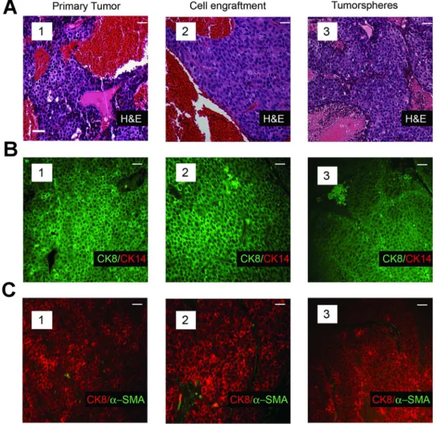
Figure 2. Tumors arising from transplant of single tumor cells recapitulate the histology and cellular composition characteristic of the parental tumors.(A), Histopathology of a primary mammary tumor from an MMTV-Neu transgenic mouse (subpanel 1), that of a tumor seeded by transplant of a single cell (subpanel 2) from the same primary tumor, and that of a tumor derived from transplant of a single cell dissociated from tumorspheres of the same transgenic strain (subpanel 3). H&E staining of tumor sections illustrates the solid and nodular cytoarchitecture characteristic of Neu-induced tumors. (B), Immunofluorescence analyses of an MMTV-Neu tumor (subpanel 1), that of a tumor seeded by transplant of a single cell (subpanel 2) from the same primary tumor, and that of a tumor (subpanel 3) seeded by transplant of a single tumorsphere-derived cell stained with antibodies to CK8 and CK14. (C), Immunofluorescence analyses of an MMTV-Neu tumor (subpanel 1), that of a tumor seeded by transplant of a single cell (subpanel 2) from the same primary tumor, and that of a tumor (subpanel 3) seeded by transplant of a single tumorsphere-derived cell stained with antibodies to CK8 anda-SMA. Note that distinct sections of the same tumor were used for the analyses shown in panels a–c. Scale bar (inset) represents 40mm in all panels.
p53-null mice [18,19,27]; as well as the mT and MMTV-beta-catenin models reported in this study. TIC frequencies in the MMTV-Wnt-1 model as determined by two different research groups varied by more than 40 fold [17,19]. TIC frequencies in two related Neu models similarly varied by 30 fold – between 0.03% (MMTV-mutant Neu) [20] and 0.9% (MMTV-proto-Neu) [19]. Indeed TIC frequencies reported by the same group in two different studies published years apart using the p53-null model varied by 13 fold [18,27].
The TIC frequencies reported previously did not exceed 1% of the bulk tumor cell population in any of the mouse models and generally were much lower than this value [17–19,27]. Our measures of TIC frequency in tumors of the three models that we examined averaged 30%. The TIC fraction in the tumors arising
in the MMTV-Neu (N202) and MMTV-b-catenin transgenic strains substantially exceeds those reported previously in the same (MMTV-Neu) [19] or related (MMTV-Wnt-1) [17,19] models by 30 fold or more. Indeed unpurified single tumor cells from tumors of each of the three models we examined initiated tumor growth at high efficiency after orthotopic transplant into immune-competent syngeneic mice demonstrating that TIC autonomously initiate tumor growth and implying that TIC must be therapeutically targeted to achieve durable breast cancer remission. The single cell seeded tumors recapitulated the cellular composition of the tumors from which they were isolated and these engrafted tumors could be serially transplanted demonstrating their capacity for differen-tiation and self-renewal. The high TIC frequency in the mouse models we analyzed may be due to the high expression of the
Figure 3. Single transplanted tumor cells isolated from the mammary tumors of MMTV-mT transgenic mice recapitulate the histology and cellular composition characteristic of their parental tumors.(A), Histology of a primary MMTV-mT tumor (subpanel 1), that of a tumor resulting from transplanting a single tumor cell from the same tumor (subpanel 2), and that of a tumor that arose from transplantating a single tumorsphere-derived cell from a tumor of the same transgenic strain (subpanel 3). (B), Immunofluorescence analyses of an MMTV-mT tumor (subpanel 1), that of a tumor seeded by transplant of a single cell from the same primary tumor (subpanel 2), and that from a tumor resulting from transplant of a single tumorsphere-derived cell from a tumor of the same transgenic strain stained with antibodies to CK8 and CK14 (subpanel 3). (C), Immunofluorescence analyses of an MMTV-mT tumor (subpanel 1), that of a tumor seeded by transplant of a single cell from the same primary tumor (subpanel 2), and that of a tumor resulting from transplant of a single tumorsphere-derived cell from a tumor of the same transgenic strain stained with antibodies to CK8 anda-SMA (subpanel 3). Note that distinct sections of the same tumor were used for the analyses shown in panels a–c. Scale bar (inset) represents 40mm in all panels.
various oncogenes under the control of the MMTV promoter. High oncogene expression has previously been shown to expand the repertoire of hematopoietic target cells that are transformed by MLL-AF9[49]. The MMTV promoter may similarly effect high expression of various oncogenes to mammary epithelial cell types that are ordinarily not susceptible to transformation by these oncogenes when expressed at lower levels.
The wide discrepancies in TIC frequency in tumors of the same models reported by various investigators and us likely result from differences in the experimental procedures used to prepare and fractionate dispersed tumor cells, and perhaps to the sensitivity of the tumor cell transplantation assay as practiced in individual laboratories. Unlike previous measurements of TIC frequency in mouse mammary tumors we did not use fluorescence activated cell sorting (FACS) to separate hematopoietic, endothelial and stromal cells from tumor cells prior to their transplantation, whereas all previous studies employed FACS to enrich for tumor cells [19,27,43]. The high pressure of FACS may reduce TIC viability as reported previously [27,43]. Moreover TIC engraftment may be aided by non-tumor derived cells such as stromal cells [50]; the depletion of such cells from the bulk tumor cell population in other studies may have led to underestimates of TIC frequency.
We used Matrigel as a vehicle to enhance TIC engraftment, whereas none of the other studies used this agent. Matrigel reportedly facilitates the engraftment of mouse mammary epithelial stem and progenitor cells and may have similarly stimulated the engraftment of TIC and progenitor-like tumor cells in our transplantation experiments [51]. In this regard we observed both large (1–2 g wet weight) and small tumors (100– 200 mg wet weight) among recipient mice transplanted with limiting dilutions of primary tumor cells. However, the small tumors, which may have been seeded by progenitor-like tumor cells with limited proliferative potential, constituted only a minor fraction of all the tumors we identified and were not scored in our analyses of TIC frequency or analyzed further.
The procedure we used to prepare dispersed tumor cells differed from those of previous studies; indeed none of the published studies used the same conditions to dissociate tumor cells from tumor tissue. Previous work employed only collagenase [18], collagenase and hyaluronidase [20], and collagenase, hyaluroni-dase and trypsin [19]. The duration of treatment of minced tumor tissue with these enzymes also varied ranging between 1 and 5 hours. We used both collagenase and trypsin to dissociate cells from tumor fragments and incubated the tissue for a period of 30 min following a protocol by Pullan and Streuli [52].
It is conceivable that the conditions we used to prepare tumor cells may have led to the selective recovery of TICs after filtering
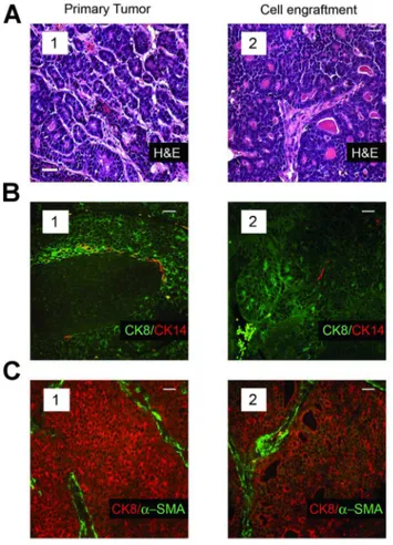
Figure 4. Single transplanted tumor cells isolated from the mammary tumors of MMTV-b-catenin transgenic mice recapit-ulate the histology and cellular composition characteristic of their parental tumors. (A), H&E staining of an MMTV-b-catenin primary tumor (subpanel 1) and a tumor obtained after transplanting a single tumor cell originating from the same tumor (subpanel 2). (B), Immunofluorescence analyses of an MMTV-b-catenin tumor (subpanel 1) and that of a tumor seeded by transplant of a single cell from the same primary tumor stained with antibodies to CK8 and CK14 (subpanel 2). (C), Immunofluorescence analyses of an MMTV-b-catenin tumor (subpanel 1) and that of a tumor seeded by transplant of a single cell from the same primary tumor stained with antibodies to CK8 anda -SMA (subpanel 2). Note that distinct sections of the same tumor were used for the analyses shown in panels a–c. Scale bar (inset) represents 40mm in all panels.
doi:10.1371/journal.pone.0058151.g004
Figure 5. Single tumorsphere-derived cells from tumors of the MMTV-Neu strain seed the growth of tumors after transplantation into syngeneic FVB/N mice.Haematoxylin-stained mammary whole mounts isolated 4 weeks post-transplant of varying numbers of tumorsphere-derived cells reveal tumor-like masses in the fat pads of recipient mice. The mass of the tumor nodules directly correlated with the number of injected tumor cells. The images were photographed at a magnification of 6.4.
the bulk tumor cells through a 40mm sieve to remove undigested tumor fragments and tumor cell aggregates. The yield of tumor cells from a 1–2 gram tumor arising in the MMTV-Neu (N202) strain approximated , 2–36108 cells prior to the removal of
tumor cell aggregates by filtration through a 40mm sieve;,95%
of these cells were viable. After filtration through the sieve,20%
of the cells were recovered in the filtrate;,95% of these cells were
viable. The latter cell yield is in keeping with that reported previously from the mammary glands of mice using the Pullan and Streuli protocol [52]. If the frequency of TIC in the Neu-induced tumors was as high as 1% as reported previously [19], then we would expect a 5-fold enrichment in the TIC fraction in the filtrate yielding a ,5% TIC frequency. However, our measurements
yielded a 6 fold higher TIC frequency in the filtrate, which
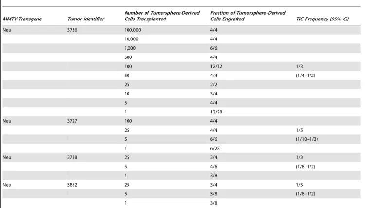
Table 2.Tumor-initiating cell frequencies in tumorpshere preparations.
MMTV-Transgene Tumor Identifier
Number of Tumorsphere-Derived Cells Transplanted
Fraction of Tumorsphere-Derived
Cells Engrafted TIC Frequency (95% CI)
Neu 3736 100,000 4/4
10,000 4/4
1,000 6/6
500 4/4
100 12/12 1/3
50 4/4 (1/4–1/2)
25 2/2
10 3/4
5 4/4
1 12/28
Neu 3727 100 4/4
25 4/4 1/5
5 6/6 (1/10–1/3)
1 6/28
Neu 3738 25 3/4 1/3
5 4/6 (1/8–1/2)
1 3/8
Neu 3852 25 3/4 1/3
5 3/8 (1/8–1/2)
1 3/8
doi:10.1371/journal.pone.0058151.t002
Table 3.Tumor-initiating cell frequencies in adherent tumor cell preparations.
MMTV-Transgene Tumor Identifier
Number of Adherent Tumor Cells Transplanted
Fraction of adherent tumor
cells engrafted TIC Frequency (95% CI)
Neu 001 100,000 1/4 1/392,318 (1/2,765831–1/55,648)
10,000 0/4
1,000 0/4
100 0/4
10 0/4
Neu 002 50,000 4/4 1/20,332
10,000 1/4 (1/54,184
1,000 0/4
-100 0/4 1/7,629)
10 0/4
Neu 406 10,000 0/4 N/A
1,000 0/4
100 0/4
10 0/4
averaged ,30%. We also note that the mouse mammary
tumorspheres were dissociated by titruation, and were not filtered through sieves, yet their resident TIC frequency approximated that of the primary tumors from which they were established. Thus whereas we cannot rule out the possibility that the means we used to isolate dispersed tumor cells enriched for TIC, we doubt that the high TIC frequencies we observed in the tumors we examined was due solely to this factor. It is also conceivable that the protocols used to dissociate the tumors, the quality of the enzymes employed in each study, and the duration of exposure of tumor tissue to these reagents used in previous studies may have led to the selective loss of TIC and may account for the striking differences in TIC frequencies reported by others and us. Resolution of the latter will require further study.
It is also not clear whether the extent of progression of the tumors analyzed in the various studies influenced TIC frequency. Multiple different TICs may be present during the early stages of tumorigenesis, but clonal evolution may occur during tumor progression resulting in the selection of a TIC population with an increased propensity to self-renew and a decreased capacity to differentiate [47,53]. The mammary tumors we investigated may have progressed to a greater extent than those studied by others and this parameter may account for the higher TIC fraction that we observed. Evidence for a relationship between tumor pro-gression and TIC frequency is suggested by recent findings demonstrating a correlation between the frequency of tumor cell engraftment and tumor grade in human breast tumors and melanomas [22,54]. Hence differences in the sensitivity of the transplantation assays, the quality of the transplanted tumor cell preparations, the use of Matrigel to facilitate tumor cell engraftment, and/or perhaps the extent of tumor progression may account for the disparate TIC frequencies reported by us and others in the mammary tumors of the same or similar models.
The question arises as to whether the mammary tumors occurring in the transgenic mice we examined follow the cancer stem cell model especially in light of their high TIC fraction. All the mouse breast cancer models studied previously with the sole exception of the MMTV-Neu transgenic mice have been suggested to follow this model [17–20]. Consistent with the cancer stem cell hypothesis tumors arising in either the MMTV-mT or MMTV-Wnt-1/b-catenin transgenic strains are heterogeneous comprising cells that express markers of either the luminal or myoepithelial lineages, the two principal cell lineages in the mouse mammary gland. By contrast, the tumors of the MMTV-Neu model are seemingly homogeneous comprising predominantly luminal-lineage restricted cells [24,25]. Accordingly it has been suggested that the mT and Wnt-1 induced tumors originate from stem cells or bipotent progenitor cells, whereas the Neu-induced tumors may emerge from luminal-restricted progenitor cells [31,55].
In keeping with the cellular heterogeneity of the tumors in the mouse models, the use of antibodies to mammary epithelial stem cell surface markers together with FACS have permitted sorting mammary tumor cells into tumorigenic and non-tumorigenic fractions. A very minor tumorigenic cell population can be separated from the bulk, non-tumorigenic tumor cell fraction using FACS in the MMTV-Wnt-1 model, analogous to the MMTV-b -catenin model we studied [17,19]. By contrast, Vaillant et al reported that whereas the tumors from the MMTV-Neu model, identical to that which we investigated, also comprised a relatively minor TIC fraction (,1%), the TIC could not be similarly
separated from the bulk tumor cells using antibodies to CD61, CD29 and CD24, leading these investigators to suggest that these tumors do not follow the cancer stem cell model [19]. Whether
other combinations of cell surface markers might permit separa-tion of TIC from the bulk tumor cell populasepara-tion in tumors of the MMTV-Neu transgenic strain has not been reported.
In light of the very high TIC frequency in the mouse mammary tumors we studied, the unavailability of markers that might facilitate fractionation of TIC from non-tumorigenic cells in the MMTV-Neu model [19] and the finding that FACS compromises TIC survival [43], we did not attempt to sort the bulk tumor cell populations of the models we studied into tumorigenic and non-tumorigenic fractions. Instead we asked whether other means might be used to determine whether the tumors we studied conformed to the cancer stem cell model. To this end we enquired whether conditions that favor normal stem cell self-renewal or differentiation might influence TIC frequency. A central tenet of the cancer stem cell model is that quasi-stable epigenetic differences distinguish TIC from their differentiating non-tumor-igenic descendants [9,47].
We found that the TIC frequency of the mammary tumors in the MMTV-Neu model could be maintained by culturing freshly isolated tumor cells in chemically-defined, serum-free medium, but that propagating these cells for a similar period and passage number in serum-containing medium reduced the TIC fraction by roughly 4 orders of magnitude. It is noteworthy that only 1% of primary tumor cells form tumorspheres, and that this fraction of sphere-forming cells does not vary during serial passage [56]. Hence the vast majority of the dispersed tumor cells do not survive plating in serum-free medium and because cell proliferation during sphere formation subsides substantially after 3–4 days in culture under the growth conditions we employed, there is no net expansion of the total tumor cell population during serial passage of bulk tumorsphere cultures. The growth factors in serum-free medium (FGF-2 and EGF) may sustain self-renewal of TIC and/ or limit their differentiation into highly proliferative progenitor-like cells. By contrast, the plating efficiency of primary tumor cells varied between 10%–30% in serum-containing medium and the cells proliferated resulting in net expansion of the starting cell population by ,10-fold during their initial serial passage. The
latter findings are consistent with the possibility that serum factors initiate a differentiation program in TIC resulting in the transient proliferation of their progenitor like descendants, which culmi-nates in their aberrant differentiation and consequent loss of proliferative and tumorigenic capacity. The latter hypothesis is consistent with the observation that normal mammary epithelial stem/progenitor cells can be propagated as mammospheres in serum-free medium, and undergo a differentiation program when propagated in serum-containing medium [36–38,48]. However, we have no evidence that the differentiation of TICs leads to their loss of tumorigenicityin vitroand cannot rule out the possibility that TICs simply do not survive in serum-containing medium.
It seems unlikely that TIC were diluted by the expansion of non-tumorigenic cells in serum-containing medium because the net increase in total cell number was roughly 10-fold, whereas the decline in TIC frequency was 10,000 fold or greater. The possibility that fibroblasts present in the primary tumor cell preparations overgrew the epithelial tumor cells during their propagation in serum-containing medium seems unlikely because the vast majority of the cells growing in serum-containing medium appeared to be epithelial in morphology independent of their passage history. Because the tumor cells were culturedin vitroin either growth condition for only 12 days it also seems unlikely that genetic changes account for their wide difference in TIC frequency.
mouse models reported by different laboratories, or whether the various models studied follow the cancer stem cell model. Whereas resolving these matters have therapeutic implications and conse-quently needs to be critically examined, the high TIC frequency in mouse mammary tumors and the capacity to maintain TIC-rich or TIC-depleted tumor cell populations in vitro may afford an opportunity to identify TIC biomarkers and TIC-targeted therapeutic agents, especially in view of the technical limitations inherent in sourcing human breast TIC to this end. Indeed we have used the mouse mammary tumorspheres and corresponding normal mouse mammospheres to identify TIC-selective com-pounds [56,57].
Conclusions
Our results demonstrate that mouse mammary tumors, in-cluding those arising in transgenic strains thought to conform to the cancer stem cell model, can comprise a very high TIC frequency that approach 50% in some tumors. Moreover, we show that a similarly high TIC frequency can be maintained by propagating the tumor cells in serum-free medium as non-adherent tumorspheres. By contrast, culturing the tumor cells for an identical period in serum-containing medium resulted in a substantial reduction in TIC frequency. These various tumor cell sources provide a rich source of tumorigenic or non-tumorigenic tumor cells for comparative analyses.
Supporting Information
Figure S1 Tumors arising from transplant of tumor cells propagated in serum-containing medium recapitulate the histology the parental tumor. (A), Histology of a primary mammary tumor from an MMTV-Neu transgenic mouse. (B), Histology of a tumor seeded by transplant of tumor cells from the tumor shown in panel A that had been propagatedin vitroin serum-containing medium. H&E staining of tumor sections illustrates the cytoarchitecture characteristic of Neu-induced tumors. Scale bar (inset) represents 40mm in all panels.
(TIF)
Acknowledgments
We thank Drs. Mickie Bhatia, John Dick, Sheila Singh, Gil Smith and Derek van der Kooy for reading the manuscript and providing helpful suggestions for its improvements.
Author Contributions
Conceived and designed the experiments: JAH JW RC. Performed the experiments: NAK AG-G RMH LK SR DWG. Analyzed the data: NAK AG-G RMH LK SR DWG JAH JW RC. Wrote the paper: JAH.
References
1. Clarke MF, Dick JE, Dirks PB, Eaves CJ, Jamieson CH, et al. (2006) Cancer stem cells – perspectives on current status and future directions: AACR Workshop on cancer stem cells. Cancer Res 66: 9339–9344.
2. Bonnet D, Dick JE (1997) Human acute myeloid leukemia is organized as a hierarchy that originates from a primitive hematopoietic cell. Nat Med 3: 730– 737.
3. Singh SK, Hawkins C, Clarke ID, Squire JA, Bayani J, et al. (2004) Identification of human brain tumour initiating cells. Nature 432: 396–401. 4. O’Brien CA, Pollett A, Gallinger S, Dick JE (2007) A human colon cancer cell
capable of initiating tumour growth in immunodeficient mice. Nature 445: 106– 110.
5. Eramo A, Lotti F, Sette G, Pilozzi E, Biffoni M, et al. (2008) Identification and expansion of the tumorigenic lung cancer stem cell population. Cell Death Differ 15: 504–514.
6. Ricci-Vitiani L, Lombardi DG, Pilozzi E, Biffoni M, Todaro M, et al. (2007) Identification and expansion of human colon-cancer-initiating cells. Nature 445: 111–115.
7. Clay MR, Tabor M, Owen JH, Carey TE, Bradford CR, et al. (2010) Single-marker identification of head and neck squamous cell carcinoma cancer stem cells with aldehyde dehydrogenase. Head Neck 32: 1195–1201.
8. Al-Hajj M, Wicha MS, Benito-Hernandez A, Morrison SJ, Clarke MF (2003) Prospective identification of tumorigenic breast cancer cells. Proc Natl Acad Sci U S A 100: 3983–3988.
9. Dick JE (2008) Stem cell concepts renew cancer research. Blood 112: 4793– 4807.
10. Krivtsov AV, Twomey D, Feng Z, Stubbs MC, Wang Y, et al. (2006) Transformation from committed progenitor to leukaemia stem cell initiated by MLL-AF9. Nature 442: 818–822.
11. Cozzio A, Passegue E, Ayton PM, Karsunky H, Cleary ML, et al. (2003) Similar MLL-associated leukemias arising from self-renewing stem cells and short-lived myeloid progenitors. Genes Dev 17: 3029–3035.
12. Prince ME, Sivanandan R, Kaczorowski A, Wolf GT, Kaplan MJ, et al. (2007) Identification of a subpopulation of cells with cancer stem cell properties in head and neck squamous cell carcinoma. Proc Natl Acad Sci U S A 104: 973–978. 13. Ishizawa K, Rasheed ZA, Karisch R, Wang Q, Kowalski J, et al. (2010)
Tumor-initiating cells are rare in many human tumors. Cell Stem Cell 7: 279–282. 14. Somervaille TC, Cleary ML (2006) Identification and characterization of
leukemia stem cells in murine MLL-AF9 acute myeloid leukemia. Cancer Cell 10: 257–268.
15. Kelly PN, Dakic A, Adams JM, Nutt SL, Strasser A (2007) Tumor growth need not be driven by rare cancer stem cells. Science 317: 337.
16. Williams RT, den Besten W, Sherr CJ (2007) Cytokine-dependent imatinib resistance in mouse BCR-ABL+, Arf-null lymphoblastic leukemia. Genes Dev 21: 2283–2287.
17. Cho RW, Wang X, Diehn M, Shedden K, Chen GY, et al. (2008) Isolation and molecular characterization of cancer stem cells in MMTV-Wnt-1 murine breast tumors. Stem Cells 26: 364–371.
18. Zhang M, Behbod F, Atkinson RL, Landis MD, Kittrell F, et al. (2008) Identification of tumor-initiating cells in a p53-null mouse model of breast cancer. Cancer Res 68: 4674–4682.
19. Vaillant F, Asselin-Labat ML, Shackleton M, Forrest NC, Lindeman GJ, et al. (2008) The mammary progenitor marker CD61/beta3 integrin identifies cancer stem cells in mouse models of mammary tumorigenesis. Cancer Res 68: 7711– 7717.
20. Cicalese A, Bonizzi G, Pasi CE, Faretta M, Ronzoni S, et al. (2009) The tumor suppressor p53 regulates polarity of self-renewing divisions in mammary stem cells. Cell 138: 1083–1095.
21. Quintana E, Shackleton M, Sabel MS, Fullen DR, Johnson TM, et al. (2008) Efficient tumour formation by single human melanoma cells. Nature 456: 593– 598.
22. Pece S, Tosoni D, Confalonieri S, Mazzarol G, Vecchi M, et al. (2010) Biological and molecular heterogeneity of breast cancers correlates with their cancer stem cell content. Cell 140: 62–73.
23. Cardiff RD, Wagner U, Henninghausen L (2001) Mammary cancer in humans and mice: a tutorial for comparative pathology. Vet Pathol 38: 357–358. 24. Rosner A, Miyoshi K, Landesman-Bollag E, Xu X, Seldin DC, et al. (2002)
Pathway pathology: histological differences between ErbB/Ras and Wnt pathway transgenic mammary tumors. Am J Pathol 161: 1087–1097. 25. Lin EY, Jones JG, Li P, Zhu L, Whitney KD, et al. (2003) Progression to
malignancy in the polyoma middle T oncoprotein mouse breast cancer model provides a reliable model for human diseases. Am J Pathol 163: 2113–2126. 26. Herschkowitz JI, Simin K, Weigman VJ, Mikaelian I, Usary J, et al. (2007)
Identification of conserved gene expression features between murine mammary carcinoma models and human breast tumors. Genome Biol 8: R76. 27. Zhang M, Atkinson RL, Rosen JM (2010) Selective targeting of
radiation-resistant tumor-initiating cells. Proc Natl Acad Sci U S A 107: 3522–3527. 28. Guy CT, Webster MA, Schaller M, Parsons TJ, Cardiff RD, et al. (1992)
Expression of the neu protooncogene in the mammary epithelium of transgenic mice induces metastatic disease. Proc Natl Acad Sci U S A 89: 10578–10582. 29. Guy CT, Cardiff RD, Muller WJ (1992) Induction of mammary tumors by
expression of polyomavirus middle T oncogene: a transgenic mouse model for metastatic disease. Mol Cell Biol 12: 954–961.
30. Li Y, Welm B, Podsypanina K, Huang S, Chamorro M, et al. (2003) Evidence that transgenes encoding components of the Wnt signaling pathway preferen-tially induce mammary cancers from progenitor cells. Proc Natl Acad Sci U S A 100: 15853–15858.
31. Jeselsohn R, Brown NE, Arendt L, Klebba I, Hu MG, et al. (2010) Cyclin D1 kinase activity is required for the self-renewal of mammary stem and progenitor cells that are targets of MMTV-ErbB2 tumorigenesis. Cancer Cell 17: 65–76. 32. Maglione JE, McGoldrick ET, Young LJ, Namba R, Gregg JP, et al. (2004)
33. Reynolds BA, Weiss S (1992) Generation of neurons and astrocytes from isolated cells of the adult mammalian central nervous system. Science 255: 1707–1710. 34. Youn BS, Sen A, Behie LA, Girgis-Gabardo A, Hassell JA (2006) Scale-up of breast cancer stem cell aggregate cultures to suspension bioreactors. Biotechnol Prog 22: 801–810.
35. Smalley MJ, Titley J, O’Hare MJ (1998) Clonal characterization of mouse mammary luminal epithelial and myoepithelial cells separated by fluorescence-activated cell sorting. In Vitro Cell Dev Biol Anim 34: 711–721.
36. Stingl J, Eaves CJ, Kuusk U, Emerman JT (1998) Phenotypic and functional characterization in vitro of a multipotent epithelial cell present in the normal adult human breast. Differentiation 63: 201–213.
37. Dontu G, Abdallah WM, Foley JM, Jackson KW, Clarke MF, et al. (2003) In vitro propagation and transcriptional profiling of human mammary stem/ progenitor cells. Genes Dev 17: 1253–1270.
38. Kurpios NA, MacNeil L, Shepherd TG, Gludish DW, Giacomelli AO, et al. (2009) The Pea3 Ets transcription factor regulates differentiation of multipotent progenitor cells during mammary gland development. Dev Biol 325: 106–121. 39. Shepherd TG, Kockeritz L, Szrajber MR, Muller WJ, Hassell JA (2001) The pea3 subfamily ets genes are required for HER2/Neu-mediated mammary oncogenesis. Curr Biol 11: 1739–1748.
40. Hu Y, Smyth GK (2009) ELDA: extreme limiting dilution analysis for comparing depleted and enriched populations in stem cell and other assays. J Immunol Methods 347: 70–78.
41. Siegel PM, Dankort DL, Hardy WR, Muller WJ (1994) Novel activating mutations in the neu proto-oncogene involved in induction of mammary tumors. Mol Cell Biol 14: 7068–7077.
42. Siegel PM, Muller WJ (1996) Mutations affecting conserved cysteine residues within the extracellular domain of Neu promote receptor dimerization and activation. Proc Natl Acad Sci U S A 93: 8878–8883.
43. Alexander CM, Puchalski J, Klos KS, Badders N, Ailles L, et al. (2009) Separating stem cells by flow cytometry: reducing variability for solid tissues. Cell Stem Cell 5: 579–583.
44. Maglione JE, Moghanaki D, Young LJ, Manner CK, Ellies LG, et al. (2001) Transgenic Polyoma middle-T mice model premalignant mammary disease. Cancer Res 61: 8298–8305.
45. Kordon EC, Smith GH (1998) An entire functional mammary gland may comprise the progeny from a single cell. Development 125: 1921–1930. 46. DeOme KB, Faulkin LJ Jr., Bern HA, Blair PB (1959) Development of
mammary tumors from hyperplastic alveolar nodules transplanted into gland-free mammary fat pads of female C3H mice. Cancer Res 19: 515–520. 47. Shackleton M, Quintana E, Fearon ER, Morrison SJ (2009) Heterogeneity in
cancer: cancer stem cells versus clonal evolution. Cell 138: 822–829. 48. Smalley MJ, Titley J, Paterson H, Perusinghe N, Clarke C, et al. (1999)
Differentiation of separated mouse mammary luminal epithelial and myoe-pithelial cells cultured on EHS matrix analyzed by indirect immunofluorescence of cytoskeletal antigens. J Histochem Cytochem 47: 1513–1524.
49. Chen W, Kumar AR, Hudson WA, Li Q, Wu B, et al. (2008) Malignant transformation initiated by Mll-AF9: gene dosage and critical target cells. Cancer Cell 13: 432–440.
50. Kuperwasser C, Chavarria T, Wu M, Magrane G, Gray JW, et al. (2004) Reconstruction of functionally normal and malignant human breast tissues in mice. Proc Natl Acad Sci U S A 101: 4966–4971.
51. Vaillant F, Lindeman GJ, Visvader JE (2011) Jekyll or Hyde: does Matrigel provide a more or less physiological environment in mammary repopulating assays? Breast Cancer Res 13: 108.
52. Pullan SE, Streuli CH (1996) Epithelial Cell Culture; Harris A, editor. Cambridge: Cambridge University Press. 97–121 p.
53. Nowell PC (1976) The clonal evolution of tumor cell populations. Science 194: 23–28.
54. Boiko AD, Razorenova OV, van de Rijn M, Swetter SM, Johnson DL, et al. (2010) Human melanoma-initiating cells express neural crest nerve growth factor receptor CD271. Nature 466: 133–137.
55. Li Y, Hively WP, Varmus HE (2000) Use of MMTV-Wnt-1 transgenic mice for studying the genetic basis of breast cancer. Oncogene 19: 1002–1009. 56. Kondratyev M, Kreso A, Hallett RM, Girgis-Gabardo A, Barcelon ME, et al.
(2012) Gamma-secretase inhibitors target tumor-initiating cells in a mouse model of ERBB2 breast cancer. Oncogene 31: 93–103.