1 Diyarbakır Kadın-Doğum ve Çocuk Hastalıkları Hastanesi, Diyarbakır, Türkiye 2 İstanbul Üniversitesi, İstanbul Tıp Fakültesi, Histoloji-Embriyoloji AD, İstanbul, Türkiye
Correspondence: Cenap Ekinci,
Diyarbakır Kadın-Doğum ve Çocuk Hastalıkları Hastanesi, Diyarbakır, Türkiye Eposta: [email protected] Received: 14.08.2012, Accepted: 02.09.2012
Copyright © JCEI / Journal of Clinical and Experimental Invesigaions 2012, All rights reserved R E S E A R C H A R T I C L E
In vitro efects of ellagic acid in C6 rat glioma cell cultures in terms of cytotoxicity and
proliferaion
Elajik asidin sıçan C6 glioma hücre kültürlerinde sitotoksisite ve proliferasyon üzerine in vitro
etkileri
Cenap Ekinci1, Aysun Ekinci1, Bülent Ahıshalı2, Ayhan Bilir2
ÖZET
Amaç: Elajik asit bitkilerden elde edilen polifenolik bir bi-leşiktir. Bu çalışmanın amacı, elajik asidin sıçan C6 gli -oma hücre dizilerinde proliferasyon ve yaşama yeteneği üzerine in vitro etkilerini doz ve zamana bağlı olarak araş -tırmak ve C6 glioma küremsi cisimlerinde oluşan elek -tronmikroskopik değişiklikleri değerlendirmektir.
Gereç ve yöntem: Elajik asidin sıçan C6 glioma hücre di -zisi üzerine etkileri doz ve zamana bağlı olarak iki boyutlu tümör hücre kültürü kullanılarak araştırıldı. Elajik asidin etkileri için üç boyutlu hücre kültürü küremsi modellerinde; hücre çoğalması, hücre yaşamsallığı, hücre döngüsünün sentez fazı ve hücre yapılarının 24, 48 ve 72. saatlerdeki durumu değerlendirildi.
Bulgular: İki boyutlu hücre kültürlerinde hücre yaşama yeteneği ve hücre çoğalması üzerinde elajik asidin baskı -layıcı etkisi görüldü (p<0.05). İki boyutlu hücre kültürlerin -de hücre sentez fazında artma veya azalma gözlenmedi (p>0.05). Üç boyutlu hücre kültürü küremsi modellerinde, elektronmikroskopik incelemede kontrol grubundan farklı yapısal değişiklikler bulundu.
Sonuç: İki boyutlu kültür modelleri kullanıldığında, elajik asidin sıçan C6 glioma hücre kültürleri üzerine olan et -kilerinin değerlendirilmesi sonucunda; elajik asidin hücre yaşayabilirliğini azalttığı, hücre yapısında bozulmalara neden olduğu ve hücre çoğalmasını engellediği bulundu. Anahtar kelimeler: Elajik asit, C6 glioma hücreleri, hücre kültürü, beyin tümörü, glioma
Abbreviations: DMSO; Dimethyl sulfoxide, DMEM/F12; Dulbecco’s Modiied Eagle Medium: Nutrient Mixture F-12, BrdU; Bromodeoxyuridine, PBS; Phosphate Buffer, BrDU-LI; Thymidine labeling index,
ABSTRACT
Objectives: Ellagic acid is a plant-derived polyphenolic compound. The aim of this study is to investigate in vitro effects of ellagic acid on the proliferation and viability of rat C6 glioma cell lineage depending on dose and time and also is to evaluate ultrastructural changes in C6 glio -ma spheroids.
Materials and methods: Effects of ellagic acid on rat C6 glioma cell line were investigated by using two-dimen -sional models of tumor cell culture depending on dose and time. The effects of Ellagic acid was evaluated as cell proliferation, cell viability, and synthesis phase of cell cycle, and cell structure at 24th, 48th, 72nd hours. Cell struc
-ture was evaluated at 24th and 72nd hours in three dimen
-sional cell culture spheroid models.
Results: Reducing effect of ellagic acid on cell prolifera -tion and cell viability was seen in two dimensional cultures (p<0.05). Increment or decrement were not seen in syn -thesis phase in two dimensional cultures (p>0. 05). Elec -tron micrographs in which three dimensional cell culture spheroid models was investigated, structural changes was found to be different from the control group.
INTRODUCTION
Glioblastoma multiforme which is the most com
-monly seen primary brain tumor disseminates
quickly. Treatment is surgical excision, postopera
-tive radioterapy and chemoterapy. Unfortunately the
mean survival is less than one year despite all treat
-ment. There is no deinitive treat-ment. New treat
-ment options are being waited. Ellagic acid which is found in pomegranate, strawberries, raspberries,
walnuts is phytochemical polyphenol compound.1-5
It is showed that, ellagic acid reduces growth of breast cancer, esophagus cancer, colon cancer,
leukemia and neuroblastoma in cell culture stud
-ies.6-8
Ellagic acid/chitosan composite biomaterial
leads cancer cells to apoptosis in one layer cell cul
-ture in a study with using U87 glioblastoma and rat
C6 glioma cell lineage.9 In this study we investigate
in vitro effects of ellagic acid on the proliferation and viability of rat C6 glioma cell lineage.
MATERIALS AND METHODS
Rat C6 glioma cell line (ECACC no: 558807), which was taken from American Type Culture Collection
was used in Laboratory of Istanbul University Histol
-ogy and Embriol-ogy Department for this study.
a. Ellagic acid preparation for proliferation experiment: Ellagic acid which was taken 2mg has
302.192 g/mol molecular weight and it was dis
-solved in 1324 µl DMSO (Dimethyl sulfoxide) so 5 mM ellagic acid stock solution was obtained by dilution of stock solution with DMSO. 50, 10, 1 µM application concentrations were reached by 100 µl
adding these doses to 5 ml wells containing me
-dium.
b. Experiment protocols in which ellagic acid doses were used: 100% live 500000 C6 gli
-oma cells were inoculated in 5 ml DMEM/F12 (Dul
-becco’s Modiied Eagle Medium: Nutrient Mixture F-12) medium in each wells of six-well plate. Groups were determined as control group, 1 µM, 10 µM, 50 µM and 100 µM groups. Only 100 µl DMSO was given to control group. C6 glioma cells inoculation were performed in three wells for each Ellagic acid dose and control group. All groups were inoculated separately for 24, 48 and 72 hours drug application
and incubation was performed in a moist environ
-ment with 37°C and 5% CO2 air-mixture.
At the end of 24, 48 and 72 hours cells were
collected separately from all experimental and con
-trol with 0.05% trypsin wells and these cells were
centrifuged. After discard of supernatant part, sus
-pension was made with 1ml medium and it was counted in counting chamber. Total cell counts were recorded to determine the proliferation rate. Cell suspension was mixed with 0.1% trypan blue dye with equal proportion. A drop of suspension was taken and stained dead cells and unstained live
cells were counted under microscope so live cell ra
-tio was determined (Table 1).
Table 1. Ellagic acid study groups
Doses Ellagic acid time groups
24 hour 48 hour 72 hour
Control 3 well 3 well 3 well
Ellagic Acid 1µM 3 well 3 well 3 well Ellagic Acid 10µM 3 well 3 well 3 well Ellagic Acid 50µM 3 well 3 well 3 well Ellagic Acid 100µM 3 well 3 well 3 well
c. Immunohistochemical marking with Brdu in two-dimensional cell culture: Round lamellas
were placed in 24 wells culture plates and 40000
cells were inoculated in each wells. Cell culture me
-dium 1 ml was placed in each well. Doses required for 1 µM, 10 µM, 50 µM and 100 µM ellagic acid groups are prepared from stock ellagic acid groups and 20 µl applications were done. 20 µl DMSO was applicated to control group. Three lamellae were prepared for each group. At the end of 24, 48 and 72 hours, cells were incubated with 20 µM BrdU (Bromodeoxyuridine) (2ml medium+100µl BrdU) for 1 hour at 37°C. After upper medium was retreated
and thrown, 1 ml sterile PBS (Phosphate buffer) so
-lution was applicated into the wells and stored in the incubator for 15 minutes. Cells were ixated in 70% ethanol in -20°C for 30 minutes after retreat and thrown of PBS. Lamellae were dried at room
temperature after ethanol was retreated. Dried la
-mellae for immunohistochemistry was rehydrated in
PBS for 10 minutes then it was kept in 5% H2O2
prepared in methanol for hydrogen peroxide inhibi
-tion in the dark. After applica-tion of ultra-V-block for
15 minutes in room temperature and humid envi
-ronment to avoid non-speciic antibody binding, it was kept in anti-BrdU antibody (NCL-BrdU; Zymed 1/50) for an hour. After washing with PBS it was kept in secondary antibody for 30 minutes, after
washing again it was kept in streptavidin peroxi
-dase for 30 minutes. After washing with PBS it was kept in AEC chromogenic-substrate solution (AEC Substrate System; Zymed) for 20 minutes and then
-ing was made with Mayer’s hematoxylin sections were closed with sealer. By counting marked and unmarked cells under a light microscope and BrdU index was determined.
d. Preparation of three dimensional cul-tures for electron microscopic evaluation: In -oculation was done for three dimensional cultures in agar coated cell culture dishes. 500000 cells in 5 ml DMEM/F12 medium were inoculated into the each wells coated with solidiied agar. Inoculation was done into the 6 wells for each control group, 1 µM and 100 µM ellagic acid groups. Mediums which were done in each 48 hours were waited for 7 days to form the spheroids from cells kept in incubator. While dose required for 100 µM ellagic acid group was applicated directly from ellagic acid solution, dose required for 1 µM ellagic acid group was prepared by dilution and 100 µl applications
were done. 100 µl DMSO was applicated to con
-trol group. Spheroids were collected at 24th and 72nd
hours and placed into different centrifuge tubes. After upper medium was retreated and thrown,1 ml 0.1 M phosphate buffered 2.5% gluteraldehyde solution was added and ixated under 4°C for 30 minutes. Post-ixation with 1% OsO4 for 60 minutes under 4°C was done after centrifuge and washing 2 times with phosphate buffer. After washing again with phosphate buffer for 10 minutes it was kept in 1% uranil acetate for 15 minutes. It was kept in 30% ethanol after phosphate buffer washing for 10 minutes. After that, it was kept in 50% ethanol for 10 minutes then 70% ethanol for 10 minutes then
90% ethanol twice for 10 minutes then pure propyl
-enoxide for 10 minutes then 1/1 ratio prepared pro
-pyleneoxide/epone mixture for 1 hour and inally it was kept in pure epone for 1 hour. After that it was embedded in tissue embedding capsules. It was kept in 37°C in the incubator for 18 hours. After trim
of blocks which was taken from the incubator, tar
-get sections containing spheroids in 1 µm semi-thin sections was detected. Then 70 nm thin sections on copper grids were done. It was evaluated under transmission electron microscopy after lead citrate and uranyl acetate contrasting.
e. Statistical analysis: Statistical analyses
were performed under the SPSS for Windows soft
-ware (version 12, SPSS Inc, Chicago, IL). Indepen
-dent Stu-dent’s T-test was used in all experimental results. Results are presented as mean ± standard
deviation and ranges. A two-tailed probability of 5% or less was considered statistically signiicant.
RESULTS
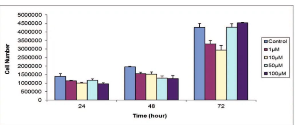
a. Results of ellagic acid kinetic cell proliferation experiments: Healthy cell proliferation pattern was
seen in C6 tumor cells in the control group. Ellagic acid reduced cell count in1 µM concentration group
at 48th and 72nd hours with related to control group
and this was statistically signiicant. Reduction in 10
µM concentration only at 72nd hour was statistically
signiicant. 50 µM concentration reduced cell count
at 48th hour and it was statistically signiicant. 100
µM concentration reduced cell count at 24th and 48th
hour with related to control group but only reduc
-tion in 48th hour was statistically signiicant (p<0.05)
(Graphic 1).
b. Cytotoxicity of ellagic acid: Application of
ellagic acid in 1µM concentration reduced live cell ratio at 24th and 48th hour with related to control
group and it was statistically signiicant (p<0.01). On the other hand this concentration increased live
cell ratio at 48th hour signiicantly. Signiicant chang
-es were not observed compared to control group in live cell ratio with 10 µM application of ellagic acid. 100 µM ellagic acid caused statistically signiicant
reduction in the live cell ratio with compared to con
-trol group at 24th hour (p<0.01) (Graphic 2).
c. Effects of different concentration of el-lagic acid on BrdU marking index (BrDU-LI): Ap -plication of 1 µM concentration of ellagic acid cause
increment at all times in BrDU-LI (Thymidine label
-ing index) values with compared to control group while this increment was statistically signiicant at
24th hour (p<0.05). BrDU-LI values were increased
at 24th and 72nd hour with compared to control group
by application of 10 µM concentration of ellagic acid
whereas increment in 24th hour was statistically
signiicant (p<0.01). Application of 50 µM concen
-tration of ellagic acid cause increment at all times in BrDU-LI values with compared to control group
whereas increment in 24th and 48th hour were sta
Graphic 1. Effects of ellagic acid on proliferation of rat C6 glioma cell depending on concentration and time
*p<0.05
Graphic 2. The ratio of viability in the control group and application ellagic acid at 1, 10, 50 ve 100 micromolar (μM) concentrations *: p<0.05, **: p<0.01.
Picture 1. Control group two-dimensional cultures at 72nd
hour indicating numerous C6 glioma cells marked by BrdU X10
Picture 2. Ellagic acid 1µM dose applied group: C6 glio -ma cells -marked by BrdU at 72nd hour X20
d. Electron microscopic evaluation of three dimensional cell cultures: Tight regulations de -pending on 3 dimensional cellular contacts of C6 glioma cells generated an appearance similar to
a typical tumor tissue to the spheroid in the con
-trol group at 24th hour. Glioma cells which form
spheroids have one nucleolus or two nucleoli and recessed and protruding euchromatic nucleus and their cytoplasms were seen electron-light in some cells and electron-dense in the other cells. Shelf-shaped mitocondria with crista were seen less in some cells and more in the other cells. Plasma
membranes of cytoplasm of adjacent cells were lo
-cated closely in the outer surface of the spheroids and gaps which have 20 to 500 nm width contain electron dense material in some parts were found between two cell membranes. Plasma membrane
gaps which did not contain electron dese mate
-rial have 4-14 nm width. Microvilli like amorphous
extensions formed by plasma membranes were in
contact with each other in the intercellular space in
-side of the spheroid. As well as, in-side spheroids in
some cells electron dense material containing vacu
-oles surrounded by membrane and wide intercellu
-lar space were seen (Picture 3).
Necrotic area which was expected in a typical
spheroid structure was not seen in this group spher
-oid. At the 24th hour application of 1 µM ellagic acid,
inside electron dense cytoplasm of tumor cells form
-ing spheroid, rich free ribosome and electron dense
matrix, mitocondria with weak crista were drew at
-tention. Connections between cells of outer surface of the spheroids were disappeared in some places and just below these cells wide intercellular spaces were occurred. In these wide intercellular spaces
extending into the inner parts of the spheroids de
-bris materials belonging to degenerated cells were found and in the cytoplasmic parts facing to these areas electron dense material containing vacuoles were seen.
At the 24th hour ellagic acid application of 100
µM concentration, degenerated cell debris contain
-ing intercellular space widen-ing were seen near the outer surface of the spheroids and this was similar
to 24th hour of 1 µM ellagic acid application results.
At the 72nd hour of 1 µM application of ellagic
acid, necrotic cells and apoptotic bodies were drew attention in the wide intercellular spaces inside the spheroids.
Near the outside face of the spheroids and in
-side of the spheroids intercellular space widening
areas were found at 72nd hour of application of 100
µM ellagic acid. In these areas microvillus like amor
-phous extensions were abundant and degenerated cell debris was not found (Picture 3, 4, 5).
three dimensional cultures at 24th hour under 1µM dose of Ellagic acid.
Picture 4. Ellagic acid 100 µM dose applied group; C6 glioma cells marked by BrdU at 72nd hour X20
Picture 5. Sferoid structure of C6 glioma cells in three dimensional cultures at 24th hour under 100m µM dose of Ellagic acid. Cell debris was observed in expanding area under the spheroid surface.
Statistical evaluation results: As a result in two dimensional cell culture model ellagic acid led to
reduction in cell proliferation with compared to con
-trol group. In S-phase evaluation ellagic acid led to increment in all concentration. Live cell ratio was
increased at 48th hour, decreased in other intervals.
DISCUSSION
In our study, we found that ellagic acid reduced cell proliferation and viability ratio in C6 glioma cells which was dose and time dependent. Statistically signiicant cell proliferation reduction was seen in
1,50, 100 µM doses of ellagic acid at 48th hour and
1,10µM doses at 72nd hour. As well as statistically
signiicant reduction in cell proliferation was seen
in 1µM dose at 72th hour, 50µM dose at 48th hour,
100µM dose at 24th hour.
Studies to clarify the mechanism of antitu
-moral effects of ellagic acid reported that drug did not cause any change in expression level of tumor suppressor gene P53 but it increase expression of
cyclin-dependent kinase inhibitor p21 (waf1/Cip1).10
On the other hand ellagic acid increases cyto
-chrome c level released from mitochondria to the cytosol and it increases caspase-3 and caspase-9 which are important proteases. It is reported that in
this way ellagic acid induced apoptosis.11
In a study on SH-SY5Y human neuroblasto
-ma cells, ellagic acid decreased cells in S phase
compared to control group and this was statisti
-cally signiicant, whereas cells in G2/M phase were slightly increased and cells in G0/G1 phase were
signiicantly increased.12,13 Mertens et al. reported
that 10 µM ellagic acid application did not effect in MOLT-4 human leukemia cell cycle compared to control group at 12th, 24th and 48th hour.14 In a study
on CaSki cervical cancer cells 10 µM ellagic acid
stops cell cycle in G1 phase.10
Li et al. reported that, 5, 10, 25 and 50 µM dos
-es of ellagic acid increased the cell ratio in G0/G1 phase of cell cycle and decreased cell ratio in G2/M phase of cell cycle in T24 human bladder cancer cells.15
In our study, effect of different concentration of ellagic acid on S phase of C6 glioma cell cycle which was expressed as BrdU-LI was increased with compared to control group in all concentrations and in time period and no decrement was seen in all concentrations and in time period. This result was in line with a study about effects of ellagic acid on Caco-2 human colon cancer cell descendants by
Larossa et al. In this study 1, 10 and 30 µM dos
-es of ellagic acid were applicated at 24th, 48th and
72nd hour and cyclin levels were investigated. It was
reported that ellagic acid reduced cyclin A and B1 and it increased cyclin E levels with related to time and dose so cell was arrested at S phase of the cell
cycle.11 This study supports our inding that is while
ellagic acid reduced cell proliferation and viability and it increased the cell ratio in S phase expressed as BrdU-LI value.
Different mutations in a particular cell’s descen
-dants may cause the diversity of polyphenol treat
-ment outcomes and the cell cycle halt in different phases. In our study, effects of ellagic acid on cell
viability and proliferation showed different statisti
-cal signiicance lines during administered dose and time interval. Effects of ellagic acid on the cell cycle
-cape from cell destruction may arise in tumor cell population.11,12,14-17
To our knowledge this study which evaluates effects of ellagic acid in three dimensional spheroid model by using electron microscopy is the irst study.
Taking into account the three-dimensional arrange
-ment of solid tumors multicellular tumor spheroids which were developed to form more suitable in vitro
systems relect three dimensional growth and orga
-nizations of tumors quite realistically, as a result in
-tercellular relations in tumor and microenvironmen
-tal conditions can be presented clearly.18 Integrins
have major role in cell-cell contact in spheroids. Integrins which bind to spheroid cell skeleton also
bind intercellular matrix. In this way, cell-cell con
-nection and spheroid integrity can be provided. It is
known that integrin release is organized by spher
-oid microenvironment.19 In our study, connections
between cells located in the outer surface of the C6 glioma spheroids were disappeared in some places and just below these cells wide intercellular spaces were occurred.
In these wide intercellular spaces extending into the inner parts of the spheroids debris materials belonging to degenerated cells were seen. In our
study, this condition with reduction in cell prolifera
-tion and cell viability caused by ellagic acid effect in
-tegrin release organized by spheroid microenviron
-ment and reduce connections of the cells in each other and external matrix.
In the way of these indings, microscopic and molecular studies are needed to ind mechanism of
effects of ellagic acid on different cell types and fur
-ther studies are needed to ind synergistic effects of
this drug in combination with different chemothera
-peutic agents.
In conclusion, ellagic acid which reduced cell proliferation and cell viability increased BrdU-LI showing S phase of the cell cycle in C6 glioma cells.
REFERENCES
1. Karakaya S. Bioavailability of phenolic compouns. Crit Rev Food Sci Nutr 2004;44(4):453-64.
2. Yang CS, Landau JM, Huang MT, Newmark HL. Inhibi -tion of carcinogenesis by dietary polyphenolic com -pounds, Ann Rev Nutr 2001:21(3):381-406.
3. Navinda P, Seeram, Lynn S, et al. In vitro antiprolifera -tive apoptotic and antioxidant activities of punicala -gin, ellagic acid and total pomegranate tannin extract are enhanced in combination with other polyphe -nols as found in pomegranate juice. J Nutr Biochem 2005:6(2);360-3.
4. Quinones-Hinojasa A, Sanai N, Smith JS, McDermott MW. Techniques to assess proliferative potential of brain tumors. J Neurooncol 2005:74(1);19-30.
5. Vekiari A, Gordon M.H, Garcia-Macias P, Labri -nea H. Extraction and determination of ellagic acid contentin chesnut bark and fruit. Food Chemistry 2008:110(4);1007-11.
6. Falsaperla M, Morgia G, Tartarone A, Ardito R, Romano G. Support ellagic acid therapy in patients with hor -mone refractory prostate cancer (HRPC) on standard chemotherapy using vinorelbine and estramustine phosphate. Eur Urol 2005:47(3);449-55.
7. Strati A, Papoutsi Z, Lianidou E, Moutsatsou P. Effect of ellagic acid on the expression of human telomer -ase reverse transcript-ase (h TERT) α+β+ transcript in estrogen receptor-positive MCF-7 breast cancer cells. Clin Biochem 2009:42(13);1358-62.
8. Alexander C, Whitley G, Stoner D, Darby M, Vale T. Intestinal epithelial cell accumulation of the can -cer preventive polyphenol ellagic cid-extensive binding to proteinamd DNA. Biochem Pharmocol 2003:66(8);907-15.
9. Sungwoo Kim, Mostafa W. Gaber, et al. The inhibi -tion of glioma growth in vitro and in vivo by chitoan/ ellagic acid composite biomaterial, Biomaterials 2009:30(27);4743-51.
10. Narayanan BA, Geoffroy O, Willingham MC, Re GG, Nixon DW. p53/p21(WAF1/CIP1) exspression and its possible role in G1 arrest and apoptosis in ellagic acid treated cancer cell, Cancer Lett 1999:136(2);215-21. 11. Larrosa M, Tomas-Barberan FA, Espin JC. The dietary
hydrlysable tannin punicalagin releases ellagic acid that induces apoptosis in human colon adenocarcino -ma Caco-2 cells by using the mitochondrial pathway. J Nutr Biochem 2006:17(5);611-25.
12. Fjaeraa C, Nanberg E. Effect of ellagic acid on proliferasyon, cell adhesion an apoptosis İn SH-SY5Y nuroblastoma cells, Biomed & Pharmacother 2009:63(2);254-61.
13. Vermeulen K, Berneman ZN, vanBockstaele DR. Cell cycle and apoptosis. Cell Prolif 2003:36(1);165-75. 14. Susanne U. Mertents-Talcott, Susan S. Percival. El
-lagic acid and quercetin interact synergistically with reveratrol in the induction of apoptosis and cause transient cel cycle arrest in human leukemia cells. Cancer Lett 2005:218(2);141-51.
15. Li TM, Chen GW, Su CC, et al. Ellagic acid in -duced p53/p21expresion, G1 arrest and apoptozis in human bladder cancer T24 cells. Anticancer Res 2005:25(8);971-9.
16. Tasaki M, Umemura T, Maeda M, et al. Safety asses -ment of ellagic acid, a food additive, in a subchronic toxicity study using F344 rats. Food Chem Toxicol 2008:46(9);1119-24.
17. Sharma G, Italia JL, Sonaje K, Tikoo K, Ravi Kumar MNV. Biodegradale in situ gelling system for subcuta -neous administration of ellagic acid and ellagic acid loaded nanoparticles: evaloation of their antioxidant potantial against cycosporine induced nephrotoxicity in rats. J Control Release 2007:118(1);27-37.
18. Carlsson J, Yuhas JM. Liquid-overlay culture o cellular spheroids: Resent results. Cancer Res 1984:95(1);1-23. 19. Santini MT, Gabriella R. Three dimensional spheroid mod