Drosophila
Histone Deacetylase-3 Controls Imaginal Disc
Size through Suppression of Apoptosis
Changqi C. Zhu1,2., Douglas J. Bornemann1,2., David Zhitomirsky1,2
, Ellen L. Miller1, Michael B. O’Connor1,2, Jeffrey A. Simon1*
1Department of Genetics, Cell Biology and Development, University of Minnesota, Minneapolis, Minnesota, United States of America,2Howard Hughes Medical Institute, Chevy Chase, Maryland, United States of America
Abstract
Histone deacetylases (HDACs) execute biological regulation through post-translational modification of chromatin and other cellular substrates. In humans, there are eleven HDACs, organized into three distinct subfamilies. This large number of HDACs raises questions about functional overlap and division of labor among paralogs. In vivo roles are simpler to address inDrosophila, where there are only five HDAC family members and only two are implicated in transcriptional control. Of these two, HDAC1 has been characterized genetically, but its most closely related paralog, HDAC3, has not. Here we describe the isolation and phenotypic characterization ofhdac3mutations. We find that bothhdac3andhdac1mutations are dominant suppressors of position effect variegation, suggesting functional overlap in heterochromatin regulation. However, all five hdac3 loss-of-function alleles are recessive lethal during larval/pupal stages, indicating that HDAC3 is essential on its own for Drosophila development. The mutant larvae display small imaginal discs, which result from abnormally elevated levels of apoptosis. This cell death occurs as a cell-autonomous response to HDAC3 loss and is accompanied by increased expression of the pro-apoptotic gene,hid. In contrast, although HDAC1 mutants also display small imaginal discs, this appears to result from reduced proliferation rather than from elevated apoptosis. The connection between HDAC loss and apoptosis is important since HDAC inhibitors show anticancer activities in animal models through mechanisms involving apoptotic induction. However, the specific HDACs implicated in tumor cell killing have not been identified. Our results indicate that protein deacetylation by HDAC3 plays a key role in suppression of apoptosis in Drosophilaimaginal tissue.
Citation:Zhu CC, Bornemann DJ, Zhitomirsky D, Miller EL, O’Connor MB, et al. (2008)DrosophilaHistone Deacetylase-3 Controls Imaginal Disc Size through Suppression of Apoptosis. PLoS Genet 4(2): e1000009. doi:10.1371/journal.pgen.1000009
Editor:R. Scott Hawley, Stowers Institute for Medical Research, United States of America
ReceivedNovember 1, 2007;AcceptedJanuary 18, 2008;PublishedFebruary 29, 2008
Copyright:ß2008 Zhu et al. This is an open-access article distributed under the terms of the Creative Commons Attribution License, which permits unrestricted use, distribution, and reproduction in any medium, provided the original author and source are credited.
Funding:This work was supported by NIH grant GM49850 to JS and MBO is an Investigator of the Howard Hughes Medical Institute.
Competing Interests:The authors have declared that no competing interests exist.
* E-mail: simon004@umn.edu
.These authors contributed equally to this work.
Introduction
Histone deacetylases (HDACs) are members of an ancient enzyme family that reverses acetylation of protein substrates. The most well-characterized HDAC substrates are the N-terminal tails of the histones. Acetylation of histone tail lysines generally correlates with gene activity, whereas HDAC-spon-sored removal of these tail modifications frequently accompanies gene silencing [1,2]. Histone acetylation state can impact gene expression through recruitment of transcriptional regulatory complexes, such as the SWI/SNF remodelling complex [3,4]. Changes in charge density resulting from histone acetylation/ deacetylation may also affect packaging of nucleosome arrays into higher-order arrangements that can impact transcription rates. A major HDAC regulatory function, then, is to promote gene silencing.
The histone deacetylase HDAC1 has been the most throughly studied HDAC at the biochemical and functional levels. Extensive analysis of HDAC1 in yeast, also known as RPD3, indicates that it can deacetylate all four core histones, that it targets hundreds of genes around the genome, and confirms its major role as a direct transcriptional repressor [1,5–8]. Biochemical studies show that
HDAC1 is typically assembled into nuclear complexes, such as the SIN3 and NURD complexes [2,9–11]. These co-repressor complexes are recruited to target genes through interactions with DNA-binding proteins. A prime example is provided by nuclear hormone receptors such as thyroid hormone receptor; the unliganded receptor recognizes target genes through its zinc finger DNA-binding domain, and it recruits a SIN3/NCoR/ HDAC1 complex, which deacetylates target chromatin and leads to gene silencing [12].
There are 11 HDAC family members in humans, defined by an approximately 350 amino acid homology region that encompasses the catalytic domain [21]. These have been classified into three major subfamilies, with class I containing HDACs 1, 2, 3 and 8, class II containing HDACs 4, 5, 6, 7, 9 and 10, and HDAC11 comprising a third distinct subtype [21] (see Figure 1A). In addition, the sirtuins represent yet another HDAC family, which are distinguished by their NAD-dependent reaction mechanism and are structurally unrelated to the family of 11 human HDACs. The large number of HDACs makes it difficult to determine which functions are shared and which can be uniquely assigned to individual family members. HDAC functional diversity is further complicated by the ability of HDACs to modify many protein substrates besides histones. Indeed, all three major HDAC subtypes are present in bacterial species [21], indicating that they likely evolved as protein deacetylases that only later acquired ability to act upon histones. In agreement with diversity of function, HDAC1 and 2 are largely nuclear, HDAC6 is cytoplasmic, and still other HDACs, including HDAC3, are found in both nucleus and cytoplasm [2,22–25]. Within the nucleus, several transcription factors, including p53, GATA-1, and YY1, are HDAC substrates [26–28]. In the cytoplasmic compartment, tubulin deacetylation by HDAC6 has been described [24]. The large number of HDAC family members and the diversity of their protein substrates predict a myriad of HDAC regulatory functions in vivo.
HDAC functions are simpler to dissect in theDrosophilasystem, where there are only five HDAC family members [21] (Figure 1A). In addition, there are only two HDACs of the class I subtype: HDAC1 (also called DmRpd3) corresponding to the nuclear HDAC1/2 of mammals, and its most closely related fly paralog, HDAC3. Furthermore, HDAC1 and HDAC3 are the only two fly HDACs implicated in transcriptional control [29]. Genetic studies using HDAC1 mutations have identified roles in many processes including heterochromatin silencing, segmentation, and ecdysone receptor function [30–33]. However, the lack of HDAC3 mutations to date has impeded understanding of its biological functions in the fly system. Here we describe isolation of HDAC3
loss-of-function alleles and present phenotypic characterization. All five HDAC3 mutations are homozygous lethal at late larval or pupal stages. The mutant larvae have abnormally small imaginal discs, which we attribute to cell-autonomous induction of apoptosis rather than defects in cell proliferation.
Results/Discussion
Isolation of Recessive LethalDrosophila hdac3Alleles
To investigate HDAC3 biological functions, we isolated a set of mutant alleles of flyhdac3. An initialhdac3allele was obtained by imprecise excision of a P transposon inserted into the neighboring gene,snr1, on the fly third chromosome (Figure 1B; see Methods). The excision yielded an approximately 1.8 kb deletion of genomic DNA that removes the 39 two-thirds of both the hdac3and snr1
coding regions. The deletion allele, termed hdac36C, breaks at codon 145 ofhdac3, within the HDAC catalytic domain. Since the severely truncated HDAC3 protein derived from this mutant allele, if stable, lacks critical portions of the catalytic domain [34,35], it likely causes severe loss of HDAC3 function. However, the usefulness of this deletion allele forhdac3functional studies is limited by the simultaneous disruption ofsnr1.
To obtain mutations that specifically disrupt HDAC3, we next performed a noncomplementation screen for EMS-induced alleles that are lethal in trans to thehdac36Cdeletion (see Methods). The recovery ofhdac3alleles, as opposed tosnr1alleles, was facilitated by use of aP[snr1+] rescue construct [36] in test crosses of the
newly isolated lethal mutations (see Methods). We isolated four newhdac3mutations from this screen, each of which is lethal in trans tohdac36C
. All fourhdac3mutant alleles cause lethality during pupal stages when either hemizygous or in transheterozygous combination with each other. Sequence analysis revealed that three of these alleles are missense mutations and one, hdac3N, converts Q57 to a stop codon (Figure 1C). Since this nonsense mutation eliminates the HDAC catalytic domain, it is likely a null allele. These results show that HDAC3 has unique functions required for fly development and viability, despite its 56% identity and 76% similarity to HDAC1 (Figure 1C).
The three-dimensional crystal structure of human HDAC8, a class I HDAC, has been determined [34,35]. Based upon high primary sequence conservation, all class I HDACs likely exhibit similar overall protein folds. Thus, we projected our threehdac3
substitutions onto the HDAC8 structure, to gain information about their possible effects on the protein (Figure 1D). The class I HDAC contains a single domain consisting of an 8-stranded parallel ß-sheet surrounded by 11 alpha-helices [34]. The catalytic site is located on one side and features a deep pocket, which is thought to bind the acetylated lysine substrate, and a Zn-binding site. All three missense substitutions are located on the opposite side, away from the catalytic site (Figure 1D). Thus, rather than specifically disrupting the catalytic center, these mutations may affect HDAC3 interactions with partner proteins, such as co-repressors, or they may cause structural destabilization. Two of these mutations affect residues that are highly conserved among HDACs; P254 is absolutely conserved among all 16 human and fly HDACs and V192 is conserved in all class I HDACs and in 14 of the 16 human and fly HDACs.
Hdac3Mutations Suppress PEV but Show Little Effect on Polycomb Silencing
DrosophilaHDAC1 has previously been implicated in two types of chromatin regulation: heterochromatin modification and gene silencing by the Polycomb system [19,20,32,33]. Indeed, the first
DrosophilaHDAC mutation was isolated in HDAC1 based upon its
Author Summary
modification of position-effect variegation (PEV) [33], a gene silencing event that occurs when a euchromatic gene is relocated to heterochromatin [37,38]. In molecular terms, HDACs may promote heterochromatin silencing by deacetylating histone H3 on lysine 9, which renders this residue available for methylation by the heterochromatin silencing machinery [39,40].
Heterochromatin silencing is conveniently assayed inDrosophila
using In(1)wm4, an X-chromosome inversion that repositions the
white eye color gene to heterochromatin [41]. This silenceswhite
expression in most cells of the eye, leading to a variegated eye color much lighter than normal (example in Figure 2A). Loss-of-function missense mutations in Drosophila hdac1 are dominant suppressors ofwm4variegation, whereashdac1null mutations have
little effect [32]. These results have been attributed to a poisoning effect of an expressed but inactive HDAC1 enzyme [32]. Similarly, we find that a null hdac1 allele, Q109stop, has little effect on wm4 variegation (Figure 2B) whereas a missensehdac1
allele which alters a catalytic pocket residue, D174G, shows significant suppression (Figure 2C).
To investigatehdac3effects upon PEV, we tested each of our five
hdac3mutations forwm4suppression. We found that heterozygosity for the hdac3 null alleles, hdac3N and hdac36C, led to strong suppression (Figure 2D and 2F) while the three missense alleles showed weaker effects, with moderate suppression byhdac3Band
hdac3I(Figure 2G and 2H) and little or no effect withhdac3J(not shown). Thus, bothhdac3andhdac1mutations can suppress PEV,
Figure 1. Loss-of-function mutations and family membership ofDrosophilahistone deacetylase-3 (HDAC3).(A) Phylogram showing relationships among the fiveDrosophila(Dm) and eleven human (Hs) histone deacetylases. The phylogram was generated from multiple sequence alignment of the catalytic domains (corresponding to amino acids 9–311 inDrosophilaHDAC1) of each HDAC, using the ClustalW program (v. 1.83). The groupings of these HDACs into Class 1, Class 2, and a third HDAC11 class has been described [21]. (B) The genomic locus of flyhdac3. Arrows depict the convergenthdac3andsnr1transcription units located at cytological locus 83A. The extent of thehdac36Cdeletion, and the location of the P element insertion inSnr1used to generate this deletion by imprecise excision, are depicted. The extents of the transcription units are drawn to scale but, for sake of simplicity, coding versus non-coding and intronic regions are not distinguished. (C) Sequence alterations ofhdacalleles. Amino acid sequences ofDrosophilaHDAC1 and HDAC3 are aligned with identical amino acids indicated by a line (|), highly similar amino acids by a colon (:) and similar amino acids by a period (.). Sequence alterations of the fivehdac3mutations isolated here are shown in red and alterations in the two
hdac1alleles used in Figure 2 are shown in blue. Brackets indicate approximate extents of the HDAC catalytic domain [21]. HDAC3 is about 50 residues shorter than HDAC1 at the C-terminus [59]. (D) Predicted locations ofhdac3substitutions projected onto the HDAC three-dimensional structure. TheDrosophilaHDAC3 sequence was aligned to the human HDAC8 sequence, whose crystal structure has been solved [34]. The structural representation was generated using the Cn3D program (version 4.1) and the HDAC8 structural coordinates ([34], PDB ID code 1W22). Altered amino acids resulting from the threehdac3missense mutations were highlighted in the alignment, mapped onto the crystal structure, and are labelled in yellow. HDAC8 was crystallized as a deacetylase dimer [34] but, for clarity, only the monomer unit is shown. The catalytic pocket is located on the right surrounding the hydroxamic acid HDAC inhibitor, depicted in gray, which was complexed with HDAC8 in the crystal structure. Locations of bound zinc (Zn) and potassium (K) ions are also shown.
doi:10.1371/journal.pgen.1000009.g001
indicating that both HDACs contribute to heterochromatin silencing. Indeed, an hdac1; hdac3 double mutant shows more complete wm4 suppression (Figure 2I) than any single mutant tested, consistent with HDAC1/HDAC3 functional overlap in heterochromatin. However, the suppressive effects of null mutations in the two genes are distinct, withhdac3nulls exerting robust dominant suppression andhdac1nulls displaying little effect. This might indicate that HDAC1 and HDAC3 affect heterochro-matin by modifying distinct molecular targets. The distinct effects of the nulls could also reflect differences in accumulation levels of the two HDACs. If HDAC3 normally accumulates close to a critical threshold for function, then null mutations could suppress PEV through haploinsufficiency. In contrast, if HDAC1 normally accumulates in excess of the amount required, then null mutations would have little dominant effect and dominant suppression might require dominant-negative or antimorphic missense alleles, such as catalytic site substitutions. Although we have not compared HDAC1 and HDAC3 expression at the protein level, in situ hybridizations suggest that HDAC1 mRNA is more abundant than HDAC3 mRNA during fly development, at least in tissues such as the central nervous system (Figure 3).
Drosophila HDAC1 has also previously been implicated in
gene silencing by the Polycomb system [19,20]. The role of HDAC1 in Polycomb silencing is largely based upon its binding to the Polycomb silencing complex, PRC2 [20,42], and enhancement of certain Polycomb group mutations by hdac1
mutations [19]. However, hdac1 single mutants have no discernible effect on expression of fly Hox genes [19], which are the most well-characterized targets of Polycomb silencing. This may reflect functional redundancy with other fly HDACs. Based upon sequence relatedness (Figure 1C), and their common roles in transcriptional control [29], HDAC3 is the best candidate to overlap with HDAC1 in Polycomb silencing. Thus, we investigated whether Hoxgene silencing is altered in
hdac1; hdac3 double mutants. Immunostaining of double
homozygoushdacnull fly embryos revealed completely normal spatial patterns of the UBX and ABD-A Hox proteins (data not shown). In addition, enhancement of dominant Polycomb phenotypes was not observed in double heterozygotes bearing both Polycomb group and hdac3 mutations. Thus, we were unable to detect a functional role for fly HDACs in Polycomb silencing. We note that the test on double mutant embryos is not
Figure 2. Suppression of position-effect variegation byhdacmutations.Panels show eye color phenotypes of adult males bearing the
In(1)wm4
X-chromosome plus wild-type third chromosomes (A) or heterozygous for the indicatedhdacmutant alleles on the third chromosome (B–I). (B) and (C) show effects ofhdac1alleles, (D) and (F–H) shows effects ofhdac3alleles, and (I) shows robust suppression by anhdac1 hdac3double mutant. (E) shows that ahdac+third chromosome balancer (TM3) has no effect on PEV in this assay. Thehdac3mutants used in these crosses were balanced using this version of TM3 and the fly in (E) was recovered as a sibling in the cross that yielded the fly in (D).
definitive due to possible roles of maternally-providedhdacgene products.
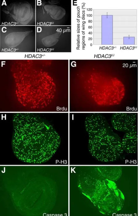
HDAC3 Mutants Have Small Imaginal Discs with Elevated Levels of Apoptosis
Although HDAC1 and HDAC3 appear to functionally overlap in regulating heterochromatin, HDAC3 clearly has a nonredun-dant role in development since allhdac3allelic combinations cause death during late third instar larval and early pupal stages. To investigate the cause of this recessive lethality, wandering third instar larvae bearing transheterozygous combinations of hdac3
point mutations, or point mutations in trans to the smallhdac36C
deficiency, were examined for developmental and tissue defects. We found that imaginal discs in these mutants are significantly reduced in size compared to wild-type (Figure 4A–4D). The pouch region of the wing disc appears particularly small and quantitative measurements revealed reductions to about 30% of normal size (Figure 4E). The small disc phenotype was also observed inhdac36C
homozygotes bearing aP[snr1+]transgene to coversnr1loss.
The small disc phenotype could result from decreased prolifer-ation or increased levels of apoptosis during development. To assess proliferation, discs fromhdac3mutant larvae were immunostained with anti-BrdU to detect DNA replication and with anti-phospho-histone H3 to detect mitotic cells. Although the discs are smaller than wild-type, the relative densities of signals with these S- and M-phase markers did not appear significantly altered (Figure 4F–4I). To track apoptosis,hdac3mutant discs were immunostained with antibody against activated caspase-3. In contrast to wild-type wing discs, which show little apoptosis (Figure 4J),hdac3 mutant wing discs display robust and widespread apoptosis (Figure 4K). In general, apoptosis in thehdac3mutants is more prominent in the
wing blade versus the notal portion of the disc and the precise patterns of apoptotic cells can vary considerably from disc to disc (Figure S1A and S1B). Thus, the main cause of reduced imaginal disc size inhdac3mutants is abnormally elevated apoptosis rather than reduced cell proliferation.
HDAC3 Loss InducesHidExpression and Apoptosis through a Cell-Autonomous Mechanism
TheDrosophilacell death regulatory pathway contains numerous components that are conserved in mammalian systems ([43,44] for reviews). These include initiator and effector caspases, inhibitor of apoptosis proteins (IAPs) which bind caspases, and pro-apoptotic proteins which interfere with IAP function. In the fly pathway, control is frequently exerted at the level of pro-apoptotic protein expression, where up-regulation of factors such as HID, RPR and GRIM promotes caspase-dependent cell death [45]. These correspond to SMAC/DIABLO regulatory factor in mammals [46]. To determine if HDAC3 loss leads to caspase activation via this conserved pathway, we performed in situ hybridizations to track levels ofhidmRNA. As shown in Figure 5A and 5B, there is a dramatic increase in hid mRNA accumulation in hdac3 mutant wing discs compared to wild-type.
In addition to these pro-apoptotic proteins, there are also cell-extrinsic cues that influence apoptosis through death receptor signaling. For example, tumor necrosis factor-alpha (TNF-a)
signaling is implicated in apoptotic control in both flies and mammals [43,47]. To address whether cell-extrinsic mechanisms are involved in apoptotic induction in hdac3 mutants, we first performed in situ hybridization to assess levels of TNF-a (eiger)
mRNA, which revealed no significant change (Figure 5C and 5D). Second, we generated somatic clones ofhdac3mutant cells within
Figure 3. Comparisons ofhdac1andhdac3mRNA expression during fly development.Panels show fly embryos and tissues after in situ hybridizations performed usinghdac1(A–H) orhdac3(I–P) probes. Panels (A–E) and (I–M) show successive stages during embryogenesis, (F) and (N) show larval brain and central nervous systems, (G) and (O) show wing imaginal discs, and (H) and (P) show ovarioles with successive stages of oogenesis.
doi:10.1371/journal.pgen.1000009.g003
imaginal discs and immunostained these discs for activated caspase. The tight correspondence between territories of hdac3
mutant cells, as visualized by GFP-negative regions, and regions of caspase activation (Figure 5E–5H) indicates that HDAC3 loss triggers apoptosis through a cell-autonomous mechanism. Taken together, these results suggest that HDAC3 impacts the cell-intrinsic apoptotic control pathway at a position upstream ofhid
mRNA accumulation. Further studies will be needed to identify
the precise protein substrate(s) targeted by HDAC3 in this pathway. One possibility is thathid transcription is up-regulated through hyper-acetylation of histones associated with hid gene chromatin. Alternatively, there may be non-histone proteins targeted by HDAC3 whose acetylation state can regulate the apoptotic pathway. The observation that fly and vertebrate HDAC3 are found in both the nucleus and cytoplasm [22,23,25] is consistent with either possibility.
Figure 4. Reduced size inhdac3mutant wing discs is due to ectopic apoptosis.(A) Third instar larval wing imaginal disc from ahdac3+/2 heterozygote stained with anti-Armadillo antibody and photographed at the same magnification as the wing disc from ahdac3I/hdac3J
mutant larva shown in (B). (C)hdac3+/2leg disc. (D)hdac3I
/hdac3Jmutant leg disc. (E) Comparison of pouch region sizes of wing imaginal discs fromhdac3+/2 heterozygotes andhdac3I/hdac3J
mutants. Eight individual wing discs of each genotype were stained with anti-Armadillo antibody, photographed, pouch regions were framed, and their two-dimensional areas were computed and analyzed by Student t-test. Error bars are standard deviation and p ,0.001. (F–K) show comparisons ofhdac3+/2wing discs (F, H and J) tohdac3I/hdac3J
mutant discs (G, I and K). (F and G) Discs were labelled and immunostained for bromodeoxyuridine (BrdU) to track cells transiting S phase. (H and I) Discs were immunostained with anti-phospho-histone H3 antibody to reveal mitotic cells. (J and K) Discs were immunostained with anti-activated caspase-3 antibody to reveal apoptotic cells. Very little apoptosis occurs inhdac3+/2heterozygous wing discs whereas robust ectopic apoptosis is seen in large portions ofhdac3I/hdac3J
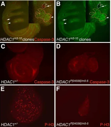
Analysis of Apoptosis and Proliferation in HDAC1 Mutant Wing Discs
To compare effects of hdac3 versus hdac1 loss-of-function in imaginal discs, we analyzed disc size, apoptosis, and proliferation in wing discs from hdac1 mutant larvae. Initially, we sought to analyze whole wing discs from the severe loss-of-function hdac1
mutants,m3-10andm5-5. However, these homozygous mutants, and them3-10/m5-5transheterozygote, failed to survive beyond the end of the second larval instar, precluding analysis of mature larvae. To circumvent this problem, we pursued two approaches: 1) we analyzed mosaic wing discs bearing m3-10 mutant clones and 2) we used a hypomorphic hdac1 allele, l(3)04556, which contains a P element insertion into the 59-UTR [30,32].
As shown in Figure 6A and 6B,hdac1m3-10wing disc clones are significantly smaller than the hdac3 clones described above (Figure 5). In the example outlined at higher magnification (Figure 6A), several features are illustrated. First, the m3-10
homozygous mutant clone is much smaller than its wild-type twinspot. Second, a subset of cells in the clone stain with anti-activated caspase-3, indicating some degree of apoptotic induction. However, numerous m3-10 clones of similar size which lacked detectable caspase-3 were also observed. This variability, together with the dramatic difference in hdac1 versus hdac3 clone sizes, suggests that apoptosis alone is unlikely to account for the small sizes ofhdac1clones. Since previous studies have shown thathdac1
knockdown impairs cell cycle progression in cultured fly cells [29], we suspected that the observed hdac1 clone sizes may result primarily from defects in proliferation rather than abnormal apoptosis.
To further address these possibilities, we analyzed whole discs isolated from a strong hypomorphic allelic combination,
hdac1P[04556]/hdac1m5-5. These transheterozygous larvae live until pupal stages. Figure 6C–6F illustrates that wing discs from this
hdac1 strong hypomorph are dramatically reduced in size
compared to wild-type. Thesehdac1 discs are also smaller than thehdac3mutant discs described above (Figure 4). When stained for activated caspase-3, thishdac1mutant shows sporadic isolated cells and small clusters of cells undergoing apoptosis (Figure 6D). This limited distribution differs from the robust and widespread apoptosis seen inhdac3mutant discs (Figure 4K and Figure S1A and S1B). When thehdac1mutant is stained for a mitotic marker (Figure 6F), fewer mitotic cells are detected at a reduced density compared to heterozygous control discs (Figure 6E). Taken together, the results suggest thathdac1mutant imaginal discs are reduced in size primarily because of a proliferation defect whereas the major defect inhdac3discs is ectopic apoptosis.
HDAC Inhibition and Apoptotic Control in Cancer
There is significant interest in deciphering the molecular consequences of HDAC loss because HDAC chemical inhibitors
Figure 5. Induction of apoptosis inhdac3mutant wing discs is cell-autonomous and accompanied byhidmRNA induction.(A–D) show in situ hybridizations to track the indicated mRNAs in imaginal discs fromhdac3+/2heterozygotes (A and C) versus fromhdac3I
/hdac3Jmutants (B and D). Hybridization probes detecthidmRNA (A and B) orTNF-amRNA (C and D). (E–H)hdac3Ihomozygous mutant clones were generated in a field ofhdac3+/hdac3I
heterozygous wing disc tissue.hdac3Imutant clones are GFP-negative. (F) Apoptotic cells detected with anti-activated caspase-3 antibody (red) are confined tohdac3I
homozygous mutant tissue. The green channel of the optic section (F) is shown alone in (E) to display GFP-negative regions. (G and H) Examples ofhdac3Imutant clones at higher magnification.
doi:10.1371/journal.pgen.1000009.g005
show potential as anticancer agents in preclinical studies with cell and animal models [48,49]. Consistent withhdac3genetic loss-of-function described here, chemical HDAC inhibitors induce tumor cell killing through robust activation of apoptosis [50,51]. Remarkably, HDAC inhibitors promote apoptotic cell death selectively in tumor cells versus in non-transformed cells [50]. However, the molecular mechanisms that connect HDAC inhibition to mammalian cell death are not yet clear. Previous studies have described up-regulation of cell-extrinsic death receptor pathways as well as effects upon components in intrinsic pathways that operate through mitochondria to trigger apoptosis [50]. Although the sum of the evidence suggests multiple inputs, the cell autonomy of the response in the fly system (Figure 5) suggests that cell-intrinsic regulation of pro-apoptotic factors makes a key contribution. Besides pro-apoptotic factors such as hid/Smac/Diablo, the Bcl2 class of cell-intrinsic regulators are commonly used in the mammalian pathway [52]. However, relatively little is known about regulatory mechanisms of the Bcl2 factors Buffy and Debcl inDrosophila[44].
It is also currently unclear which of the 11 human zinc-dependent HDACs must be inhibited to trigger tumor cell apoptosis. Many of the commonly used HDAC inhibitors such as trichostatin (TSA) are broad spectrum antagonists that affect all family members [50]. Indeed, encouraging preclinical results have been obtained with suberoylanilide hydroxamic acid, which affects all eleven human HDACs, and with valproic acid, which inhibits eight of the eleven including all class I enzymes [48–50,53–55]. Our data show that loss of HDAC3 alone is sufficient to trigger robust apoptosis in fly tissues, indicating that this conserved HDAC plays a substantial role among family members in apoptotic control. Further studies will be needed to assess if specific inhibition of human HDAC3 makes a critical contribution to tumor cell killing.
Division of Labor and Functional Roles of HDAC1 and HDAC3
A recent transcription profile microarray study using cultured fly cells suggests that HDAC1 and HDAC3 are the only two fly HDACs with major functions in transcriptional control [29].
Figure 6. Analysis ofhdac1mutant wing discs and clones for effects upon apoptosis and proliferation.(A,B)hdac1m3-10homozygous mutant clones were generated in a field ofhdac1+/hdac1m3-10
heterozygous wing disc tissue. (A) Wing disc showing a GFP-negativehdac1m3-10
mutant clone (arrow) and its wild- type twinspot clone marked by strong GFP expression (arrowhead). These clones are highlighted at higher magnification in the inset (upper right corner). A subset of cells within thehdac1m3-10
mutant clone (arrow) stain with anti-activated caspase-3 antibody (red). This mutant clone is much smaller than its wild-type twinspot clone (arrowhead). (B) The green channel images of the optic sections in (A) are shown alone in (B) to better display GFP-negative regions. Large central regions of the disc that appear unstained are out of the focal plane. (C) Control wing disc from a heterozygoushdac1+/2late wandering third instar larva stained with anti-activated caspase-3 antibody shows very few apoptotic cells. (D)hdac1P[04556]/m5-5mutant wing disc (left) and haltere disc (right) showing scattered apoptotic cells (red) stained with anti-activated
caspase-3 antibody. (E,F) Wing discs were immunostained with anti-phospho-histone H3 antibody (red) to reveal mitotic cells. The density of mitotic cells in the heterozygoushdac1+/2disc (E) is greater than in thehdac1P[04556]/m5-5
disc (F). Panels C–F also illustrate thathdac1mutant wing discs are greatly reduced in size compared to control wing discs. All images besides the insets in A,B were taken at the same magnification.
Thus, HDAC3, the closest fly paralog by sequence, is also likely to be the most functionally related fly family member to HDAC1. Our isolation ofhdac3mutations provided the opportunity to begin to assess this HDAC1/HDAC3 relationship in vivo. Our results show that both hdac3 and hdac1 mutations can suppress PEV (Figure 2), indicating roles for both HDACs in heterochromatin regulation. The fact that anhdac1;hdac3double mutant displays an enhanced effect (Figure 2I) suggests that both HDACs make significant contributions to this process. The simplest molecular explanation is that both enzymes directly deacetylate histone residues that then become methylated in heterochromatin. However, RNA interference experiments using cultured fly S2 cells suggest that HDAC1 is the predominant histone-modifying enzyme, with little unique contribution detected from HDAC3, at least in this cell type [29]. Thus, HDAC3 function at heterochro-matin could reflect deacetylation of either histone or non-histone substrates.
In a developmental context, requirements for HDAC3 function distinct from HDAC1 could occur at times or in cell types that accumulate HDAC3 but not HDAC1. However, the spatial distributions of hdac3 and hdac1 mRNAs are both widespread (Figure 3), their temporal profiles during development are similar [25], and we have not detected individual tissues or cell types wherehdac3product accumulates withouthdac1. In general,hdac1
mRNA levels appear more abundant than those ofhdac3especially in the CNS (Figure 3). This is consistent with the report that the genome-wide transcriptional response to HDAC1 knockdown in cultured fly cells is more robust and involves a larger number of affected genes as compared to HDAC3 knockdown [29]. Thus, HDAC1 may be needed for certain processes that do not require HDAC3. Indeed, both our in vivo results (Figure 6), and fly S2 cell studies [29], support a preferential role for HDAC1, as opposed to HDAC3, in controlling cell proliferation.
The most significant HDAC3 function revealed by our genetic approach is in control of cell death (Figures 4 and 5). Since HDAC3 loss is by itself sufficient to trigger ectopic apoptosis, neither HDAC1 nor other fly HDACs can substitute for this requirement, at least in imaginal disc tissue. These results contrast with findings on apoptosis from an HDAC knockdown study using cultured fly cells. Although treatment with a broad-specificity HDAC inhibitor, trichostatin, did induce apoptosis in S2 cells, neither HDAC1 nor HDAC3 knockdown, nor the double knockdown, affected cell viability [29]. One possible explanation for this discrepancy is that the degree of hdac3 loss-of-function produced by our mutations in vivo is more severe than the degree achieved by RNA interference. Alternatively, the conflicting results may reflect tissue differences in the response to HDAC loss; S2 cells are derived from embryonic neuronal cells whereas the most dramatic induction of apoptosis is seen in a larval epithelial tissue, imaginal discs. Indeed, we note that there is little apoptotic induction in nervous system tissue of the same larvae that display robust induction in discs (Figure S1). Further studies will be needed to determine the mechanisms and pathways by which HDACs control apoptosis in various tissues during normal development as well as in mammalian cell and animal models for cancer.
Materials and Methods
Isolation and Characterization ofHdac3Mutant Alleles
Thehdac36Cdeletion was generated by imprecise excision of a P element, P[PZ ry+]Snr101319, inserted into the neighboring snr1
gene. After introduction of a P transposase source, potential local deficiencies were recovered as ry2 progeny that failed to
complement lethality of the original P insertion. PCR analysis of genomic DNA from 23 independent balanced lethal lines, using
snr1 and hdac3 primers, identified a single line bearing an
approximately 1.8 kb local deletion. Gel-isolation and sequencing of the altered PCR product defined a deletion that removed about two-thirds of thehdac3coding region (Figure 1C) plus allsnr1DNA 39to the original P insertion.
The fourhdac3point mutations were isolated based upon failure to complement recessive lethality ofhdac36C. Homozygous males of genotypeP(ry+t7.2
= neoFRT)82B ry506 were treated with EMS as
described [56] and mutagenized third chromosomes were balanced over TM6B, Tb Sb. Males bearing the mutagenized chromosomes were mated to females of genotype w2; hdac36C
P[Snr1+ w+]/TM6B,Tb and progeny were scored for absence of
viable non-Tubby pupae. The presence of the P[Snr1+] rescue
construct [36], kindly provided by Andrew Dingwall and recombined onto the hdac36C chromosome, facilitated recovery of hdac3lethal mutations. Four lethal mutations were recovered from 5000 matings, siblings were used to establish balanced lethal stocks, and test crosses confirmed that each mutation is lethal in trans tohdac36C. The four mutant lesions were identified by PCR sequencing of genomic DNA isolated from adult flies heterozygous for each mutation.
Tests for Suppression of Position Effect Variegation
To test the four EMS-inducedhdac3alleles, females of genotype
In(1)wm4/In(1)wm4; P(ry+t7.2
= neoFRT)82B ry506/P(ry+t7.2
= -neoFRT)82B ry506 were mated to males of genotype P(ry+t7.2
=
-neoFRT)82B ry506 hdac3/TM3, Sb and the non-Stubble male
progeny were scored for eye color phenotypes. These progeny are heterozygous for thehdac3 allele and essentially isogenic for the remainder of the third chromosome to minimize background effects. Additionalhdac mutants were tested by mating In(1)wm4;
FRT82Bfemales to males of genotypehdac1m5-5/TM3,hdac1m3-10/ TM3,hdac36C/TM3 orhdac1m3-10hdac36C/TM3. The latter stock contains a recombinant third chromosome bearing mutations in bothhdac1andhdac3. Thesehdac1alleles were isolated previously as members of complementation groupl(3)64Cc in a deficiency non-complementation screen [57].
Generation ofHdac3andHdac1Somatic Clones
Forhdac3clones, flies of genotypeFRT82B hdac3I/TM6B,Tbor
FRT82B hdac3J/ TM6B,Tbwere crossed toyw hsFlp;FRT82B ubi-GFP flies at 25uC. For hdac1 clones, flies of genotype FRT2A hdac1m3-10/TM6B,Tbwere crossed tohsFlp;FRT2A ubi-GFPflies. Progeny of these crosses were heat shocked as first instar larvae at 37uC for 1.5 hours to induce expression of FLP recombinase and mitotic recombination. Larvae were then reared at 25uC for 72 hours prior to dissection and immunostaining. Somatic clones homozygous for the hdac mutations were detected as regions lacking GFP expression.
Immunostaining of Larval Tissues
Wandering third instar larvae were dissected in PBS (137 mM NaCl, 2.7 mM KCl, 4.3 mM Na2HPO4 and 1.4 mM KH2PO4,
pH 7.4), and tissues were fixed with 3.7% formaldehyde in PBS for one hour at room temperature and then washed three times in PBS plus 0.1% Triton X-100 (PBT). Mouse anti-armadillo and mouse anti-BrdU antibodies were obtained from the Develop-mental Studies Hybridoma Bank (University of Iowa) and were used at 1/100 dilution. Rabbit anti-phosphorylated histone H3 (pSer10, Sigma) was used at 1/400 dilution. Rabbit anti-activated caspase-3 antibody was obtained from Idun Pharmaceuticals and used at 1/2000 dilution. Primary antibody incubations were
performed overnight at 4uC after dilution into PBT. Anti-rabbit and anti-mouse fluorescent conjugated secondary antibodies (Molecular Probes) were diluted 1/200 in PBT and incubated with tissue samples at room temperature for 2 hours. Confocal images were taken with a Zeiss Axioplan2 confocal microscope.
BrdU Incorporation
Larval central nervous systems and imaginal discs were dissected in PBS, transferred to M3 complete medium containing 0.4 mg/ml BrdU (Roche), and incubated at 25uC for 30 minutes to allow incorporation. Tissues were then fixed as above for immunostaining, incubated in 2N HCl for 30 minutes at room temperature, and then washed three times in PBT before addition of anti-BrdU monoclonal antibody.
In Situ Hybridization
Antisense probes were prepared by in vitro transcription with digoxygenin uridine triphosphate using linearized plasmids bearing the relevant cDNA inserts as template. Thehid, TNF-a
andhdac3probes were generated using cDNAs obtained from the
Berkeley Drosophila Genome Project (http://www.fruitfly.org/ DGC/) and thehdac1probe was derived from a full-length cDNA clone, pBS-Rpd3, obtained from Pierre Spierer. Sense probes of similar lengths, which were prepared and hybridized in parallel, yielded no signals above background. Whole mount in situ hybridizations and signal detection were performed as described [58].
Supporting Information
Figure S1 Apoptotic induction in hdac3 mutant larval tissues. All panels show tissues immunostained with anti-activated caspase-3
antibody. (A–B) Examples of wing imaginal discs from hdac3I/ hdac3J mutants to illustrate variable patterns of apoptosis. Apoptotic cells occur more frequently in the wing blade region than in the portion that will give rise to notum. (C and D) Portions of brain lobe (br), eye (ed), and antennal (ad) imaginal discs from hdac3+/2heterozygous (C) and hdac3I/hdac3J mutant (D) larvae. Very few cells are labeled either in developing brain lobes (br) or eye and antenna imaginal discs of hdac3+/2heterozygous third instar larvae. Apoptotic induction in the hdac3 mutant is seen in portions of the eye and antennal discs (arrows and arrow heads) but not in the brain lobes. (E and F) Haltere (hd) and leg (ld) imaginal discs from hdac3+/2heterozygous (E) and hdac3I/hdac3J mutant (F) larvae. Very few apoptotic cells are seen in control discs from heterozygotes whereas robust apoptosis is evident throughout the leg disc (arrows) from the hdac3 mutant.
Found at: doi:10.1371/journal.pgen.1000009.s001 (5.18 MB TIF)
Acknowledgments
We thank Tom Hays for providing thehdac1fly mutants used in this study, Andrew Dingwall for supplying fly transformants bearing the P[snr1+] rescue construct, and Pierre Spierer for providing thehdac1cDNA. We also thank David Umulis for help with computing wing disc pouch region areas. We are grateful to Tom Neufeld and members of the Simon and O’Connor labs for useful discussions.
Author Contributions
Conceived and designed the experiments: CZ DB MO JS. Performed the experiments: CZ DB DZ EM MO JS. Analyzed the data: CZ DB MO JS. Contributed reagents/materials/analysis tools: CZ DB MO JS. Wrote the paper: CZ DB MO JS.
References
1. Kurdistani SK, Grunstein M (2003) Histone acetylation and deacetylation in yeast. Nat Rev Mol Cell Biol 4: 276–284.
2. Ng HH, Bird A (2000) Histone deacetylases: silencers for hire. Trends Biochem Sci 25: 121–126.
3. Agalioti T, Lomvardas S, Parekh B, Yie J, Maniatis T, et al. (2000) Ordered recruitment of chromatin modifying and general transcription factors to the IFN-beta promoter. Cell 103: 667–678.
4. Hassan AH, Neely KE, Workman JL (2001) Histone acetyltransferase complexes stabilize swi/snf binding to promoter nucleosomes. Cell 104: 817–827. 5. Robyr D, Suka Y, Xenarios I, Kurdistani SK, Wang A, et al. (2002) Microarray
deacetylation maps determine genome-wide functions for yeast histone deacetylases. Cell 109: 437–446.
6. Kadosh D, Struhl K (1998) Histone deacetylase activity of Rpd3 is important for transcriptional repression in vivo. Genes Dev 12: 797–805.
7. Rundlett SE, Carmen AA, Suka N, Turner BM, Grunstein M (1998) Transcriptional repression by UME6 involves deacetylation of lysine 5 of histone H4 by RPD3. Nature 392: 831–835.
8. Suka N, Suka Y, Carmen AA, Wu J, Grunstein M (2001) Highly specific antibodies determine histone acetylation site usage in yeast heterochromatin and euchromatin. Mol Cell 8: 473–479.
9. Zhang Y, Sun ZW, Iratni R, Erdjument-Bromage H, Tempst P, et al. (1998) SAP30, a novel protein conserved between human and yeast, is a component of a histone deacetylase complex. Mol Cell 1: 1021–1031.
10. Zhang Y, Ng HH, Erdjument-Bromage H, Tempst P, Bird A, et al. (1999) Analysis of the NuRD subunits reveals a histone deacetylase core complex and a connection with DNA methylation. Genes Dev 13: 1924–1935.
11. Wade PA, Gegonne A, Jones PL, Ballestar E, Aubry F, et al. (1999) Mi-2 complex couples DNA methylation to chromatin remodelling and histone deacetylation. Nat Genet 23: 62–66.
12. Xu L, Glass CK, Rosenfeld MG (1999) Coactivator and corepressor complexes in nuclear receptor function. Curr Opin Genet Dev 9: 140–147.
13. Tsai CC, Kao HY, Yao TP, McKeown M, Evans RM (1999) SMRTER, a Drosophila nuclear receptor coregulator, reveals that EcR-mediated repression is critical for development. Mol Cell 4: 175–186.
14. Nomura T, Khan MM, Kaul SC, Dong HD, Wadhwa R, et al. (1999) Ski is a component of the histone deacetylase complex required for transcriptional repression by Mad and thyroid hormone receptor. Genes Dev 13: 412– 423.
15. Huang Y, Myers SJ, Dingledine R (1999) Transcriptional repression by REST: recruitment of Sin3A and histone deacetylase to neuronal genes. Nat Neurosci 2: 867–872.
16. Roopra A, Sharling L, Wood IC, Briggs T, Bachfischer U, et al. (2000) Transcriptional repression by neuron-restrictive silencer factor is mediated via the Sin3-histone deacetylase complex. Mol Cell Biol 20: 2147–2157. 17. Chen G, Fernandez J, Mische S, Courey AJ (1999) A functional interaction
between the histone deacetylase Rpd3 and the corepressor groucho in Drosophila development. Genes Dev 13: 2218–2230.
18. Lai EC (2002) Keeping a good pathway down: transcriptional repression of Notch pathway target genes by CSL proteins. EMBO Rep 3: 840–845. 19. Chang YL, Peng YH, Pan IC, Sun DS, King B, et al. (2001) Essential role of
Drosophila Hdac1 in homeotic gene silencing. Proc Natl Acad Sci U S A 98: 9730–9735.
20. Tie F, Furuyama T, Prasad-Sinha J, Jane E, Harte PJ (2001) The Drosophila Polycomb Group proteins ESC and E(Z) are present in a complex containing the histone-binding protein p55 and the histone deacetylase RPD3. Development 128: 275–286.
21. Gregoretti IV, Lee YM, Goodson HV (2004) Molecular evolution of the histone deacetylase family: functional implications of phylogenetic analysis. J Mol Biol 338: 17–31.
22. Takami Y, Nakayama T (2000) N-terminal region, C-terminal region, nuclear export signal, and deacetylation activity of histone deacetylase-3 are essential for the viability of the DT40 chicken B cell line. J Biol Chem 275: 16191–16201. 23. Barlow AL, van Drunen CM, Johnson CA, Tweedie S, Bird A, et al. (2001)
dSIR2 and dHDAC6: two novel, inhibitor-resistant deacetylases in Drosophila melanogaster. Exp Cell Res 265: 90–103.
24. Hubbert C, Guardiola A, Shao R, Kawaguchi Y, Ito A, et al. (2002) HDAC6 is a microtubule-associated deacetylase. Nature 417: 455–458.
25. Cho Y, Griswold A, Campbell C, Min KT (2005) Individual histone deacetylases in Drosophila modulate transcription of distinct genes. Genomics 86: 606–617. 26. Ito A, Kawaguchi Y, Lai CH, Kovacs JJ, Higashimoto Y, et al. (2002) MDM2-HDAC1-mediated deacetylation of p53 is required for its degradation. Embo J 21: 6236–6245.
27. Huo X, Zhang J (2005) Important roles of reversible acetylation in the function of hematopoietic transcription factors. J Cell Mol Med 9: 103–112. 28. Yao YL, Yang WM, Seto E (2001) Regulation of transcription factor YY1 by
29. Foglietti C, Filocamo G, Cundari E, De Rinaldis E, Lahm A, et al. (2006) Dissecting the biological functions of Drosophila histone deacetylases by RNA interference and transcriptional profiling. J Biol Chem 281: 17968–17976. 30. Mannervik M, Levine M (1999) The Rpd3 histone deacetylase is required for
segmentation of the Drosophila embryo. Proc Natl Acad Sci U S A 96: 6797–6801.
31. Perrimon N, Lanjuin A, Arnold C, Noll E (1996) Zygotic lethal mutations with maternal effect phenotypes in Drosophila melanogaster. II. Loci on the second and third chromosomes identified by P-element-induced mutations. Genetics 144: 1681–1692.
32. Mottus R, Sobel RE, Grigliatti TA (2000) Mutational analysis of a histone deacetylase in Drosophila melanogaster: missense mutations suppress gene silencing associated with position effect variegation. Genetics 154: 657–668.
33. De Rubertis F, Kadosh D, Henchoz S, Pauli D, Reuter G, et al. (1996) The histone deacetylase RPD3 counteracts genomic silencing in Drosophila and yeast. Nature 384: 589–591.
34. Vannini A, Volpari C, Filocamo G, Casavola EC, Brunetti M, et al. (2004) Crystal structure of a eukaryotic zinc-dependent histone deacetylase, human HDAC8, complexed with a hydroxamic acid inhibitor. Proc Natl Acad Sci U S A 101: 15064–15069.
35. Somoza JR, Skene RJ, Katz BA, Mol C, Ho JD, et al. (2004) Structural snapshots of human HDAC8 provide insights into the class I histone deacetylases. Structure 12: 1325–1334.
36. Zraly CB, Marenda DR, Nanchal R, Cavalli G, Muchardt C, et al. (2003) SNR1 is an essential subunit in a subset of Drosophila brm complexes, targeting specific functions during development. Dev Biol 253: 291–308.
37. Reuter G, Spierer P (1992) Position effect variegation and chromatin proteins. Bioessays 14: 605–612.
38. Karpen GH (1994) Position-effect variegation and the new biology of heterochromatin. Curr Opin Genet Dev 4: 281–291.
39. Rea S, Eisenhaber F, O’Carroll D, Strahl BD, Sun ZW, et al. (2000) Regulation of chromatin structure by site-specific histone H3 methyltransferases. Nature 406: 593–599.
40. Czermin B, Schotta G, Hulsmann BB, Brehm A, Becker PB, et al. (2001) Physical and functional association of SU(VAR)3–9 and HDAC1 in Drosophila. EMBO Rep 2: 915–919.
41. Tartof KD, Hobbs C, Jones M (1984) A structural basis for variegating position effects. Cell 37: 869–878.
42. Kuzmichev A, Nishioka K, Erdjument-Bromage H, Tempst P, Reinberg D (2002) Histone methyltransferase activity associated with a human multiprotein complex containing the Enhancer of Zeste protein. Genes Dev 16: 2893–2905. 43. Hay BA, Huh JR, Guo M (2004) The genetics of cell death: approaches, insights
and opportunities in Drosophila. Nat Rev Genet 5: 911–922.
44. Hay BA, Guo M (2006) Caspase-dependent cell death in Drosophila. Annu Rev Cell Dev Biol 22: 623–650.
45. Bergmann A, Yang AY, Srivastava M (2003) Regulators of IAP function: coming to grips with the grim reaper. Curr Opin Cell Biol 15: 717–724.
46. Shi Y (2002) Mechanisms of caspase activation and inhibition during apoptosis. Mol Cell 9: 459–470.
47. Lavrik I, Golks A, Krammer PH (2005) Death receptor signaling. J Cell Sci 118: 265–267.
48. Johnstone RW (2002) Histone-deacetylase inhibitors: novel drugs for the treatment of cancer. Nat Rev Drug Discov 1: 287–299.
49. Minucci S, Pelicci PG (2006) Histone deacetylase inhibitors and the promise of epigenetic (and more) treatments for cancer. Nat Rev Cancer 6: 38–51. 50. Bolden JE, Peart MJ, Johnstone RW (2006) Anticancer activities of histone
deacetylase inhibitors. Nat Rev Drug Discov 5: 769–784.
51. Marks PA, Jiang X (2005) Histone deacetylase inhibitors in programmed cell death and cancer therapy. Cell Cycle 4: 549–551.
52. Jiang X, Wang X (2004) Cytochrome C-mediated apoptosis. Annu Rev Biochem 73: 87–106.
53. Insinga A, Monestiroli S, Ronzoni S, Gelmetti V, Marchesi F, et al. (2005) Inhibitors of histone deacetylases induce tumor-selective apoptosis through activation of the death receptor pathway. Nat Med 11: 71–76.
54. Drummond DC, Noble CO, Kirpotin DB, Guo Z, Scott GK, et al. (2005) Clinical development of histone deacetylase inhibitors as anticancer agents. Annu Rev Pharmacol Toxicol 45: 495–528.
55. Egger G, Liang G, Aparicio A, Jones PA (2004) Epigenetics in human disease and prospects for epigenetic therapy. Nature 429: 457–463.
56. Lewis EB, Bacher F (1968) A method of feeding ethyl methanesulfonate (EMS) to Drosophila males. Dros Inf Serv 43: 193.
57. Gepner J, Li M, Ludmann S, Kortas C, Boylan K, et al. (1996) Cytoplasmic dynein function is essential in Drosophila melanogaster. Genetics 142: 865–878. 58. Ross JJ, Shimmi O, Vilmos P, Petryk A, Kim H, et al. (2001) Twisted gastrulation is a conserved extracellular BMP antagonist. Nature 410: 479–483. 59. Johnson CA, Barlow AL, Turner BM (1998) Molecular cloning of Drosophila melanogaster cDNAs that encode a novel histone deacetylase dHDAC3. Gene 221: 127–134.