Oficial Journal of the Brazilian Psychiatric Association Volume 35 • Number 1 • February/2013
Psychiatry
Revista Brasileira de Psiquiatria
Abstract
Invasion of the central nervous system (CNS) by microorganisms is a severe and frequently fatal event during the course of many infectious diseases. It may lead to deafness, blindness, cerebral palsy, hydrocephalus, cognitive impairment or permanent neurological dysfunction in survivors. Pathogens can cross the blood-brain barrier by transcellular migration, paracellular migration and in infected macrophages. Pathogens may breach the blood-brain barrier and be recognized by antigen-presenting cells through the binding of Toll-like receptors. This induces the activation of nuclear factor kappa B or mitogen-activated protein kinase pathways and subsequently induces leukocyte iniltration and proliferation and the expression of numerous proteins involved in inlammation and the immune response. Many brain cells can produce cytokines, chemokines and other pro-inlammatory molecules in response to bacteria stimuli; as a consequence, polymorphonuclear cells are attracted and activated, and release large amounts of superoxide anion and nitric oxide, leading to peroxynitrite formation and oxidative stress. This cascade leads to lipid peroxidation, mitochondrial damage and blood-brain barrier breakdown, contributing to cellular injury during neuronal infection. Current evidence suggests that bacterial CNS infections can play a role in the etiopathogenesis of behavioral disorders by increasing pro-inlammatory cytokines and bacterial virulence factors. The aim of this review is to summarize the current knowledge of the relevant pathophysiologic steps in CNS infections.
© 2013 Associação Brasileira de Psiquiatria. Published by Elsevier Editora Ltda. All rights reserved.
Pathophysiology of bacterial infection of the
central nervous system and its putative role in
the pathogenesis of behavioral changes
Tatiana Barichello,
1Jaqueline S. Generoso,
1Graziele Milioli,
1Samuel G. Elias,
1Antônio Lúcio Teixeira
21Laboratory of Experimental Microbiology and National Institute of Science and Technology (INCT) for Translational
Medicine, Universidade do Extremo Sul Catarinense, Brazil.
2Laboratory of Medical Investigation, Universidade Federal de Minas Gerais, Brazil.
Received on October 16, 2012; accepted on November 9, 2012
DESCRIPTORS: Bacterial Infection; Behavior;
Central Nervous System; Inlammation;
Meningitis. SPECIAL ARTICLE
Corresponding author: Prof. Tatiana Barichello, PhD. - Laboratório de Microbiologia Experimental, PPGCS,
Universidade do Extremo Sul Catarinense, 88806-000 Criciúma, SC, Brazil. Fax: +55 48 3443 4817. E-mail: [email protected] © 2013 Associação Brasileira de Psiquiatria. Published by Elsevier Editora Ltda. All rights reserved.
Introduction
Bacterial infection of the central nervous system (CNS) is a life-threatening condition with a high rate of mortality. Even with protective barriers, pathogens can reach the CNS.1 The
replication of these microorganisms within the CNS occurs in association with the release of compounds such as
peptido-glycans, cell wall fragments, lipoteichoic acids (from Gram
positive bacteria)2, lipopolysaccharides (from Gram negative
bacteria) and exotoxins, which are highly immunogenic and may elicit an increased inlammatory response in the host.1-4
The host’s inlammatory response against the infection has
been increasingly recognized as playing a central role in neurological morbidity and mortality. CNS infections may present clinically as meningitis, meningoencephalitis and, more rarely, myelitis.5
Cognitive outcomes after bacterial meningitis include impairment of memory, decreased psychomotor performance
and impairment of attention/executive functions.6 In
neona-tal meningitis, the main pathogens are Streptococcus agalac-tiae and Escherichia coli K1, which together are responsible for at least two thirds of all meningitis deaths in Europe.7,8
Listeria monocytogenes,9 also causes meningitis, usually in
immunocompromised individuals and in pregnant women.10
E. coli K1 infection includes cognitive involvement in 50%
of patients.11 The main bacterial agents causing meningitis
in young children and adults are Streptococcus pneumoniae
and Neisseria meningitidis, with the former being responsible
for two-thirds of cases in Europe and the United States.2,12-14
Pneumococcal meningitis leads to cognitive impairment in
30 to 52% of surviving patients,2 including learning dificul
-ties, cognitive slowness, short-term memory deicits and
poor academic performance.15 The aim of this review is to
summarize the current knowledge of the relevant pathophysi-ologic steps of CNS infections: (a) bacterial crossing through
the blood-brain barrier; (b) bacterial survival and growth
within the CNS; (c) bacterial induction of CNS inlammation;
and (d) the role of bacterial infection in the development of behavioral disorders.
Microbial traversal of the blood-brain barrier
The CNS is protected by a bony skull, the leptomeninges, the blood-brain barrier (BBB) and the blood-cerebrospinal
luid barrier.1 The BBB is composed of brain microvascular
endothelial cells, astrocytes and pericytes. It maintains
the neural microenvironment by regulating the flux of
molecules into and out of the brain, and protects the brain
from microorganisms and toxins that come from the blood.3
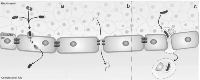
Pathogens can cross the BBB by transcellular migration, para- cellular migration and by “hitch-hiking” inside infected
macrophages. Using transcellular migration, pathogens can
cross the BBB without any evidence of intercellular tight-junction disruption or detection of microorganisms between endothelial cells.16 S. agalactiae, S. pneumoniae, E. coli
and N. meningitidis reach the CNS through this mechanism3
(Figure 1a). The paracellular traversal mechanism involves the penetration of the pathogen between barrier cells, with or without evidence of tight-junction disruption. Borrelia sp
and Treponema pallidum cross the BBB through this
mecha-nism (Figure 1b). Other pathogens can cross the BBB inside
infected macrophages. L. monocytogenes enters host cells during internalization of phagosomal vacuoles, and produces phospholipase C and the pore-forming cytolysin listeriolysin O to escape the phagosome and replicate within the host cyto-sol.17Mycobacterium tuberculosis also crosses the BBB while
residing in the phagosomes of macrophages3,18 (Figure 1c).
Mechanisms of innate immunity in the CNS
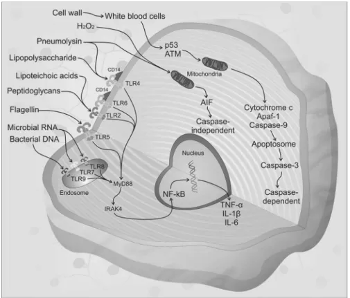
The innate immune response represents the first line of defense against invading microorganisms. Bacterial compounds such as peptidoglycans, cell wall fragments, lipopolysaccharides and lipoteichoic acid, collectively known
as pathogen-associated molecular patterns (PAMPs) are highly immunogenic and elicit strong inlammatory responses in the
host. These compounds are recognized by antigen-presenting cells through binding to pattern recognition receptors (PRRs), including the Toll-like receptors (TLRs). Eleven members of the TLR family have been described to date in humans. TLRs
are either expressed at the cell surface for extracellular
ligand recognition or localized to endosomal compartments for the recognition of pathogen-associated nucleic acids.19
Microglial cells express all TLRs identiied to date, astrocytes express TLR 2, 3 and 9, neurons express TLR 3, 7, 8 and 9 and oligodendrocytes express TLR 2 and 3.19 Most TLRs transduce
their signal through a common intracellular adapter protein
known as myeloid differentiation factor 88 (MyD88).20 MyD88
is associated with Interleukin-1 (IL-1) and receptor-associated
kinase-4 (IRAK-4), which is a serine/threonine kinase that
plays an essential role in signal transduction by Toll/IL-1 receptors (TIRs).21 Subsequently, IRAK interacts with the
tu-mor necrosis factor (TNF) receptor-associated factor (TRAF) family, providing a link to NF-κB-inducing kinase, resulting in the nuclear translocation of NF-κB.22 NF-κB comprises
a closely associated family of transcription factors, which
play a key role in the expression of genes implicated in the
development of accessory cell and lymphocyte populations.23
NF-κB is also a transcriptional activator of various genes
involved in neuronal pathogenesis and in the production of cytokines and chemokines.24,25 IL-1β production requires a
second signal that is provided by intracellular protein
com-plexes known as inlammasomes. The NLRP3 (cryopyrin) and the AIM2 (absent in melanoma 2) inlammasomes, which are activated by the exotoxin pneumolysin and bacterial DNA
in pneumococcal infection, respectively, mediate cleavage
of pro-IL-1β into mature IL-1β.26 NLRP3 has been implicated
in responses to the following bacteria: Staphylococcus au-reus, L. monocytogenes, Klebsiella pneumoniae and E. coli.27
Thus, when bacteria reach the CNS, PAMPs are recognized by
PRRs, initiating the activation of the host immune response,
which triggers the production of pro-inlammatory cytokines and chemokines and the expression of co-stimulatory mol -ecules1 (Figure 2). In response to these stimuli, leukocytes
are attracted from the blood and activated. For instance,
polymorphonuclear leukocytes cross the BBB by binding to
selectins E and P along with IL-8 (CXCL-8). TNF-α induces production of adhesion molecules ICAM-1 and ICAM-2, which allow extravasation of the leukocyte along chemoattractant
concentration gradients.28 Leukocytes work to eliminate the
invading pathogen through a rapid and robust production
of reactive oxygen species (ROS). They release high amounts of superoxide anion (O2-) and nitric oxide (NO), generating peroxynitrite (ONOO-).29 The resulting oxidative stress
leads to activation of cytokines, enhancing leukocyte
activa-tion, lipid peroxidaactiva-tion, mitochondrial damage and metal -loproteinase activation.4,30 In humans with meningococcal
meningitis, TNF-α levels in the serum are correlated with
fatal outcomes.31 TNF-α is released into serum before IL-6
in meningococcal septic shock. High serum levels of IL-6 are also associated with unfavorable outcomes. In
ad-dition, IL-1 was exclusively detected in patients who also had high serum levels of IL-6, TNF-α, LPS and rapid fatal
outcomes.32 However, in the CSF of patients with meningitis,
higher concentrations of TNF-α were observed than patients with septic shock/bacteremia and the CSF/blood glucose was inversely correlated with TNF-α, IL-6 and IL-1.33 Patients with
pneumococcal meningitis also exhibit a cerebral production of TNF-α, IL-1β and IL-6.34
Neuronal damage in the context of bacterial
infection of the CNS
Brain injury and neuronal death are not mediated simply by the presence of a viable pathogen, but they also occur as a consequence of the host’s reaction to bacterial compounds.35
Bacterial cell wall compounds, mainly polysaccharide cap-sules, are most likely the most important virulence factor.
Bacterial cell wall compounds trigger a host inlammatory
response from leukocytes and activate the tumor suppressor
protein p53 and the ataxia telangiectasia-mutated (ATM)
kinase, triggering caspase dependent-apoptosis.36 The
pres-ence of the capsule also prevents the binding of iC3b (a
complement factor that stimulates phagocytosis) and Fc
(which stimulates receptor-mediated phagocytose) to the bacterial cell surface, affecting antiphagocytic activity.37,38
Pathogens such as S. pneumoniae, N. meningitidis, E. coli
K1 and H. inluenzae use this pathogenic mechanism.37,39,40
The virulence of other bacteria is based on the produc-tion of enzymes such as coagulase, proteolytic enzymes, hyaluronidase, neuraminidase and catalase.37,41 Hemolysin
and cytolysin from Streptococcus have the ability to cause
inlammatory activation, and these same enzymes produced
by S. agalactiae induce chemokines and ICAM-1 in brain
microvascular endothelium cells.36 Teichoic acids present in
Gram+ bacteria can induce TNF-α and NO production, as well as the expression of ICAM-1.42 Exotoxins are also produced
by some bacteria as part of their growth and metabolism,39,41
while endotoxins are part of the external portion of Gram
(-) bacteria.39S. pneumoniae bacterial compounds like
pep-tidoglycans and lipoteichoic acids are recognized in the CNS by TLR2.43 The exotoxin pneumolysin is recognized by TLR422
and bacterial DNA is recognized by TLR9, which is an intra
-cellular PRP activated by CpG.44 Pneumolysin and H 2O2 are
produced and released by pneumococcus, causing neuronal cell death through mitochondrial damage.45,46 This damage
leads to the release of apoptosis-inducing factor (AIF) into the cytosol. AIF induces apoptosis by a caspase-independent
pathway.36,46 When pneumococcal cell wall compounds are
released, they activate the host immune response and a
large number of polymorphonuclear cells are attracted. p53 and ATM converge on mitochondria to initiate the release of
cytocrome-c, which is necessary to form the apoptosome
(Apaf-1) and activate caspase-9, resulting in activation of
caspase-3 and caspase-dependent apoptosis.36S. agalactiae
produces lipoteichoic acid that is through to activate TLR2,
while TLR7 and/or TLR8 interact with microbial RNA.2 In
vitro, L. monocytogenes cell wall components such as
lipo-teichoic acids are recognized by TLR2 with the help of CD14
and TLR6.47-50 The protein lagellin is recognized by TLR5.49
L. monocytogenes is a classic example of a cytosol-adapted
pathogen. It can escape quickly from the phagosomes of macrophages (or other cell types) and it reproduces rapidly in the cytosol.51 This microorganism also expresses the toxin
listeriolysin O, which is responsible for forming pores in the cell and causing lymphocyte apoptosis.52E. coli produces the
endotoxin lipopolysaccharide, which is recognized by TLR4.53
Furthermore, E. coli damages the microvascular endothelial cells that constitute the BBB in the human brain.54 Another
important pathogen, S. pyogenes, produces bacterial
viru-lence factors such as, M protein, hyaluronic acid, capsule, ibronectin-binding proteins, streptolysin O and streptolysin S. The M protein binds to complement factors and other host
proteins, allowing the pathogen to avoid activation of the alternate complement pathway evade phagocytosis.55
The M protein has also been implicated in the internaliza -tion process, which involves cytoskeletal rearrangements. It is possible that these proteins provide an intraepithelial refuge for the organism, sheltered from phagocytes and humoral antibodies.56,57 Another intracellular bacterium,
Mycobacterium tuberculosis, infects the CNS and causes
different manifestations of disease, like meningitis, space-occupying lesions in the brain parenchyma, and focal disease of the spinal cord and its osseous structures.58 In previous
studies of S. agalactiae meningitis, oxidative damage was
elevated and enzymatic defense was decreased.59 TNF-α,
IL-1β, IL-6 and cytokine-induced neutrophil chemoattrac
-tant-1 (CINC-1) levels were increased in the irst hours after
neonatal meningitis induction, prior to BBB breakdown in the
hippocampus and cortex.59 In infant rats with
pneumococ-cal meningitis, the peak of pro-inlammatory cytokine and
chemokine production was also associated with a disrup-tion of the BBB.60 TNF-α and CINC-1 levels were increased
in the blood from jugular veins in comparison with arterial
blood in the irst hours after pneumococcal meningitis
induction, indicating their production in the CNS.61 Moreover,
animals that survived bacterial meningitis showed impair-ments of memory and learning, depressive-like-behavior and
anxiety-like symptoms.62-64
Behavioral disorders
Neuronal infection causes intense an inflammatory host
immune response; it is potentially fatal and contributes to
neurological symptoms.1 There are several studies describing
a probable role of cytokines and oxidative stress in behavioral
disorders.65,66 Epidemiological research over the last twenty
years has indicated that the risk for schizophrenia is enhanced
in offspring exposed to viral or bacterial infections in utero,
possibly by a disruption in the programmed maturation of the brain in prenatal and early neonatal life.67 This association
between prenatal exposure to bacterial infection and risk of schizophrenia was evaluated, for example, in the
Copenhagen Perinatal Cohort, which showed that prenatal
exposure to bacterial infections in pregnant women were
correlated with an increased risk of schizophrenia in their offspring.68 It has been proposed that this association might
be mediated through transplacental passage of maternally produced cytokines in response to these bacterial infec-tions.68,69 Individuals with meningitis during childhood had a
increased risk for psychosis in adulthood when compared to their siblings without childhood meningitis.70 The
im-mune system may signal the CNS through the action of cytokines, which have also been implicated in the
develop-ment of schizophrenia. For instance, IL-1β is capable of
inducing the differentiation of mesencephalic progenitor cells into dopaminergic neurons.71,72 IL-6 is highly
effec-tive in decreasing the survival of fetal brain serotonergic neurons73 and is increased in patients with schizophrenia.74
There was a signiicant association between maternal IL-8
levels during the second trimester of pregnancy and the risk of schizophrenia in the offspring.75 Cytokines in the
CNS are also implicated to depressive disorders.65 Animals
subjected to pneumococcal meningitis and sepsis models had increased levels of pro-inflammatory cytokines in parallel with depressive-like behavior.62,76 Interestingly,
treatment with imipramine reversed these depressive-like
symptoms and decreased TNF-α levels in the cortex 10 days
after pneumococcal meningitis induction.77 Impairment
of learning and memory has also been demonstrated in bacterial meningitis in a neonatal animal model. Neonatal Wistar rats subjected to S. agalactiae meningitis showed aversive memory impairment in adulthood and a decrease
of the brain-derived neurotrophic factor (BDNF) levels in the hippocampus and cortex.58 Furthermore, the decreased
BDNF levels in the hippocampus were correlated with memory
impairment in adult animals subjected to pneumococ-cal meningitis in the neonatal period.69 Other neuronal
disorders are also related to bacterial infection.
Obsessive-compulsive disorder or “tic” disorders are examples of
pediatric neuropsychiatric disorders that have been associ-ated with autoimmune disease secondary to S. pyogenes,
a group A β-hemolytic streptococcal infection.78,79 The
involvement of the host immune response is suggested by
quantitative changes in circulating levels of TNF-α, IL-1β
and IL-6 levels in these patients. Pathogenic mechanisms include antibodies targeting the streptococcal epitope
GlcNAc cross-reacting with neuronal molecules like tubu -lin, lysoganglioside and dopamine receptors.80 Cytokines
have an important role in the pathophysiology of brain infections, and they are implicated in a variety of common diseases that have been associated with production of cyto-kines, such as psychiatric disorders.4,65
Conclusion
Understanding the interactions between the complex net
-works of cytokines, other inlammatory mediators and leuko -cytes, and bacterial virulence factors may help to establish more effective therapeutic strategies for CNS infections and,
therefore, a better outcome for affected subjects. Moreover,
these diseases highlight the role of the immune system in the pathophysiology of psychiatric disorders, which represents an interesting research venue.
Acknowledgments
This research was supported by grants from CNPq, FAPESC, FAPEMIG, UNESC, NENASC project (PRONEX program CNPq/ FAPESC), INCT-TM and L´Oréal-UNESCO Brazil Fellowship for
Women in Science 2011.
Disclosures
Tatiana Barichello
Employment: Laboratório de Microbiologia Experimental e Instituto Nacional de Ciência e Tecnologia Translacional em Medicina, Programa de Pós-Graduação em Ciências da Saúde, Universidade do Extremo Sul
Catarinense, Criciúma, SC, Brazil.
Jaqueline S. Generoso
Employment: Laboratório de Microbiologia Experimental e Instituto Nacional de Ciência e Tecnologia Translacional em Medicina, Programa de Pós-Graduação em Ciências da Saúde, Universidade do Extremo Sul
Catarinense, Criciúma, SC, Brazil.
Graziele Milioli
Employment: Laboratório de Microbiologia Experimental e Instituto Nacional de Ciência e Tecnologia Translacional em Medicina, Programa de Pós-Graduação em Ciências da Saúde, Universidade do Extremo Sul
Catarinense, Criciúma, SC, Brazil.
Samuel G. Elias
Employment: Laboratório de Microbiologia Experimental e Instituto Nacional de Ciência e Tecnologia Translacional em Medicina, Programa de Pós-Graduação em Ciências da Saúde, Universidade do Extremo Sul
Catarinense, Criciúma, SC, Brazil.
Antônio Lúcio Teixeira
Employment: Laboratório Interdisciplinar de Investigação Médica, Faculdade de Medicina, Universidade Federal de Minas Gerais, Belo
Horizonte, MG, Brazil.
* Modest
** Signiicant
*** Signiicant. Amounts given to the author's institution or to a colleague for
research in which the author has participation, not directly to the author.
References
1. Sellner J, Tauber MG, Leib SL. Pathogenesis and pathophysiology of bacterial CNS infections. Handb Clin Neurol. 2010;96:1-16. 2. Mook-Kanamori BB, Geldhoff M, van der Poll T, van de Beek D. Pathogenesis and pathophysiology of pneumococcal meningitis. Clin Microbiol Rev. 2011;24:557-91.
3. Kim KS. Mechanisms of microbial traversal of the blood-brain barrier. Nat Rev Microbiol. 2008;6:625-34.
4. Barichello T, Generoso JS, Collodel A, Moreira AP, Almeida SM. Pathophysiology of acute meningitis caused by Streptococcus pneumoniae and adjunctive therapy approaches. Arqu Neuropsiquiatr. 2012;70:366-72.
5. Somand D, Meurer W. Central nervous system infections. Emerg Med Clin North Am. 2009;27:89-100, ix.
6. Hoogman M, van de Beek D, Weisfelt M, de Gans J, Schmand B. Cognitive outcome in adults after bacterial meningitis. J Neurol, Neurosurg, Psychiatry. 2007;78:1092-6.
7. Holt DE, Halket S, de Louvois J, Harvey D. Neonatal meningitis in England and Wales: 10 years on. Arch Dis Child Fetal Neonatal Ed. 2001;84:F85-9.
8. Heath PT, Okike IO, Oeser C. Neonatal meningitis: can we do better? Adv Exp Med Biol. 2011;719:11-24.
9. Roos KL, van de Beek D. Bacterial meningitis. In: PJ Vinken, GW Bruyn, editors. Handbook of clinical neurology. 2010;96:51-63. 10. Pamer EG. Immune responses to Listeria monocytogenes. Nat
Rev Immunol. 2004;4:812-23.
11. Feigin RD. Bacterial meningitis in the newborn infant. Clin Perinatol. 1977;4:103-16.
12. Arda B, Sipahi OR, Atalay S, Ulusoy S. Pooled analysis of 2,408 cases of acute adult purulent meningitis from Turkey. Med Princ Pract. 2008;17:76-9.
13. van de Beek D, de Gans J, Spanjaard L, Weisfelt M, Reitsma JB, Vermeulen M. Clinical features and prognostic factors in adults with bacterial meningitis. New Eng J Med. 2004;351:1849-59. 14. Grandgirard D, Leib SL. Meningitis in Neonates: Bench to
15. Kihara M, de Haan M, Were EO, Garrashi HH, Neville BG, Newton CR. Cognitive deicits following exposure to pneumococcal meningitis: an event-related potential study. BMC Infect Dis. 2012;12:79.
16. Kim KS. Microbial translocation of the blood-brain barrier. Int J Parasitol. 2006;36:607-14.
17. Lopez-Castejon G, Corbett D, Goldrick M, Roberts IS, Brough D. Inhibition of calpain blocks the phagosomal escape of Listeria monocytogenes. PloS one. 2012;7:e35936.
18. Bobadilla K, Sada E, Esther Jaime M, González Y, Ramachandra L, Rojas RE, et al. Human phagosome processing of Mycobacterium tuberculosis antigens is modulated by IFN-gamma and IL-10. Immunol. 2012 Aug; [epub].
19. Hanke ML, Kielian T. Toll-like receptors in health and disease in the brain: mechanisms and therapeutic potential. Clin Sci (Lond). 2011;121:367-87.
20. Kronfol Z, Remick DG. Cytokines and the brain: implications for clinical psychiatry. The Am J Psychiatry. 2000;157:683-94. 21. Ichiyama T, Isumi H, Yoshitomi T, Nishikawa M, Matsubara T,
Furukawa S. NF-kappaB activation in cerebrospinal luid cells from patients with meningitis. Neurol Res. 2002;24:709-12. 22. Malley R, Henneke P, Morse SC, Cieslewicz J, Lipsitch M,
Thompson CM, et al. Recognition of pneumolysin by Toll-like receptor 4 confers resistance to pneumococcal infection. Proc Natl Acad Sci. 2003;100:1966-71.
23. Tato CM, Hunter CA. Host-pathogen interactions: subversion and utilization of the NF-kappa B pathway during infection. Infect Immun. 2002;70:3311-7.
24. Koedel U, Bayerlein I, Paul R, Sporer B, Pister HW. Pharmacologic interference with NF-kappaB activation attenuates central nervous system complications in experimental Pneumococcal meningitis. J Infect Dis. 2000;182:1437-45.
25. Kastenbauer S, Koedel U, Weih F, Ziegler-Heitbrock L, Pister HW. Protective role of NF-kappaB1 (p50) in experimental pneumococcal meningitis. Eur J Pharmacol. 2004;498:315-8. 26. Koppe U, Suttorp N, Opitz B. Recognition of Streptococcus
pneumoniae by the innate immune system. Cell Microbiol. 2012;14:460-6.
27. Davis BK, Wen H, Ting JP. The inlammasome NLRs in immunity, inlammation, and associated diseases. Annu Rev Immunol. 2011;29:707-35.
28. Granert C, Raud J, Xie X, Lindquist L, Lindbom L. Inhibition of leukocyte rolling with polysaccharide fucoidin prevents pleocytosis in experimental meningitis in the rabbit. J Clin Investig. 1994;93:929-36.
29. Klein M, Koedel U, Pister HW. Oxidative stress in pneumococcal meningitis: a future target for adjunctive therapy? Prog Neurobiol. 2006;80:269-80.
30. Sellner J, Täuber MG, Leib SL. Chapter 1 - Pathogenesis and pathophysiology of bacterial CNS infections. In: Karen LR and Allan RT, editors. Handbook of Clinical Neurology. Elsevier; 2010. p. 1-16.
31. Waage A, Halstensen A, Espevik T. Association between tumour necrosis factor in serum and fatal outcome in patients with meningococcal disease. Lancet. 1987;1:355-7.
32. Waage A, Brandtzaeg P, Halstensen A, Kierulf P, Espevik T. The complex pattern of cytokines in serum from patients with meningococcal septic shock. Association between interleukin 6, interleukin 1, and fatal outcome. J Exp Med. 1989;169:333-8. 33. Waage A, Halstensen A, Shalaby R, Brandtzaeg P, Kierulf
P, Espevik T. Local production of tumor necrosis factor alpha, interleukin 1, and interleukin 6 in meningococcal meningitis. Relation to the inlammatory response. J Exp Med. 1989;170:1859-67.
34. Moller K, Tofteng F, Qvist T, Sahl C, Sonderkaer S, Pedersen BK. Cerebral output of cytokines in patients with pneumococcal meningitis. Cri Care Med. 2005;33:979-83.
35. Scheld WM, Koedel U, Nathan B, Pister HW. Pathophysiology of bacterial meningitis: mechanism(s) of neuronal injury. J Infect Dis. 2002;186(Suppl 2):S225-33.
36. Mitchell L, Smith SH, Braun JS, Herzog KH, Weber JR, Tuomanen EI. Dual phases of apoptosis in pneumococcal meningitis. The Journal of infectious diseases. 2004; 190: 2039-46.
37. Mitchell AM, Mitchell TJ. Streptococcus pneumoniae: virulence factors and variation. Clin Microbiol Infect. 2010;16:411-8. 38. Jonsson S, Musher DM, Chapman A, Goree A, Lawrence EC.
Phagocytosis and killing of common bacterial pathogens of the lung by human alveolar macrophages. J Infect Dis. 1985;152:4-13.
39. Raymond J. [Neisseria meningitidis: characterisation and epidemiology]. Archives de pediatrie: organe oficiel de la Societe francaise de pediatrie. 2012;19(Suppl 2):S55-60. 40. Gilsdorf JR, Marrs CF, Foxman B. Haemophilus inluenzae:
genetic variability and natural selection to identify virulence factors. Infect Immun. 2004;72:2457-61.
41. Xu SX and McCormick JK. Staphylococcal superantigens in colonization and disease. Front Cell Infect Microbiol. 2012;2:52. 42. Freyer D, Manz R, Ziegenhorn A, Weih M, Angstwurm K, Döcke WD, et al. Cerebral endothelial cells release TNF-alpha after stimulation with cell walls of Streptococcus pneumoniae and regulate inducible nitric oxide synthase and ICAM-1 expression via autocrine loops. J Immunol. 1999;163:4308-14.
43. Mitchell D, Yong M, Schroder W, Black M, Tirrell M, Olive C. Dual stimulation of MyD88-dependent Toll-like receptors induces synergistically enhanced production of inlammatory cytokines in murine bone marrow-derived dendritic cells. J Infect Dis. 2010;202:318-29.
44. Hemmi H, Takeuchi O, Kawai T, Kaisho T, Sato S, Sanjo H, et al. A Toll-like receptor recognizes bacterial DNA. Nature. 2000;408:740-5.
45. Braun JS, Hoffmann O, Schickhaus M, Freyer D, Dagand E, Bermpohl D, et al. Pneumolysin causes neuronal cell death through mitochondrial damage. Infect Immun. 2007;75:4245-54. 46. Braun JS, Sublett JE, Freyer D, Mitchell TJ, Cleveland JL, Tuomanen EI, et al. Pneumococcal pneumolysin and H(2)O(2) mediate brain cell apoptosis during meningitis. J Clin Invest. 2002;109:19-27.
47. Flo TH, Halaas O, Lien E, Ryan L, Teti G, Golenbock DT, et al. Human toll-like receptor 2 mediates monocyte activation by
Listeria monocytogenes, but not by group B streptococci or lipopolysaccharide. J Immunol. 2000;164:2064-9.
48. Seki E, Tsutsui H, Tsuji NM, Hayashi N, Adachi K, Nakano H, et al. Critical roles of myeloid differentiation factor 88-dependent proinlammatory cytokine release in early phase clearance of
Listeria monocytogenes in mice. J Immunol. 2002;169:3863-8. 49. Janot L, Secher T, Torres D, Maillet I, Pfeilschifter J, Quesniaux VF, et al. CD14 works with toll-like receptor 2 to contribute to recognition and control of Listeria monocytogenes infection. J Infect Dis. 2008;198:115-24.
50. Hayashi F, Smith KD, Ozinsky A, Hawn TR, Yi EC, Goodlett DR, et al. The innate immune response to bacterial lagellin is mediated by Toll-like receptor 5. Nature. 2001;410:1099-103. 51. Lam GY, Czuczman MA, Higgins DE, Brumell JH. Interactions of Listeria monocytogenes with the autophagy system of host cells. Advances in immunology. 2012;113:7-18.
52. Carrero JA, Unanue ER. Mechanisms and immunological effects of apoptosis caused by Listeria monocytogenes. Advances in immunology. 2012;113:157-74.
54. Khan NA, Iqbal J, Siddiqui R. Escherichia coli K1-induced cytopathogenicity of human brain microvascular endothelial cells. Microb Pathog. 2012;53(5-6):269-75.
55. Cole JN, Barnett TC, Nizet V, Walker MJ. Molecular insight into invasive group A streptococcal disease. Nat Rev Microbiol. 2011;9:724-36.
56. Gordon N. Sydenham's chorea, and its complications affecting the nervous system. Brain Dev. 2009;31:11-4.
57. Osterlund A, Engstrand L. Intracellular penetration and survival of Streptococcus pyogenes in respiratory epithelial cells in vitro. Acta Otolaryngol. 1995;115:685-8.
58. Drevets DA, Leenen PJ, Greenield RA. Invasion of the central nervous system by intracellular bacteria. Clin Microbiol Rev. 2004;17:323-47.
59. Barichello T, Lemos JC, Generoso JS, Cipriano AL, Milioli GL, Marcelino DM, et al. Oxidative stress, cytokine/chemokine and disruption of blood-brain barrier in neonate rats after meningitis by Streptococcus agalactiae. Neurochem Res. 2011;36:1922-30. 60. Barichello T, Pereira JS, Savi GD, Generoso JS, Cipriano AL, Silvestre C, et al. A kinetic study of the cytokine/chemokines levels and disruption of blood-brain barrier in infant rats after pneumococcal meningitis. J Neuroimmunol. 2011;233:12-7. 61. Barichello T, Generoso JS, Silvestre C, et al. Circulating
concentrations, cerebral output of the CINC-1 and blood-brain barrier disruption in Wistar rats after pneumococcal meningitis induction. Eu J Clin Microbiol Infect Dis. 2012;31:2005-9. 62. Barichello T, Dos Santos I, Savi GD, Simões LR, Generoso JS,
Comim CM, et al. Depressive-like-behavior and proinlamatory interleukine levels in the brain of rats submitted to pneumococcal meningitis. Brain Res Bull. 2010;82:243-6.
63. Barichello T, Lemos JC, Generoso JS, Carradore MM, Moreira AP, Collodel lA, et al. Evaluation of the brain-derived neurotrophic factor, nerve growth factor and memory in adult rats survivors of the neonatal meningitis by Streptococcus agalactiae. Brain Res Bull. 2012 Jun [epub].
64. Barichello T, Silva GZ, Generoso JS, Savi GD, Michelon CM, Feier G, et al. Time-dependent behavioral recovery after pneumococcal meningitis in rats. J Neural Transm. 2010;117:819-26.
65. Marques AH, Cizza G, Sternberg E. [Brain-immune interactions and implications in psychiatric disorders]. Rev Bras Psiquiatria. 2007;29(Suppl 1):S27-32.
66. Brietzke E, Kapczinski F. TNF-alpha as a molecular target in bipolar disorder. Prog Neuropsychopharmacol Biol Psychiatry. 2008;32:1355-61.
67. Brown AS, Susser ES. In utero infection and adult schizophrenia. Ment Retard Dev Disabil Res Rev. 2002;8:51-7.
68. Sorensen HJ, Mortensen EL, Reinisch JM, Mednick SA. Association between prenatal exposure to bacterial infection and risk of schizophrenia. Schiz Bull. 2009;35:631-7.
69. Gilmore JH, Jarskog LF. Exposure to infection and brain development: cytokines in the pathogenesis of schizophrenia. Schiz Res. 1997;24:365-7.
70. Abrahao AL, Focaccia R, Gattaz WF. Childhood meningitis increases the risk for adult schizophrenia. World J Biol Psychiatry. 2005;6(Suppl 2):44-8.
71. Potter ED, Ling ZD, Carvey PM. Cytokine-induced conversion of mesencephalic-derived progenitor cells into dopamine neurons. Cell Tissue Res. 1999;296:235-46.
72. Ling ZD, Potter ED, Lipton JW, Carvey PM. Differentiation of mesencephalic progenitor cells into dopaminergic neurons by cytokines. Exp Neurol. 1998;149:411-23.
73. Jarskog LF, Xiao H, Wilkie MB, Lauder JM, Gilmore JH. Cytokine regulation of embryonic rat dopamine and serotonin neuronal survival in vitro. Int J Dev Neurosci. 1997;15:711-6.
74. Kunz M, Cereser KM, Goi PD, Fries GR, Teixeira AL, Fernandes BS, et al. Serum levels of IL-6, IL-10 and TNF-alpha in patients with bipolar disorder and schizophrenia: differences in pro- and anti-inlammatory balance. Rev Bras Psiquiatria. 2011;33:268-74. 75. Brown AS, Hooton J, Schaefer CA, Zhang H, Petkova E, Babulas
V, et al. Elevated maternal interleukin-8 levels and risk of schizophrenia in adult offspring. Am J Psychiatry. 2004;161:889-95.
76. Comim CM, Cassol-Jr OJ, Constantino LC, Petronilho F, Constantino LS, Stertz L, et al. Depressive-like parameters in sepsis survivor rats. Neurotox Res. 2010;17:279-86.
77. Barichello T, Milioli G, Generoso JS, Cipriano lAL, Costa CS, Moreira AP, et al. Imipramine reverses depressive-like parameters in pneumococcal meningitis survivor rats. J Neural Transm. 2012;119:653-60.
78. Marconi D, Limpido L, Bersani I, Giordano A, Bersani G. [PANDAS: a possible model for adult OCD pathogenesis]. Riv Psichiatria. 2009;44:285-98.
79. Tan J, Smith CH, Goldman RD. Pediatric autoimmune neuropsychiatric disorders associated with streptococcal infections. Can Fam Phys. 2012;58:957-9.