Functional analysis of
post-cordectomy larynx reconstructed
with vestibular fold flap
Summary
Hilton Ricz1, Rui C. M. Mamede2, Lílian Aguiar-Ricz3
S
everal reports of techniques for larynx reconstruction after partial vertical laryngectomy are available in the literature, some of them using structures of the larynx itself such as the vestibular fold, but few have emphasized analysis of laryngeal function after reconstruction. Thus, the objective of the present study was to assess laryngeal function in patients submitted to total or complete cordectomy (type IV) followed by reconstruction with vestibular fold flap. Study design: Cohort transversal. Material and Method: Ten patients, nine males and one female aged 45 to 75 years (mean age: 64.5 years), with glottis carcinomas treated by total or complete cordectomy (type IV) and reconstructed w i th vesti bul ar fol d fl ap w ere submi tted to videolaryngostroboscopy for assessment of laryngeal permeability, flap positioning, laryngeal closure, arytenoid movement, characteristics of speech sound source (vibrating or frictional) and, when the source was vibrating, location and structures of the sound source. Voice quality was evaluated by perceptual acoustic assessment and by objective computer analysis. The function of lower airway protection during swallowing was analyzed by endoscopic evaluation of swallowing.Results: There was no need to maintain tracheostomy during the late postoperative period since the reconstructed laryngeal lumen remained pervious. The function of airway protection during swallowing was preserved in all patients, with full coaptation of laryngeal structures in 30% of the cases and one patient presented immobility of the operated hemilarynx in midline position. Vibrating sound source was detected in 90% of the cases and was located in the glottic region in seven patients. The vestibular fold flap participated in the composition of the vibrating sound source in all cases. Computerized analysis revealed the following mean values: fundamental frequency, 177.5 Hz, jitter 1.11% and shimmer 7.04%. Using GRBAS scale we observed normal voice in one patient and four patients with discrete dysphonia.
Conclusions: Laryngeal reconstruction with vestibular fold flap after cordectomy was able to maintain laryngeal function, providing normal voice according to perceptive auditory or acoustic analysis (fundamental frequency, 205 Hz, jitter 0.13% and shimmer 1.16%), with full coaptation of laryngeal structures in 30% of the cases, vibrating sound source in the glottic region in 70% and participation of the flap as a vibrating structure in 90%, as well as the maintenance of the laryngeal functions of breathing and airway protection during swallowing.
Key words: partial vertical laryngectomy, cordectomy, vestibular fold flap, laryngoplasty.
« « « « « « « « Rev Bras Otorrinolaringol.
V.70, n.6, 727-33, nov./dec. 2004
1 Assistant Professor, Department of Ophthalmology, Otorhinolaryngology and Head and Neck Surgery, Medical School, Ribeirão Preto, University of Sao Paulo. 2 Associate Professor, Department of Ophthalmology, Otorhinolaryngology and Head and Neck Surgery, Medical School, Ribeirão Preto, University of Sao Paulo.
3 Speech and Hearing Therapist, Division of Speech and Hearing Therapy, Hospital das Clínicas, Medical School, Ribeirão Preto, University of Sao Paulo. Address correspondence to: Hilton Marcos Alves Ricz – Departamento de Oftalmologia, Otorrinolaringologia e Cirurgia de Cabeça e Pescoço da Faculdade de Medicina de
Ribeirão Preto da Universidade de São Paulo – Av. Bandeirantes, 3900, Monte Alegre 14048-900 Ribeirão Preto, SP Tel (55 16) 602-2801/ 602-2353 – Fax (55 16) 602-2353 – E-mail: [email protected]
Article submited on March 22, 2004. Article accepted on October 26, 2004. ORIGINAL ARTICLE
INTRODUCTION
Partial laryngectomy in all its variations causes deficit in glottic coaptation, which anatomically can be corrected by reconstruction of the resected area. To that end, many techniques are reported in the literature, most of them using flaps from neighboring areas and some use structures of the larynx itself, such as the vestibular fold. Réthi1 was the first
one to use displacement of the vestibular fold mucosa, replacing the space left by the vocal fold resection. He used to associate one flap of neck region skin to fill in the space left by the advance of the vestibular fold. In 1971, Sala & Pivotti2 advocated displacement of the vestibular fold based
on the upper pedicle, whereas Friedman & Toriumi3 reported
it based on the posterior pedicle. After making minor modifications to the technique described by Sala & Pivotti2,
other authors used and recommended vestibular fold flap4-6.
The present study aimed at assessing laryngeal functions in patients submitted to cordectomy owing to squamous cell carcinoma of glottic region whose larynges were reconstructed with vestibular fold flap.
MATERIAL AND METHOD
We analyzed patients with squamous cell carcinoma of the glottic region seen at Hospital das Clínicas, Medical School, Ribeirão Preto, University of Sao Paulo, surgically treated by laryngofissure and total or complete (type IV) cordectomy, as classified by the European Laryngological Society (2000), whose larynges were reconstructed by ves-tibular fold flap. There were 10 patients, nine were male and one was female, aged between 45 and 75 years, mean age of 64.5 years.
All patients were classified by TNM UICC system as T1a N0 M0 when indicated for surgical management. In order to select and characterize the sample, we reviewed the medical charts and laryngeal images recorded and stored, to collect information about initial lesion and its classification. The analyzed patients presented minimum 19 and maximum 51 months of postoperative follow-up with mean of 32.3 months.
We excluded from the sample patients that required resection of part of the opposite vocal fold or the wedge of thyroid cartilage, arytenoid cartilage or the vestibular fold. We also excluded patients that required other laryngeal surgical interventions.
The surgical technique was the same in all cases, and we performed surgical resection of the affected vocal fold, with surgical margins. The upper limit of resection was at the level of the floor of the ventricle, the lower one at subglottic level, and the lateral limit included the internal perichondrium of thyroid cartilage.
A monopediculated flap, with upper pedicle, made with two longitudinal incisions, an anterior and a posterior
one, and displacement of vestibular fold at the level of the perichondrium up to the aryepiglottic fold, was used for the reconstruction. The displacement of the flap, constituted as such, allowed positioning of the vestibular fold at the same level as the contralateral vocal fold.
Patients w ere submitted to functional laryngeal analysis, w hich comprised videolaryngostroboscopy, swallowing endoscopic assessment and vocal recording for auditory perceptual analysis and objective computerized assessment.
Based on the image recordings, using videolaryngos-troboscopy with rigid laryngoscope type Hopkins with 70o
angle, we observed parameters that provided data about laryngeal permeability and sound source. Laryngeal open-ing duropen-ing inspiration and absence of stenosis, anterior syn-echia, granuloma, laryngocele, vocal fold immobility or other abnormalities were also collected to determine laryngeal permeability. Permeability was confirmed by absence of late postoperative tracheostomy and information provided by the patients.
The position of the flap in relation to the opposite vocal fold during phonation, glottic closure, arytenoid movement and type, location and composition of sound source provided information about phonation. The flap w ould be w ell positioned if it w ere at the same plane as the op p osi te vocal fol d; w e coul d have found difference of level of position in all its extension or only at the anterior or posterior thirds, w hich w as considered surgical technique failure. Glottic closure w as not complete if there w as any type of chink during p h o n ati o n . Aryten o i d m o vem en t w as d ef i n ed as symmetrical or asymmetrical.
Sound source was classified as vibrating, or if there was vibration of structures that participated in glottic closure, it was considered frictional. If vibrating, its location was determined as subglottic, glottic, supraglottic or mixed. Predominant structures in the sound source composition on each side of the larynx w ere marked: vestibular fold, epiglottis, aryepiglottic fold, remaining vocal fold, vestibular fold flap, arytenoid mucosa or fibrosis.
We also assessed vocal quality using auditory perceptual analysis and GRBAS scale, and objective computerized analysis with acoustic measures of fundamen-tal frequency, jitter and shimmer, using software Dr. Speech 3.0®.
RESULTS
We did not detect tumor recurrence in the larynx of thestudied patients. One patient (10%) presented neck lymphatic metastasis 15 months after treatment of primary tumor, and was submitted to neck dissection. Fifteen months after the surgery, when the present study was conducted, there were no signals of tumor recurrence.
Patients in the present study did not depend on tracheostomy at late postoperative period to breathe. Tracheostomy was maintained at early postoperative period as airflow for a period that ranged from 2 to 15 days, mean of 6.2 days.
Based on videolaryngostroboscopic images to assess laryngeal permeability we can notice that nine patients presented the organ with intact permeability (90%). One patient presented immobility of operated hemilarynx at midline position. This affection did not hinder patient’s breathing, given that he did not report respiratory difficulties and did not require tracheostomy to conduct everyday physical activities.
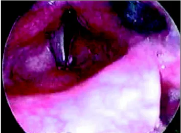
Using videolaryngostroboscopic data, we could ob-serve that in six patients (60%) the flap was at the same plane as the opposite vocal fold (Figure 1), in two (20%) it was higher and in two (20%) only the anterior third was on the upper plane, classified as not leveled.
Laryngeal closure w as considered complete in 30% of the cases (3 patients) and incomplete in 70% (Figure 2). In seven patients w ho had incomplete closure the follow ing abnormalities w ere present: anterior-posterior chink in two cases (28.6%), medium-posterior chink in one case (14.3%), posterior chink in three (42.8%), and hourglass chink in 14.3%. Arytenoid movement w as symmetrical in 90% of the cases. In the case of asymmetry, left arytenoid
was immovable.
We found one patient (10%) w ith frictional sound source, that is, there w as no vibration in laryngeal structures and the sound w as produced by the friction of the air current passing through the laryngeal structures. In nine patients (90%) the sound source w as vibrating and it was localized in the glottic region in seven patients, and in the supraglottic region in one. One patient presented mixed sound source, w ith vibration of glottic and supraglottic structures.
There was a vibrating predominant structure in each side of the larynx. On the non-operated side, in tw o patients, predominant vibration w as noticed in the vesti-bular fold and in seven, in the remaining vocal fold. On the operated side, the vibrating structure w as the vestibular fold flap in 9 patients. In Chart 1, we can see the description of patients including sound source and structures that comprised it.
Auditorilly-perceived vocal quality was classified according to GRBAS scale, showing one patient with normal voice (10%), and four with mild dysphonia (40%). The remaining 50% showed moderate (30%) or severe (20%) dysphonia. Results are presented in Chart 2. We should point out that patient 3 presented abnormal resonance because he had complete cleft palate. Cleft palate produces hypernasality, even with total palatine obturating splint. The results of computed acoustic analysis are distributed in Chart 1, in which we can observe that 2 patients did not undergo the assessment.
During sw allow ing, w e observed that laryngeal sensitivity w as preserved in all patients; there w as no laryngeal penetration, laryngotracheal aspiration or accumulation of contrast in the vallecula and piryform sinuses.
Figure 1. View of the larynx during inspiration. We can observe the
abduction flap at the same level as the opposite vocal fold.
Figure 2. View of the larynx during phonation. We can observe the
DISCUSSION
Cordectomy is a term used to designate vocal fold resections, both for open and endoscopic approach. In 2000, the European Laryngological Society proposed a classification of endoscopic cordectomy by dividing them as: subepithelial (Type I), subligamentar (Type II), transmuscular (type III), total or complete (type IV), extended to the contralateral vocal fold (Type Va), extended to the arytenoid (Type Vb), extended to the vestibular fold (type Vc), and extended to the subglottis (type Vd). According to the authors, this classification could also be applied to open resections.
Endoscopic cordectomy has been widely diffused and oncological results are similar to those reported with open surgery 8, with good vocal quality when they are types I
and II 9. However, in wider resections (cordectomies III, IV
and V), severe dysphonia is a predictable result 9. It can be
minimized when using reconstruction that provides vibrating tissue and allows glottic coaptation, such as what was conducted in this study. This fact was confirmed by the recent study by Remacle et al.10, Zeitels et al.11 and Zeitels et al.12,
who were concerned about post-laser therapy dysphonias and suggested phonosurgical techniques to improve laryngeal function and vocal quality, similarly to what was proposed in our study.
In our patients it was not necessary to maintain tracheostomy in late postoperative period, even in the patient that developed vocal fold immobility at midline position. It shows that the proposed technique preserved the organ as the airflow tract. Depending on the technique and the surgical reconstruction used, many authors do not report the same success rate 13-15. It is known that thick
flaps, such as myocutaneous ones, tend to generate small areas for breathing 16, however, given that the thickness of
the lowered vestibular fold was not affected, we would not expect airflow blockages.
Brasnu et al.5 observed 18.1% of granulomas and 3.7%
of anterior synechiae in patients submitted to vertical partial laryngectomy, reconstructed with vestibular fold flaps, which was not detected in our study, probably because the whole wound area was closed and also because the analysis was conducted only in patients in which there was no anterior
Chart 1. Description of sound source and structures that comprise it observed under videolaryngostroboscopy and result of computerized analysis of patients treated with cordectomy and reconstructed with vestibular fold.
Videolaryngostroboscopy Vocal analysis
Patient Soundsource Vibrating Vibrating Degree of fundamental jitter shimmer
structureoperated structurenon- dysphonia frequency % %
side operated side
1 vibrating flap vestibular fold 1 - -
-2 vibrating flap vocal fold 0 205 hz 0.13 1.16
3 vibrating flap vocal fold 1 208 hz 3.6 23
4 vibrating flap vocal fold 1 - -
-5 vibrating flap vestibular fold 2 112 hz 0.28 4.5
6 vibrating flap vocal fold 2 139 hz 0.6 6.0
7 vibrating flap vocal fold 2 164 hz 0.3 3.2
8 vibrating flap vocal fold 1 152 hz 0.3 4.8
9 frictional absent absent 3 304 hz 1.2 5.3
10 vibrating flap vocal fold 3 136 hz 2.5 8.4
Chart 2. Description of results obtained with auditory perceptual analysis in patients treated with cordectomy and reconstructed with vestibular fold according to GRBAS scale.
G R B A S
Patient Degree Of Dysphonia Hoarseness Breathiness Asthenia Strain
1 1 1 1 1 0
2 0 0 0 0 0
3 1 1 1 1 0
4 1 1 1 0 0
5 2 2 2 1 0
6 2 2 2 1 0
7 2 2 2 1 0
8 1 1 1 0 0
9 3 3 3 3 1
commissure resection, an area known for its likelihood of complications. Vocal fold immobility observed in one of our patients has probably occurred by fibrosis at the level of the cricoarytenoid joint, since the margin of the resection of the tumor reached the vocal process.
In 40% of the cases, w e did not reach perfect positioning of the vestibular fold and the flap was positioned higher than desired. In 20% of the cases, the flap was entirely above the opposite vocal fold level and in other 20%, there was non-leveling at the anterior third. In our opinion, this technical failure was due to incomplete decrease of the flap or cicatricial retraction of the surgical bed. The anterior third of the vestibular fold is the most difficult region to position it.
Only in three cases there was complete laryngeal closure, and in two, the flap was at the same level as the opposite vocal fold, whereas in another case, there was level discrepancy only at the anterior third. According to Hirano et al.17, laryngeal closure is complete in hemilaryngectomies,
reconstructed with different techniques. The author referred that during vibration cycles, there is complete closure only in 19% of the cases. In a study conducted by Mandell et al.18, with videolaryngostroboscopy in 42 patients submitted
to vertical partial laryngectomy, with different reconstruction techniques, they observed that complete glottic closure was present in only 16.7% of the cases. The authors found pos-terior chinks in 26.2%, irregular chinks in 23.8%, anpos-terior chinks in 9.5%, and in 23.8% it was not possible to define the presence of chinks. In our patients, incomplete closure was related to the presence of posterior chink in 42.8% of the cases and medium-posterior chink in 14.3%. A case of hourglass chink was detected when the flap was positioned above the level of the vocal fold, forcing it to hyperadduct only on the medium third of the contralateral vestibular fold, given that the sound source was at supraglottic level. In one of the cases of anterior-posterior chink, there was a sound source at supraglottic level. In one of the cases of anterior-posterior chink, there was clear bowing of the flap, which did not allow coaptation of the contralateral vocal fold. The other case of anterior-posterior chink was observed in an older patient aged 75 years, probably owing to muscle hypotony, resulting from presbyphonics.
Arytenoids move symmetrically, except in one single case in which immobility resulted from the surgical procedure. Since the immobility was at midline position, it did not affect laryngeal closure. In this patient, there was posterior chink. We assessed the vibrating capabilities of the laryngeal structures and noticed in our sample that only one patient presented frictional sound source, that is, there was no vibration of the structures that participate in the glottic closure. Similar result was achieved by Hashimoto19, upon
studying the sound source in patients submitted to vertical partial laryngectomy with varied types of reconstruction, and he observed in 7.1% of the cases that this source was
frictional. The author referred that to create a vibrating sound source, there should be a balance betw een laryngeal configuration and aerodynamic forces. Behlau and Gonçal-ves20 emphasized that in all its variations, vertical partial
laryngectomy caused deficiency in glottic coaptation, which could be corrected in the reconstruction but the geometry of the glottis and the histological architecture of the sound source were definitely affected. This observation can justify the presence of frictional sound source in this study.
In our sample, the vibrating sound source was primarily located in the glottic region (77.8%). A patient presented supraglottic sound source and another one had mixed sound source. These data are anatomically related with position of the flap, because the patient with supraglottic sound source had the flap positioned superiorly to the level of the opposite vocal fold, whereas the patient with mixed sound source had difference of level of the flap in its anterior third, positioning superiorly to the glottic level. We should bear in mind that the other patient with flap positioned above the vocal fold level presented frictional sound source. Therefore, it is noticed that the position of the flap above the opposite vocal fold level excluded the vibrating sound source from the glottic region.
Hashimoto19 referred that the supraglottic region was
the preferred region for the formation of the vibrating sound source in most cases in which he conducted vertical partial laryngectomy. Camargo21 justified the presence of vibrating
sound source in the supraglottic region w ith lack of reconstruction of the surgical wound in the glottic region, associated with the presence of loosen tissues in the structures of the region, which favors its collapse towards the laryngeal lumen and vibration. Behlau et al.22, studying 69 patients
submitted to vertical partial laryngectomy with sound source at the supraglottis in 75% of the cases and at the glottis in 17%, concluded that glottic reconstruction should not be faced as sound source, but as a means to maintain laryngeal lumen permeability, creating conditions for the supraglottis to take on the function of sound source. In our patients, we tried to provide loose tissue in the glottic region by lowering the vestibular fold, so as to maintain it as a vibrating sound source. In our opinion, the vibrating sound source at the glottic level depends on the presence of an intact vocal fold on the opposite side and a flap that replaces the resected vocal fold. In this situation, we found a patient with normal vocal quality for age and gender, and it was nearly impossible to believe that the patient had only one vocal fold. In computed vocal analysis, the patient presented fundamen-tal frequency of 205Hz, jitter of 0.13% and shimmer of 1.16%. Acoustic findings in our patients showed a tendency to elevate fundamental frequency, coinciding with the data by Bertino et al.23, especially when the sound source is
fibers existing in the vestibular fold. In our patients, acoustic parameters of jitter and shimmer range respectively from 0.13 to 3.6% and 1.16 to 23%, mean of 1.11 and 7.04%. Perreti et al.9 found values of jitter with median of 1.43% in
cases of endoscopic cordectomies type I and II and medial of 2.96% in more extensive cordectomy. Shimmer showed median of 6.52% in the first group and 8.05% in the other group. These data reinforced the importance of conducting reconstruction.
In 1999, Biacabe et al.6, upon studying patients
su b m i t t ed t o v er t i cal p ar t i al l ar y n gect o m y an d reconstructed with vestibular fold flap, found in a two-year postoperative group mean fundamental frequency of 157Hz and jitter mean of 3.9%. Our results show ed that only tw o patients w ith poor results in vocal quality had high values of jitter. Another patient w ith high value had cleft palate. Excluding this patient, the jitter mean w as 0.75%.
Upon analyzing Chart I, we noticed that the patient without dysphonia had shimmer value within the normal parameters, whereas patient 10, with anterior-posterior tri-angular chink, had highly abnormal value. The patient with cleft palate had very abnormal results. Apart from this patient, the shimmer mean was 4.76%. In the study by Biacabe et al.6, the mean was 12%.
The assessment of laryngeal function during sw allow ing in patients submitted to vertical partial laryngectomy is scarce in the literature and normally it is conducted subjectively 14,15,24. In this study, the function was
investigated through endoscopic assessment of swallowing, showing that the sphincterial action of the larynx was preserved, which had already been noticed in the analysis of phonation.
In view of these results, w e could notice that laryngeal functions w ere maintained and that the voice obtained ranged from normal to mild dysphonic when the flap w as positioned appropriately, including a posterior chink in some cases. In this situation, the flap w as part of the vibrating source, which was placed at glottic level. We believe that other studies should be conducted to improve the reconstruction technique, especially concerning positioning of the flap in the vestibular fold to increase the number of normal voice patients submitted to partial laryngectomy by open resection.
CONCLUSION
We concluded that post-cordectomy laryngeal reconstruction conducted with vestibular fold flap enabled production of normal voice (fundamental frequency of 205Hz, jitter 0.13%, shimmer 1.16%), providing complete coaptation in 30% of the cases, vibrating sound source in the glottic region in 70% and participation of the flap as vibrating structure in 90% of the cases, in addition to
maintaining the laryngeal functions of breathing and protecting the airways during swallowing.
REFERENCES
1. Réthi A. Cordectomie étendue en arriere. Ann Otolaryngol Chir Cervicofac 1963; 80:539-44.
2. Sala O, Pivotti G. Cordectomie et phonation. Ann Otolaryngol Chir Cervicofac 1971; 88:511-3.
3. Fri edman M, Tori umi D M. Gl otti c reconstructi on fol l ow i ng hemilaryngectomy: false cord advancement flap. Laryngoscope 1987; 97: 882-4.
4. Fukuda H, Tsuji DH, Kawasaki Y, Kawaida M, Sakou T. Displacement of the ventricular fold following cordectomy. Auris Nasus Larynx 1990; 17:221-8.
5. Brasnu D, Laccourreye O, Weinstein G, Fligny I, Chabardes E. False cord reconstruction of the glottis follow ing vertical partial laryngectomy: a preliminary analysis. Laryngoscope 1992; 102:717-9.
6. Biacabe B, Crevier-Buchman L, Hans S, Laccourreye O, Brasnu D . Vo cal f u n cti o n af ter p arti al l aryn gecto my w i th gl o tti c reconstruction by false vocal fold flap: durational and frequency measures. Laryngoscope 1999; 109:698-704.
7. Remacle M, Eckel HE, Antonelli A, Brasnu D, Chevalier D, Friedrich G, Olofsson J, Rudert HH, Thumfart W, de Vincentiis M, Wustrow TP. Endoscopic cordectomy: a proposal for a classification by the Working Committee, European Laryngological Society. Eur Arch Otorhinolaryngol 2000; 257:227-31.
8. De Campora E, Radici M, De Campora L. External versus endoscopic approach in the surgical treatment of glottic cancer. Eur Arch Otorhinolaryngol 2001; 258:533-6.
9. Perretti G, Piazza C, Balzanelli C, Cantarella G, Nicolai P. Vocal outcome after endoscopic cordectomies for Tis and T1 glottic carcinomas. Ann Otol Rhinol Laryngol 2003; 112:174-9. 10. Remacle M, Lawson G, Medayat A, Trussat T, Jamart J. Medialization
framew ork surgery for voice improvement after endoscopic cordectomy. Eur Arch Otorhinolaryngol 2001; 258:267-71. 11. Zeitels SM, Jarboe J, Franco RA. Phonosurgical reconstruction of
early glottic cancer. Laryngoscope 2001; 111:1862-5.
12. Zeitels SM, Hillman RE, Franco RA, Bunting GW. Voice and treatment outcome from phonosurgical management of early glottic cancer. Ann Otol Rhinol Laryngol 2002(Suppl); 190:3-20. 13. Hirano M. Technique for glottic reconstruction following vertical
partial laryngectomy: a preliminary report. Ann Otol Rhinol Laryngol 1976; 85:25-31.
14. Calcaterra TC. Sternohyoid myofascial flap reconstruction of the larynx for vertical partial laryngectomy. Laryngoscope 1983; 93:422-4.
15. Brasil OOC, Pontes PAL. Laringectomias parciais verticais: recons-trução com retalho miocutâneo de platisma. Rev Paul Med 1990; 108:213-20.
16. Mamede RCM, Mello-Filho FV. Reconstrução nas laringectomias parciais. Rev Bras Cir Cab Pesc 1993; 17:157-63.
17. H i rano M, Kuri ta S, Matsuok a H . Vocal functi on fol l ow i ng hemilaryngectomy. Ann Otol Rhinol Laryngol 1987; 96:586-9. 18.Mandell DL, Woo P, Behin DS, Mojica J, Minasian A, Urk en
ML, Bi l l er H F. Vi deol aryngostroboscop y f ol l ow i ng verti cal p ar t i al l ar y n gect o m y . A n n O t o l Rh i n o l Lar y n go l 1999; 108:1061-7.
19. Hashimoto IK. Reconstruções laríngeas em laringectomias parciais verticais por carcinoma da região glótica – estudo da função fona-tória; 1995, Dissertação de mestrado, Universidade Federal de São Paulo, São Paulo.
21. Camargo ZA. Parâmetros vocais e conFigurações laríngeas na fonação de indivíduos submetidos às laringectomias parciais verticais; 1996, Dissertação de Mestrado, Pontifícia Universidade Católica, São Paulo. 22. Behlau M, Gonçalves MI, Pontes P, Brasil OC. Physiology of sound source follow ing partial vertical laryngectomy. In: VII Pacific Voice Conference – Voice Conservation, Treatment and Restoration after Laryngeal Carcinoma. San Francisco, 1994. Digest. pp. 32-3.
23. Bertino G, Bellomo A, Ferrero FE, Ferlito A. Acoustic analysis of voice quality with or without false vocal fold displacement after cordectomy. J Voice 2001; 15:131-40.