Evaluation of Epworth
Sleepiness Scale in patients with
obstructive sleep
apnea-hypopnea syndrome
Summary
Letícia Boari1, Carolina M. Cavalcanti2, Samantha R. F. D. Bannwart3, Oscimar B. Sofia4, José Eduardo L. Dolci5
1 Master studies under course, Department of Otorhinolaryngology, FCMSCSP, sponsored by CNPq. 2 Resident Physician, Department of Otorhinolaryngology, Santa Casa de Sao Paulo. 3 Resident Physician, Department of Otorhinolaryngology, Santa Casa de Sao Paulo.
4 Master studies under course, Department of Otorhinolaryngology, FCMSCSP, sponsored by CNPq. Craniomaxillofacial surgeon and Otorhinolaryngologist, CEMA. 5 Joint Professor, Department of Otorhinolaryngology, FCMSCSP.
Medical School, Santa Casa de Sao Paulo.
Address correspondence to: Rua dos Heliotrópios 133/ 94 Mirandópolis São Paulo SP 04049 -000 Fax: (55 11)5575-12 23 – E-mail: [email protected]
Study presented as poster at III Congresso Triológico de Otorrinolaringologia, held on October 08 - 11, 2003, in Rio de Janeiro. Article submited on July 29, 2004. Article accepted on March 30, 2004.
T
oday obstructive sleep apnea–hypopnea syndrome ( OSAH S) i s a p ubl i c heal th i ssue, si nce i t i ncreases cardiovascular morbidity-mortality rate and the risk of car crashes. Overnight polysomnography is the gold standard for diagnosis and follow-up of affected patients. However, because the test is expensive, time-consuming and of difficult access, others methods have been proposed. Although the Epworth Sleepiness Scale (ESS) is subjective, the questionnaire is simple, easy to be applied and free of charge. Aim: to compare Epworth Sleepiness Scale scores an d ap n ea-h y p o p n ea i n d ex ( AH I ) , m easu r ed b y polysomnography, in patients diagnosed with OSAHS. Study Design: clinical retrospective study. Method: chart analysis of 66 patients complaining from snoring, who underwent surgery (uvulopalatopharyngoplasty w ith/ w ithout nasal surgery). ESS score and AHI were evaluated before and after surgery. Results: 78% of patients with normal AHI, scored < 10 in the ESS and 65% of patients with severe AHI scored >10. There were no statistically significant results for groups presenting mild and moderate apnea. Conclusion: ESS can detect normal and severe levels of apnea, but is not able to detect mild and moderate levels. Therefore, ESS can be used in the follow-up of patients with OSAHS, however, it cannot replace polysomnography because it does not detect all levels of apnea.Key words: obstructive sleep apnea–hypopnea syndrome; Epworth Sleepiness Scale; polysomnography;
apnea-hypopnea index.
« « « « Rev Bras Otorrinolaringol.
V.70, n.6, 752-6, nov./dec. 2004
ORIGIN AL ARTICLE
INTRODUCTION
Many patients search for medial care to treat snoring, especially when it causes family/ marriage issues. Excessive noise is the major complaint, but other signs also occur. Usually, complaints such as fatigue, daytime sleepiness, behavioral and cognitive dysfunction are not taken into account by the patient, w ho believes that such symptoms result from physical/ emotional stress and/ or aging. How ever, an appropriate w ork up is able to prove that snoring may be a symptom of a sleep disorder, obstructive sleep apnea–hypopnea syndrome (OSAHS). Estimates show
that snoring prevalence in adults in the 5th and 6th decades
of life is 40-60%, and 90-95% of patients w ith OSAHS
snore1.
This syndrome is currently considered a public health issue because it increases the risk of work-related and traffic accidents, as well as cardiovascular morbidity-mortality rate. Its prevalence varies according to the population. In the United States, estimates are of 4% in men and 2% in women between 30-60 years, especially in obese patients. However, these data are underestimated, and about 95% of the patients
with sleep disorders are not diagnosed1,2. Haponik et al.3
showed in their study that none of the primary care physicians included sleep disorders in their differential diagnosis lists. On the other hand, when they became aware of the disease, 82% of the doctors started to investigate sleep disorders. Therefore, the health care provider, especially the otorhinolaryngologist, should be aware of this syndrome to diagnose and treat it promptly, preventing its severe consequences.
Currently, overnight polysomnography is the gold stan d ard f o r d i agn o si s o f O SAH S. Reco rd i n gs o f electroencephalogram, electrooculogram, electromyography, electrocardiogram, oxymetry, airflow and breathing effort provide important data to evaluate the severity of the disease. However, the test is expensive, time-consuming, of difficult access outside big cities and is not always well
accepted by patients.4,5
Although subjective, the Epworth Sleepiness Scale may contribute to the analysis of signs and symptoms. It is
easy to be applied, fast and free of charge.6
In summary, the purpose of our study was to evaluate Epworth Sleepiness Scale scores and apnea and hypopnea index (AHI), measured by polysomnography, in patients diagnosed w ith OSAHS, as w ell as to compare these parameters.
OBJECTIVE
The purpose of our study was to evaluate pre and postoperative Epworth Sleepiness Scale Scores and AHI in patients diagnosed w ith OSAHS, and compare these parameters.
METHODS
The study was conducted through chart analysis of patients complaining from snoring, from June 1999 to June 2003, at the Department of Otorhinolaryngology in Hospi-tal CEMA. The inclusion criteria were adult men and women comp l ai ni ng from snori ng, w ho underw ent uvul o-palatopharyngoplasty (UPPP). We used modified Fairbanks technique, with and without nasal approach (septoplasty and/ or partial inferior turbinectomy); pre and postopera-tive polysomnography was conducted and the questionnaire for sleep evaluation, the Epworth Sleepiness Scale (ESS) (Table 1), was applied before and after surgery. All charts contained complete physical examination performed by an otorhinolaryngologist.
The cases excluded from the study were: charts with incomplete data, patients presenting concurrent diseases (cardiopathy, thyroid disorders, diabetes mellitus and chronic lung disease), craniofacial dimorphism or retrognathia and patients who underwent UPPP in other departments. It is worth noting that the postoperative data were obtained, at least, 6 months after surgery. All surgeries were performed by the same surgical team.
Polysomnography, conducted in the Department of
Otorhinolaryngology or at In stituto Paulista do Son o, were
in accordance with the minimal standards required, which
were published on Atuali zação Otorri n olari n gológi ca em
Cirurgia do Ronco e Apnéia do Sono (Otorhinolaryngological Update in Snoring Surgery and Sleep Apnea) by SBORL,
20024.
We used the classification adopted by the American
Academy of Sleep Medicine7 to categorize the level of
dysfunction, as shown in Table 2.
Epworth Sleepiness Scale was applied, the answers were given by the patient and the physician would only interfere in cases of doubt or misinterpretation.
Therefore, our study had 66 cases that met the criteria for inclusion. We considered 10 the normal limit of the
Epworth Sleepiness Scale, as stated in its original version.9
The statistical analysis was conducted by the test for similar proportions and Chi-square independence test. We found 95% confidence interval and p=0.05.
RESULTS
The studied group included 66 snoring patients who underw ent surgery. UPPP w i thout nasal ap p roach (septoplasty and/ or turbinectomy) was performed in only 8 patients, whereas the remaining patients underwent both surgeries. There were 6 women and 60 men, mean age was 42.47 (±10.7), ranging from 17 to 67 years old.
In the preoperative period, 72.7% (48) subjects had abnormal scores (>10) in ESS, whereas only 27.2% (18) had
during the postoperative period - 74.2% (49) had normal scores and 25.7% (17) had abnormal scores (Chart 1). These results show ed statistically significant improvement (p<0.001) in ESS scores after surgery, as shown in Table 1. Moreover, during the preoperative period, ESS normal average score was 6.6 (± 2.5) and the abnormal average score was 16.0 (± 3.4). During postoperative examination, normal and abnormal average scores were 5.6 (±2.8) and 14.5 (±3.5), respectively (Table 2).
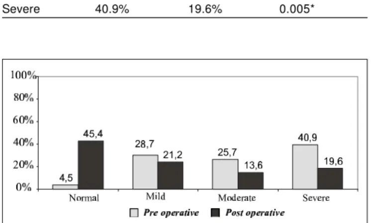
During the preoperative period, AHI found in polysomnography was: 4.5% (3) normal; 28.78% (19) mild; 25.75% (17) moderate, and 40.90% (27) severe. During the postoperative period, we found 45.45% (30) normal, 21.21% (14) mild, 13.63% (9) moderate, and 19.69% (13) severe. Comparing the results before and after surgery, there was statistical significance in normal (0.001*) and severe (0.005*) scores, as shown in Table 3 and Chart 2.
The chi-square independence test was applied to check association between both variables studied. Because we had the purpose to analyze AHI and ESS scores in a given time point, without taking into account the reliability of methods, we mixed the preoperative and postoperative groups to obtain a most significant sample. Therefore, of the 33 patients with normal AHI, 26 (78.7%) were normal according to the Epworth Sleepiness Scale and only 7 (21.%) were abnormal. This result was statistically significant. As for mild and moderate levels, there was no correlation between parameters. Statistically significant results were also found in severe AHI, and 26 (65%) out of 40 patients had abnormal scores in the ESS – Table 4.
DISCUSSION
Obstructive Sleep Apnea and Hypopnea Syndrome has been carefully studied. In 1999, the American Academy of Sleep Medicine described this syndrome as recurrent episodes of total or partial upper airways obstruction, causing
desaturation and sleep fragmentation.4 This is possibly
associ ated w i th the frequent symp tom of dayti me
sleepiness7. About 90% of patients affected with OSAHS
show daytime sleepiness, whereas the remaining patients
show changes only during overnight monitoring 8.
I t i s gen er al l y accep t ed t h at o v er n i gh t polysomnography is the gold standard for OSAHS diagnosis.
According to Atualização Otorrin olarin gológica em
Cirur-gi a do Ron co e Apn éi a do Son o by SBORL, 2002, overnight recordings are the most significant ones, including electroencephalogram, electrooculogram, electromyography of submentonian and anterior tibial, electrocardiography,
oxymetry, airflow determination and breathing effort4,5.
However, the test is expensive and of difficult access. Epworth Sleepiness Scale was developed in 1991 by Dr. John W. Murray, an Australian physician. Ever since, it has been used globally, and it was translated into many
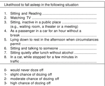
Chart 1. Epworth Sleepiness Scale.
Likelihood to fall asleep in the following situation
1. Sitting and Reading ... 2. Watching TV ... 3. Sitting, inactive in a public place ...
(e.g., waiting room, a theater or a meeting) 4. As a passenger in a car for an hour without a
break ... 5. Lying down to rest in the afternoon when circumstances
permit ... 6. Sitting and talking to someone ... 7. Sitting quietly after lunch without alcohol ... 8. In a car, while stopped for a few minutes in
traffic ... 0- would never doze off
1- slight chance of dozing off 2- moderate chance of dozing off 3- high chance of dozing off
Chart 2. Classification of the severity level of OSAHS according to the American Academy of Sleeping Medicine -1997.
APNEA/HYPOPNEA INDEX LEVEL
< 5 NORMAL
5-15 MILD
15-30 MODERATE
> 30 SEVERE
Table 1. Pre and postoperative quantitative analysis ofESS.
Epworth Pre Post p-value
Normal 27.3 % 74.3% < 0.001*
Abnormal 72.7% 25.7% < 0.001*
languages, such as German, Spanish and Japanese. Dr. Murray intended to quantify the likelihood of someone to fall asleep in 8 different common situations (see Chart 1). The scores range from 0 to 24, and 10 is the normal limit. According to the author, like any other Scale, the evaluation is not subjective but it is influenced by patient’s reading and
comprehension skills and honest answers.6
In his studies, Dr. Murray showed statistically significant results in ESS scores when comparing normal non-snoring subjects and snoring subjects. The average of the first group
was 5.9±2.2 (2-10), 10 was considered the normal limit.9
Moreover, the Scale was able to separate primary snoring subjects from patients with OSAHS; the higher the ESS score, the more severe the condition. All patients with
severe conditions scored over 10.6 However, it is worth noting
that the severity level used by Dr. Murray was based on his judgment, where AHI < 5 was normal; 5 to 25 was mild; 25 to 50 was moderate, and > 50 was severe; besides he only had 19 subjects in the severe group.
Our study show ed that, separately, there w as statistically significant increase of normal patients, in the pre and postoperative periods, considering both parameters, AHI and ESS. Moreover, corroborating the abovementioned study, there was statistically significant correlation between AHI and ESS when severe and normal groups were evaluated. Therefore, 65% of 40 severe patients scored over 10 in the ESS and 78.8% of 33 normal patients scored under 10. For mild and moderate groups, there w as no statistically significant correlation. It is worth noting that, in our study, we put the preoperative and postoperative groups together to increase the sample of each subgroup. Therefore, another study separating such groups should be conducted in the future.
According to some authors, ESS cannot detect all levels of severity nor the frequency of apnea and hypopnea;
therefore, ESS cannot replace polysomnography.8 Moreover,
Nguyen et al10 showed that the Scale is not reliable and the
score may vary up to 7 when the questionnaire is repeated within 6 months in the same group, with no interference but weight loss. However, we should consider that weight loss may have improved the condition and, consequently, interfered in ESS scores. Our study could not evaluate test reliability because the patients underwent surgery and ESS was applied before and after surgery.
The normal limit of ESS has also been questioned. Douglas proposes >12 as normal based on a British study
showing that 95% of normal people scored 11.8 If in our
study we had considered 12 as normal, we would have still found the same statistically significant results for normal and severe groups.
Today, ESS helps to diagnose sleep disorders and recommend polysomnography. Our study contributed to attest that Epworth Sleepiness Scale can be used to qualify, but not quantify, the level of disease. It can be used in the
Table 2. Pre and postoperative quantitative analysis ofESS.
Epworth p re post
Normal Abnormal Normal Abnormal
Mean 6.6 16.0 5.6 14.5
Standard Deviation 2.5 3.4 2.8 3.5
Median 6.0 15.0 5.5 13.0
Minimum 3.0 11.0 1.0 11.0
Maximum 10.0 23.0 10.0 22.0
Table 3. Pre and postoperative analysis of AHI.
AHI Pre Post p-value
Normal 4.5% 45.4% < 0.001*
Mild 28.7% 21.2% 0.387
Moderate 25.7% 13.6% 0.076
Severe 40.9% 19.6% 0.005*
Table 4. Comparison between Epworth and AHI, before and after surgery.
Pre and Post Epworth Total
Normal Abnormal
AHI Normal 26 7 33
Mild 17 16 33
Moderate 10 16 26
Severe 14 26 40
Total 67 65 132
p-value = 0.001*
Table 5. Qualitative analysis of severe, moderate, mild and normal levels in association with Epworth Sleepiness Scale.
IAH
ESS severe moderate mild normal total
mean 12.83 11.66 10.37 7.71
standard
deviation 6.17 5.46 4.82 5.01
mode 12 15 8 7
median 12 13 10 7
minimum 3 3 1 1
maximum 23 21 23 22
total 41 27 32 32 132
postoperative period of patients who are not willing to repeat polysomnography. It is worth saying that ESS should not replace polysomnography, but it can be an important tool in the follow-up period.
Further studies are needed to better characterize the normal limit and reliability, so that it can become more significant in practice.
CONCLUSION
We concluded that ESS shows correlation with AHI for severe and normal levels, but not for mild and moderate levels. Therefore, it can be used in the follow-up of the postoperative period, but should not replace polysomnography as a diagnostic tool. Other studies are required to evaluate reliability and the normal limit for ESS, based on current data for severity of the condition.
REFERENCES
1. Bittencourt LR, Togeiro SMGP, Bagnato MC. Diagnóstico da Sín-drome da apnéia e hipopnéia obstrutiva do sono. In: Stamm A. ed. Rinologia; 2002; São Paulo: Komedi; 2002: 103-11.
2. Mercado JC. I dentifying Obstructive Sleep Apnea: A Highly Prevalent and Underdiagnosed Disease. Physician Assistant 2003; 27(2): 39-45.
3. H ap o n i k EF, Frye AM, Ri ch ard s B et al . Sl eep h i sto ry i s neglected diagnostic information. J Gen Intern Med. 1996;11: 759- 61.
4. Atual i zação Otorri nol ari ngol ógi ca em Ci rurgi a de Ronco e Apnéia. Revista Brasileira de Otorrinolaringologia set/ out 2002; 68(5) sup l .3.
5. Zonato AI, Bittencourt LR, Martinho FL, Júnior JFS, Gregório LC, Tufik S. Association of Systematic Head and Neck Physical Ex am i n at i o n W i t h Sev er i t y o f O b st r u ct i v e Sl eep Ap n ea-Hypopnea Syndrome. Laryngoscope. 2003; 113(6):973-80. 6. Murray JW. Daytime Sleepiness, Snoring and Obstructive Sleep
Ap nea: The Ep w orth Sl eep i ness Scal e. Chest 1993; 103(1): 30- 6.
7. Sleep-Related Breathing Disorders in Adults: Recommendations f o r Syn d ro me D ef i n i ti o n an d Measu remen t tech n i q u es i n Clinical Research- AASM Task Force. Sleep 1999; 22(5): 667-89.
8. Douglas NJ. The Obstructive Sleep Apnea/ Hypopnea syndrome. Practical Neurology 2003; 3(1): 22-9.
9. Murray JW. A New Method for Measuring Daytime Sleepiness: The Epw orth Sleepiness Scale. Sleep 1991; 14: 540-5. 10. Ngu yen AT, Pal ayew M, Bal tazan M, Gu i l l o n S, Smal l D .