Entomophytophagy ('Sequential Predatory,
then Phytophagous Behaviour') in an Indian
Braconid
‘
Parasitoid
’
Wasp (Hymenoptera):
Specialized Larval Morphology, Biology and
Description of a New Species
A. P. Ranjith1,2☯, Donald L. J. Quicke3☯, U. K. A. Saleem1¤, Buntika A. Butcher3,
Alejandro Zaldívar-Riverón4, M. Nasser1
*
1Insect Ecology and Ethology Laboratory, Department of Zoology, University of Calicut, Kerala, Pin: 673635, India,2Department of Zoology, Malabar Christian College, Calicut, Kerala, Pin: 673001, India, 3Department of Biology, Faculty of Science, Chulalongkorn University, Phayathai Road, Pathumwan, BKK 10330, Thailand,4Colección Nacional de Insectos, Instituto de Biología, Universidad Nacional Autónoma de México, 3er. circuito exterior s/n Cd. Universitaria, Copilco, Coyoacán, A. P. 70–233, C.P. 04510, D.F., México
☯These authors contributed equally to this work.
¤ Current address: Department of Zoology, Government College Madappally, Calicut, Kerala, Pin: 686546, India
Abstract
The vast majority of braconid wasps are parasitoids of other insects. Although a few cases of pure phytophagy (primary gall production and seed predation) are known, no previous ento-mophytophagous species (i.e. ones that display entomophagy and phytophagy sequentially), has been discovered among braconids. We describe the detailed biology and specialized lar-val morphology for the first confirmed entomophytophagous braconid species. Leaf galls on Garuga pinnataRoxb. (Burseraceae) in India, induced by the psyllid,Phacopteron lentigino-sumBuckton (Hemiptera: Psylloidea, Phacopteronidae) were sampled throughout a period of several months and found to suffer a high level of attack by a new speciesBracon garugapha-gaeRanjith & Quicke which is here described and illustrated. The wasps oviposit singly into the galls without paralysing the psyllids. The larvae first attack psyllid nymphs which they seek out within the gall, kill them with a single bite and consume them. Unique dorsal abdomi-nal tubercles, with eversible tips present on the abdomiabdomi-nal segments of the larvae that are used to help maintain larval position while feeding, are illustrated. After consuming all avail-able prey, the larvae continue feeding on gall tissue until mature enough to spin cocoons and pupate. The new species illustrates, for the first time, a possible intermediate stage in the evo-lution of pure phytophagy within the Braconidae. Interestingly, the two unrelated seed preda-torBraconspecies are also associated with Burseraceae, perhaps indicating that this plant family is particularly suited as a food for braconine wasps.
a11111
OPEN ACCESS
Citation:Ranjith AP, Quicke DLJ, Saleem UKA, Butcher BA, Zaldívar-Riverón A, Nasser M (2016) Entomophytophagy ('Sequential Predatory, then Phytophagous Behaviour') in an Indian Braconid
‘Parasitoid’Wasp (Hymenoptera): Specialized Larval Morphology, Biology and Description of a New Species. PLoS ONE 11(6): e0156997. doi:10.1371/ journal.pone.0156997
Editor:Renee M. Borges, Indian Institute of Science, INDIA
Received:August 26, 2015
Accepted:May 23, 2016
Published:June 29, 2016
Copyright:© 2016 Ranjith et al. This is an open access article distributed under the terms of the
Creative Commons Attribution License, which permits unrestricted use, distribution, and reproduction in any medium, provided the original author and source are credited.
Data Availability Statement:All relevant data are within the paper and its Supporting Information files.
Funding:The authors have no support or funding to report.
Introduction
Braconid wasps represent one of the most diversified groups of insects comprising 46 subfami-lies with nearly 1000 genera and 15,000 described species, the vast majority of which are para-sitoids of other insects [1] and have been extensively used as experimental models of host–
parasite associations [2–7]. Phytophagy in the Braconidae was first discovered in the subfamily Doryctinae with various species in a small number of genera being found to be primary gall formers [8–12]. Gall induction in the Doryctinae appears to be phylogenetically associated with parasitism of gall formers, but nothing is known of the transitional stages. Since then, pri-mary gall formation has also been demonstrated in the genusMesostoa(Mesostoinae) [13] whilst purely phytophagous seed predation is known in two Neotropical species ofBracon
(Braconinae) [14,15].
Members of the Braconinae, which is one of the largest of the subfamilies, are mostly ecto-parasitoids that develop on concealed hosts that are usually paralysed as a result of venom injected by the female at the time of oviposition [16]. It is dominated by the genusBracon
which has more than 850 described species though molecular data strongly indicate that the genus is paraphyletic and possibly even polyphyletic [17]. Not surprisingly the host range of
‘Bracon’, is also by far the largest in the subfamily, and includes Coleoptera, Lepidoptera, Dip-tera [16], Hemiptera [18] as well as phytophagous Hymenoptera [19–21]. Almost all of these hosts share a moderate degree of concealment, usually in living plant tissues, and typically include inhabitants of tree bark, stems of annual and biennial plants, galls, seed heads or ves-sels, as well as leaf rollers, leaf miners and case-bearers [16].
Here we describe the biology of a new Indian braconid wasp species,Bracon garugaphagae
sp. nov., whose larvae are initially predators of leaf gall-inducing psyllids (Hemiptera: Psylloi-dea) onGaruga pinnataRoxb. (Burseraceae). After the primary gall makers are consumed, they complete the greater part of their larval development consuming gall tissue. This is the first known instance of entomophytophagy [22] in the family. The wasp’s larval morphology includes several unique adaptations to this way of life.
Materials and Methods
Ethics statement
Necessary permits to conduct sampling of leaf galls were obtained from the Government of Kerala, India.
Study site
This study was carried out in two sites in Malappuram district, Kerala, south India; viz. Kottak-kal (10°99’N, 76°00’E) and Vettichira (10°93’N, 76°02’E). The sites have a tropical climate and during the study period the average annual temperature was 26°C and annual rainfall was 2842 mm [23].
Sampling and data collection
Weekly field surveys were carried out from August 2014 to January 2015. Leaf galls induced by the psyllid,Phacopteron lentiginosumBuckton (Phacopteronidae), on shrubs ofGaruga pin-nataRoxb. were collected at different developmental stages and dissected under a stereomicro-scope (Olympus).The number of gall-inducing nymphs and the presence or absence of braconid eggs, and early and mature larvae were recorded. Digital photographs of dissected galls and braconid wasp developmental stages were taken in situ with a Canon IXUS 255 HS
Species description
Alcohol-preserved specimens were processed with hexamethyldisilazane and later card-mounted. Images of the holotype and the egg, larva and pupa of the braconid wasp were taken with a Leica DFC 295 camera attached to a Leica S8 APO Stereozoomtrinocular microscope (Leica, Heerburg, Switzerland). Image stacks were combined into a single image and measure-ments of the holotype were done using Leica Application Suite V4.2. Images were edited using Photoshop CS8 (Version 6.1) (Adobe Inc.).
Morphological terminology employed in the description follows van Achterberg [24,25] except wing venation nomenclature which follows Quicke [7]. Terms for sculpturing follow Eady [26] and Harris [27].
Scanning electron microscopy and light microscopy of larvae
Larvae of different stages were dried with hexamethyldisilazane, mounted on entomological minuten pins and glued with epoxy resin on to standard electron microscopy stubs, sputter-coated with gold and studied under a JEOL JSM-5410LVmicroscope.
Larval head capsules were prepared from the gold-coated specimens as well as from uncoated material by macerating in 0.2M aqueous KOH to remove soft tissues, washing in dilute acetic acid, followed by dehydration through to xylene and mounting in Permount1.
Molecular protocol
A specimen was sequenced for the barcoding 5’fragment of the mitochondrial cytochrome oxi-dase gene and the nuclear 28S rDNA D2–D3 region following the methods of Zaldivar-Riverón et al. [28,29]. Sequences were deposited in GenBank with accession numbers CNIN2004 KT343804 and CNIN2004 KT343805.
Data analysis
All statistical analyses were carried out using the software package R [30].
Nomenclatural Acts
The electronic edition of this article conforms to the requirements of the amended Interna-tional Code of Zoological Nomenclature (ICZN), and hence the new names contained herein are available under that Code from the electronic edition of this article. This published work and the nomenclatural acts it contains have been registered in ZooBank, the online registration system for the ICZN. The ZooBank Life Science Identifiers (LSIDs) can be resolved and the associated information viewed through any standard web browser by appending the LSID to the prefix“http://zoobank.org/”. The LSID for this publication is: urn:lsid:zoobank.org:pub: FF0DA8B1-FB69-4203-8D37-0A6BDA5A2B64. The electronic edition of this work was pub-lished in a journal with an ISSN, and has been archived and is available from the following digi-tal repositories: PubMed Central, LOCKSS
Results
Biology
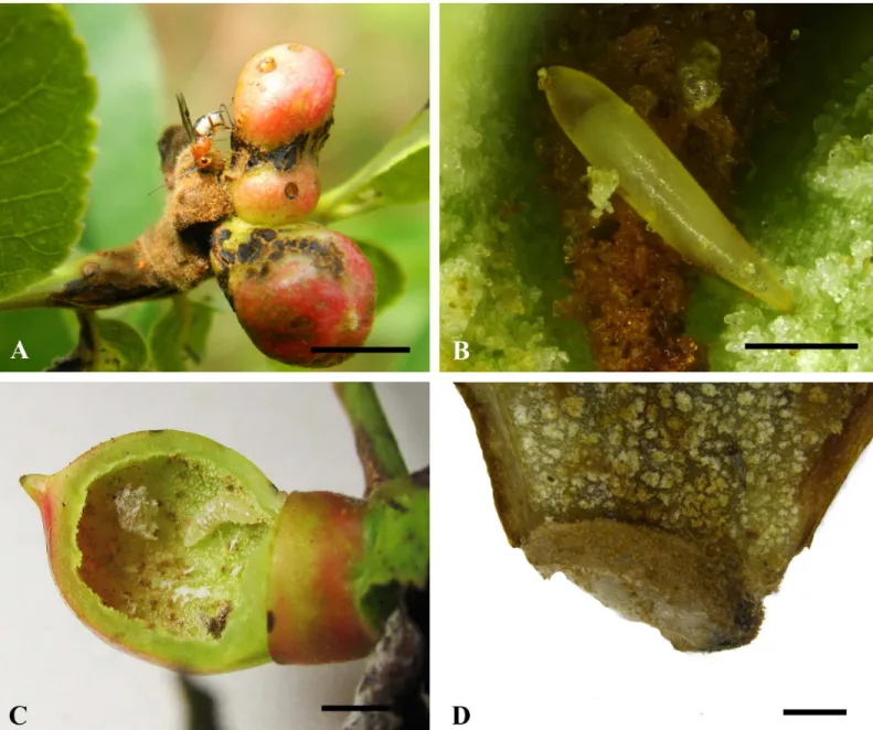
garugaphagaewere observed in galls ranging from 8–22 mm in diameter (n = 779). We did not manage to determine the precise number of instars. Maximum larval length was 5.3 mm (Fig 1C) (n = 779), at which stage larvae started to spin a cocoon (Fig 1D). The spindle-shaped, pale brown cocoons (3.5–3.7 mm long; n = 13) are constructed at the base of the gall chamber with the head end facing towards the gall floor. Adult wasps emerge from the gall by chewing an exit hole with their mandibles.
Fig 1. Biology ofBracon garugaphagaeRanjith & Quicke sp. nov.1A, Adult female wasp ovipositing into gall induced by the psyllid,Phacopteron
lentiginosumon leaf ofGaruga pinnata. 1B, Braconid egg in situ.1C, Larva in situ in cut open gall. 1D, Cocoon attached to inner wall of gall. Scale bars: (1A)
8 mm, (1B) 300μm, (1C) 5 mm, (1D) 1 mm.
doi:10.1371/journal.pone.0156997.g001
Larval morphology
Larvae of all sizes possess unique, dorsal, chimney-like tubercles on abdominal segments 1–9 (Figs2A, 2B, 2C,3A–3E and 3F). The apex of each tubercle is formed of a pair of soft, eversible membranous lobes (Figs2B, 2Cand3F) which help maintain the larva in position while feed-ing. The larval cuticle is extensively denticulate and the spiracular system open at all stages. The late instar larvae are covered with a white, waxy substance (Fig 4A and 4B). The larval head has well-developed papilliform antennae (Figs2D, 2E,3B, 3C and 3D) and labial and maxillary palps. The mandibles are strongly recessed, heavily sclerotised and possess two robust ancillary teeth near the base (Fig 2F). Most other cephalic structures are relatively weakly sclerotised (epistome, hypostome labial sclerite). Hypostomal spur virtually absent.
Larval behaviour
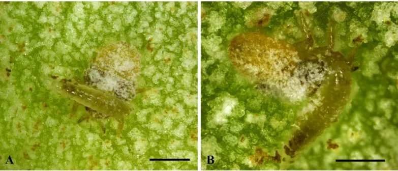
Only oneBraconlarva was observed inside each gall.Bracon garugaphagaepreferentially attacks the third and fourth instar nymphs of the psyllid, by first coiling around the host and biting the host just below the head with their tridentate mandibles (Fig 4A and 4B), which results in the death of the host. Following feeding on the psyllid nymphs,Bracon garugaphagae
larvae exhibit phytophagy which we observed directly in opened galls and confirmed by exam-ining gut contents from mature larvae using both light and scanning electron microscopy. Plant tissue in the larval gut had been chewed into small irregular fragments; only rarely did we find any recognizable pieces of psyllid cuticle
Incidence
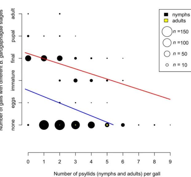
The incidence ofB.garugaphagaesp. nov. larvae in galls was monitored from August 2014 to January 2015. The larvae were first observed in galls which were 1–2 weeks old. The percentage incidence of braconid larvae in immature galls of size 8–12 mm in diameter was 6.5% (n = 779) and in mature galls of size 15–22 mm the incidence was 22%. Some of the galls (n = 53) were observed without any gall-inducing nymphs, but with the presence ofB.garugaphagaelarva.
Fig 5shows the relationship between theBraconstages found in sampled galls and the
num-ber of psyllids present, both nymphs and adults. Two regression analyses were carried out, one with all galls included, even those that had noBraconindividuals present, and one with only galls containing aBraconindividual. Both relationships were highly significantly negative (all data: GLM with Poisson errors and log link-total data: null deviance = 674.62 on 778 degrees of freedom, residual deviance = 558.65 on 777 degrees of freedom, p<0.0001; only whenBracon
present: null deviance = 30.195 on 248 degrees of freedom, residual deviance: 22.693 on 247 degrees of freedom, p<0.0001). Logistic regression of presence or absence ofBraconin galls
(n = 779) versus number of psyllids present, showing highly significant negative relationship (GLM with binomial errors: null deviance = 976.24 on 778 degrees of freedom, residual devi-ance = 920.01 on 777 degrees of freedom, p<5e-12). Gall size and the presence or absence of
Braconwere not correlated (GLM with binomial errors, p = 0.699), but there was a significant positive correlation between gall size and the number of psyllids (GLM with Poisson errors and log link: null deviance = 190.78 on 778 degrees of freedom, residual deviance = 188.82 on 777 degrees of freedom, p = 0.0046).
Systematic part
Bracon garugaphagaesp. nov. Ranjith & Quicke, 2015 (Fig 6)
Distribution. Known only from Kerala, south India. Description: Female
Length of body 3.6 mm (3.6–5.8 mm in paratypes), of fore wing 2.8 mm (2.8–4 mm in para-types), and of antenna 2.8 mm (2.8–3.8 mm in paratypes).
Head. Antenna with 24 flagellomeres (24–28 in paratypes). Terminal flagellomere strongly acute. Median flagellomeres normal in dorsal view. First flagellomere 1.2 times length of sec-ond and third flagellomeres respectively, first flagellomere 2.3 times as long as wide. Mandible twisted, only a single tooth visible in anterior view (Fig 6B). Inter-tentorial distance: tentorio-ocular distance = 1.5:1.0 (1.31–1.7: 0.82–1.07 in paratypes). Inter-tentorial distance: height of clypeus = 3.4: 1.0 (2.62–3.54: 0.81–1.04 in paratypes). Face slightly rugose in anterior half with smooth posterior half, sparsely setose laterally, smooth area laterally bordered by indistinct longitudinal groove (Fig 6B). Height of eye: shortest distance between eyes: width of head = 1.0: 1.1: 2.2(1.0–1.68: 1.1–1.83: 2.2–3.67 in paratypes). Oculo-antennal groove well-developed. Frons shiny. Stemmaticum triangular forming equilateral triangle. Shortest distance between posterior ocelli: transverse diameter of posterior ocellus: shortest distance between posterior ocellus and eye = 1.45: 1.0: 3.45 (1.45–1.81: 1–2.53: 3.45–7.06 in paratypes).
Mesosoma 1.6 (1.3–1.6 in paratypes) times longer than maximum height, largely smooth, shiny. Mesoscutum sparsely setose laterally (Fig 6C). Pronotum smooth. Notauli only indicated anteriorly (Figs6Cand7C). Scutellar sulcus narrow, divided by eight carinae. Scutellum smooth (Figs6Cand7C). Median area of metanotum large, smooth, slightly bulged in lateral view, without carina anteriorly. Propodeum smooth with a strong medial longitudinal carina (propodeal carina weak in paratypes), sparsely setose laterally (Fig 6D).
Fore wing, length of veins 3RSb: 3RSa: r-rs = 4.8: 1.7: 1.0 (4.8–5.1: 1.7–2: 1–1.3 in paratypes). Length of veins 2RS: 3RAa: rs-m = 1.6: 1.75: 1.0 (1.6–2.01: 1.75–2.14: 1–1.02 in paratypes). Vein 2-M 1.6 times 3RSa. Vein 1-M straight. Vein (RS+M)a strongly curved posteriorly (Fig 6F). Vein rs-m without bulla. Vein 1RS forming an angle of 70° with vein C+SC+R. Vein m-cu 0.46 times 1-M. Vein 1cu-a interstitial. Hind wing vein R1 = 1.4 (1.4–1.7 times in paratypes) times length of 1r-m. Apex of vein C+SC+R with one hamulus. Base of hind wing with medium sized glabrous area distal to vein cu-a on posterior half of cell.
Claws with pointed basal lobe. Lengths of fore femur: tibia: tarsus = 1.0: 1.1: 1.1 (1–1.3: 1.1–
1.4: 1.1–1.3 in paratypes). Fore tibia with transverse apical row of thickened bristles. Lengths of hind femur: tibia: basitarsus = 1.8: 2.45: 1.0 (1.8–3.44: 2.45–4.52: 1–1.75 in paratypes).
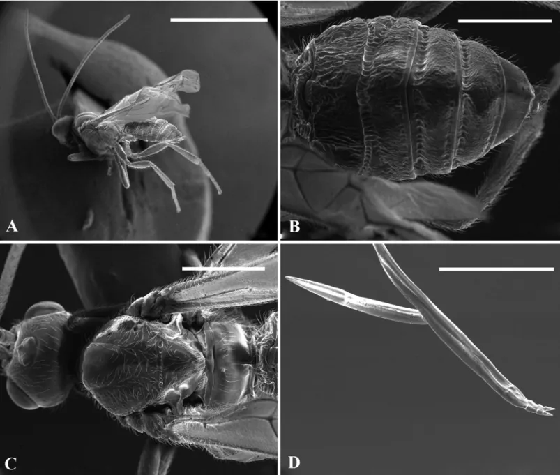
Metasoma largely sculptured (moderately sculptured in paratypes) and dull with seven exposed, sparsely setose, tergites (Fig 6E). First metasomal tergite as long as wide, median area largely smooth and shiny, dorso-lateral carina strong and lamelliform. Second tergite rugose, 2.8 times wider than medially long, with large triangular mid basal area formed posteriorly into a mid-longitudinal carina that extends 0.3 times length of tergite, with a pair of sub lateral grooves. Second metasomal suture sinuate medially, strongly crenulate; third tergite rugose, 3.8 times wider than medially long, without sublateral grooves and with antero-lateral areas defined. Tergite 4–7 rugose, sparsely setose. Hypopygium sharply pointed, reaching end of metasomal tergites (Fig 6A). Ovipositor sheaths 2.2 times longer than hind tibia. Ovipositor slender, darkened sub apically (Fig 6A), dorsal valve with a distinct nodus and ventral valves with three normal teeth (Fig 7D).
Fig 2. Stereozoom and Light microscopic images of larva ofBracon garugaphagaeRanjith & Quicke sp. nov.2A–C, Mature larva showing dorsal abdominal tubercles with eversible tips.2D, Head capsule and anterior thorax of living mature larva showing denticulate cuticle. 2E, 2F, cl. Scale bars: (2A) 500μm, (2B) 300μm, (2C, 2D) 250μm.
Head brownish yellow except malar area, palps, basal region of mandible dark yellowish, tips of mandible black, antenna dark brown, compound eye greyish brown, mesosoma largely dark yellow except lateral mesoscutum brownish yellow, area near to medial longitudinal carina of propodeum dark brown, yellowish laterally, metasoma dark brown except lateral areas of tergites 1–6, medial area of first tergite yellow, mid basal area of second tergite dark yellow, tergites 2–6 blackish medially, hypopygium dark sub apically and ventrally, ovipositor sheath brown, legs mostly yellowish except fore telotarsi, hind coxa medially, hind femur, tibia and tarsus brown–black, wings hyaline, pterostigma dark brown, vein 1-SR+M and r-m less pigmented.
Male: Similar to female, somewhat smaller, 4–4.2 mm and antenna 2.8–3.1 mm, flagellum with 24 segments, body slightly yellower than females, and metasomal tergites less strongly sculptured (Fig 7A and 7B).
Material examined. Holotype: female,INDIA, Kerala, Malappuram, Kottakkal, 10°99’N, 76°00’E, emerged from leaf galls onGaruga pinnataRoxb. 20.ix.2014, leg. U.K.A. Saleem (Department of Zoology, University of Calicut, Kerala, India).
Paratypes: (12 females, 25 males),INDIA, Kerala, Malappuram, Kottakkal, 10°99’N, 76° 00’E, emerged from leaf galls onGaruga pinnataRoxb. 1.x.2011, leg. U.K.A. Saleem (4 females, 20 males); same data except 20.ix.2014 (5 females, 4 males);INDIA, Kerala, Malappuram, Vet-tichira, 10°93’N, 76°02’E, emerged from leaf galls onGaruga pinnataRoxb. 21.i.2015, leg. A.P. Ranjith (3 females, 1 male).
All specimens were reared from leaf galls. The holotype and all paratypes are deposited in Department of Zoology, University of Calicut. Male and female pairs will be deposited in the Forest Research Institute, Dehradun, India (FRI), Natural History Museum, London, UK, the
Fig 3. Scanning electron microscope images of larva ofBracon garugaphagaeRanjith & Quicke sp. nov.3A–B, Final instar larva oblique dorsal view and detail of head capsule. 3C, Putative 2ndinstar larva detail of head capsule, 3D, Same, in anterior view, 3E, Dorsal, chimney-like tubercles on abdominal segments 1–9, 3F, Soft, eversible membranous lobes on the apex of tubercle. Scale bars: (A) 500μm,
(B, D, E) 100μm, (C) 50μm, (F) 10μm.
doi:10.1371/journal.pone.0156997.g003
Fig 4. Behaviour ofBracon garugaphagaeRanjith & Quicke sp. nov.4A–B, Braconid larva feeding on immature psyllids. Scale bars: (4A–B) 500μm.
Smithsonian Institution, Washington DC, USA and Muséum National d'Histoire Naturelle, Paris, France.
Notes. This new species can be distinguished from the two other known phytophagous
Braconspecies, both of which are Neotropical, in having 24 flagellomeres (58 inB. phytopha-gousand 49 inB.zuleideae), median flagellomeres normal in dorsal view (diamond-shaped in
B.phytophagus), face slightly rugose anteriorly and smooth posteriorly (smooth inB. phytopha-gousandB.zuleideae), scutellar sulcus moderately wide (narrow inB.phytophagus), propo-deum with a strong medial longitudinal carina (propopropo-deum smooth without carina inB.
phytophagousandB.zuleideae), fore wing vein 1RS forming an angle of 70° with vein C+SC+R (80° inB.phytophagousand 100°B.zuleideae). Further, the Neotropical species are generally larger and have the ovipositor exceedingly thin and largely unsclerotised except for the apical part which is nearly black and presumably very hard.
Fig 5. Relationship between the number of galls with different stages ofBracon garugaphagaeRanjith & Quicke sp. nov. and the number of live psyllids present in each gall.Symbol size indicates numbers of galls; numbers of galls with nymphal and adult psyllids are shown in black and yellow respectively. Regression lines are shown for all data (blue) and only data whenBraconpresent (red). Slopes of both relationships are highly significantly different from zero.
doi:10.1371/journal.pone.0156997.g005
Bracon garugaphagaesp. nov. can be distinguished fromB.psyllivorusAchterberg (reared from psyllid-induced leaf galls) in having the frons with a distinct medial longitudinal suture, impressed notauli, scutellum without antero-medial puncture, scutellar sulcus divided by eight carinae (five inB.psyllivorus), metanotum not tuberculate in lateral view, metasomal tergites 3–5 with distinct longitudinal strip and apex of ovipositor with distinct dorsal nodus and ven-tral serrations.
Fig 6. Bracon garugaphagaeRanjith & Quicke sp. nov., female, holotype;6A, Habitus in lateral view, 6B, Head in frontal view, 6C, Mesosoma in dorsal view, 6D, Propodeum and first metasomal tergite in dorsal view, 6E, Metasomal tergite in dorsal view, 6F, Wings. Scale bars: (6A) 1 mm, (B) 100μm (6C, D, F) 200μm (6E) 500μm.
doi:10.1371/journal.pone.0156997.g006
Considering the size of the subfamily, very few braconines have sequence data available in GenBank. The closest BLAST search matches (conducted on 25/07/2015) for both sequenced gene fragments were members of the Braconinae but only 93% and 98% similar for CO1 and 28S respectively.Bracon phytophaguswas not sequenced for CO1 but its 28S sequence is only 94% similar (differing in 29 positions) showing, as expected based on morphology, that the seed-predating New World species are only distantly related to the new Indian species. No DNA data are available forB.psyllivorusfor comparison.
Discussion
Relationships of
Bracon garugaphagae
sp. nov
The new species belongs to the large spectrum ofBraconspecies of size between 3–6 mm and differs markedly from the two other known phytophagousBraconspecies both of which are Neotropical [14,15] notably in having the strongly sculptured metasomal tergites. DNA sequence data further indicates that they are only distantly related. Instead, the new species appears to be most closely related toB.psyllivorus, which also attacks gall-forming Psylloidea viz:B.psyllivorusreared from the leaf galls induced byPauropsylla gibberulosaLi andP. braco-naeLi (Hemiptera: Triozidae) [18]. Apart from the host record, no further biological details are known forB.psyllivorus[18], and the possibility that it is also entomophytophagous cannot be excluded.
Biology
Braconine wasps are almost entirely idiobiont ectoparasitoids of various concealed Coleoptera, Diptera, Hymenoptera and Lepidoptera with one small group of genera, the Aspidobraconina, being idiobiont endoparasitoids on exposed butterfly pupae [7,31]. Psyllids have only been recorded as hosts of two other species of braconid wasp. Chadwick and Nikitin [32] recorded an unidentifiedBraconsp. from a psyllid host in Australia, but no further details were pro-vided. Recently, Li et al. [18] described a Chinese species,B.psyllivorus, as a parasitoid/predator of the psyllidsP.gibberulosaLi andP.braconaeLi (Hemiptera: Triozidae) that produce galls on the fig tree (Ficus hainanensisMerr. & Shun.; Moraceae).
The negative relationship between the number of psyllid individuals in galls and the devel-opmental stage ofB.garugaphagaeindicates that the latter sequentially predate the psyllid nymphs before turning to phytophagy. The dietary shift seems likely to be because the psyllids do not provideB.garugaphagaewith sufficient food to complete development. We do not know whetherB.garugaphagaeis obligately or only facultatively phytophagous during its late larval stages. In contrast to the previous explanation, the absence ofB.garugaphagaelarvae from galls with higher numbers (7–8 nymphs) of psyllids, may simply be that because the para-sitoid larvae have to compete with psyllid nymphs for plant tissue the adult female parapara-sitoids selectively avoid such galls.
Predatory behaviour is known in several parasitoid wasp taxa including cryptine ichneumo-noids in spider egg sacs [7]. However, entomophytophagy whilst well-known in several euryto-mid chalcidoids, which develop initially as parasitoids and complete their life cycle as
predatory behaviour. One Neotropical species ofCompsobraconoidesis predatory onAzteca
ants and their brood [37], an AfrotropicalTrigastrothecaspecies also consumes ant broods in ant plant domatia [38]; and an AustralianBraconspecies consumes broods of gall-forming fer-gussoninid flies [32]. All these braconid species that prey on broods are contained within a sin-gle swollen plant structure, either an ant domatium or an insect-induced gall. It is also likely that a similar biology prevails in another braconine,Ficobracon brusiAchterberg & Weiblen, which has been reared from figs ofFicus wassaRoxb., but it is not clear whether it is a parasit-oid/predator of the pollinating agaonid wasps (Chalcidoidea) or other fig inquilines or even whether it might be at least partly phytophagous [39]. Several other members of Braconinae, Doryctinae and Mesostoinae have also been associated with plant galls as inquilines or parasit-oids on gall inducers [11,39,40] and the possibility that some of these are also partly phytoph-agous cannot be excluded.
Final instar larval cephalic structures, notably the long and relatively heavily sclerotised mandibles with two/three robust accessory teeth are very different from those of purely ecto-parasitoid braconines. The latter have a very robust, almost square mandibular base and a short blade furnished with a series of small, closely spaced (comb-like) teeth [41]. The final instar larval mandibles of the entirely phytophagousBracon phylacteophagusare even more heavily sclerotised, have even more closely-spaced robust ancillary teeth and the hypostomal spur is very well developed. In contrast, the mandibles of the gall-forming Mesostoinae are short-bladed, robust and lack ancillary teeth [42]. Clearly there are multiple possible types of larval head capsule adaptations for feeding on plant gall tissue.
All three known phytophagousBraconspecies, viz.,B.phytophagus,B.zuleideaeandB. gar-ugaphagaeare associated with members of the plant family Burseraceae [14,15]. The Neotropi-cal species do not seem to induce gall tissue in the seeds they feed upon and their larval mandibles are far more highly modified and hardened than in the Indian species described here. The Neotropical species are not reliant on another insect having previously damaged the seed, and instead the apex of their ovipositors are extremely heavily sclerotised almost certainly as an adaptation to penetrating a much harder seed-coat. No such ovipositor modification is displayed byB.garugaphagaewhich only has to penetrate relatively softer gall tissue to reach its host.
Supporting Information
S1 Video. Showing the phytophagous feeding behaviour ofBracon garugaphagaeRanjith & Quicke sp. nov. (MP4).This video was recorded by A. P. Ranjith on June 2015 at the Insect Ecology and Ethology Laboratory, University of Calicut, with a Leica S8 APO stereozoomtrino-cular microscope.
(MP4)
Acknowledgments
APR is grateful to Kiran Kishore and Sruthy Vasudevan for contributing much effort to the lengthy gall dissections. APR and MN are grateful to the University of Calicut for facilities pro-vided and MN is grateful to SAP and UGC for their support. Chris Raper kindly stacked the light microscopy larval head capsule images for us.
Author Contributions
Conceived and designed the experiments: APR DLJQ UKAS MN. Performed the experiments:
materials/analysis tools: APR DLJQ UKAS BAB AZR MN. Wrote the paper: APR DLJQ MN. Described the species: APR DLJQ.
References
1. Wharton RA. Bionomics of the Braconidae. Ann Rev Ent. 1993; 30: 121–143. doi:10.1146/annurev. en.38.010193.001005
2. Jervis MA, Kidd NAC. Host-feeding strategies in hymenopteran parasitoids. Bio Rev. 1986; 61: 395– 434. doi:10.1111/j.1469-185X.1986.tb00660.x
3. Godfray HC. Parasitoids: behavioural and evolutionary ecology. Princeton University Press, Prince-ton, NJ. 1994.
4. Pennacchio F, Strand MR. Evolution of developmental strategies in parasitic Hymenoptera. Ann Rev Ent. 2006; 51: 233–258. doi:10.1146/annurev.ento.51.110104.151029PMID:16332211
5. Heimpel GE, de Boer JG. Sex determination in the Hymenoptera. Ann Rev Ent. 2008; 53: 209–230. doi:10.1146/annurev.ento.53.103106.093441PMID:17803453
6. Desneux N, Barta RJ, Hoelmer KA, Hopper KR, Heimpel GE. Multifaceted determinants of host speci-ficity in an aphid parasitoid. Oecologia. 2009; 160: 387–398. doi:10.1007/s00442-009-1289-xPMID: 19219460
7. Quicke DLJ. The Braconid and Ichneumonid Parasitic Wasps: Biology, Systematics, Evolution and Ecology. Wiley Blackwell, Oxford. 2015.
8. de Macêdo MV, Monteiro RT. Seed predation by a braconid wasp,Allorhogassp. (Hymenoptera). J New York Ent Soc. 1989; 97: 359–362.
9. Marsh PM. Description of a phytophagous doryctine braconid from Brazil (Hymenoptera: Braconidae). Proc Entomol Soc Wash. 1991; 93: 92–95.
10. Marsh PM. The Doryctinae of Costa Rica (excluding the genusHeterospilus). Mem Amer Ent Inst. 2002; 70: 1–319.
11. Wharton RA, Hanson PE. Gall wasps in the family Braconidae (Hymenoptera). In: Raman A, Schaefer WC, Whiters TM, editors. Biology, Ecology, and Evolution of Gall-inducing Arthropods. New Hamp-shire: Science Publishers; 2005. pp. 495–505.
12. Zaldívar-Riverón A, Mori M, Quicke DLJ. Systematics of the cyclostome subfamilies of braconid para-sitic wasps (Hymenoptera: Ichneumonoidea): a simultaneous molecular and morphological Bayesian approach. Mol Phyl Evol. 2006; 38: 130–145. doi:10.1016/j.ympev.2005.08.006PMID:16213168 13. Austin AD, Dangerfield PC. Biology ofMesostoa kerriAustin and Wharton (Insecta: Hymenoptera:
Bra-conidae: Mesostoinae), an endemic Australian wasp that causes stem galls onBanksia marginataCav. Aust J Bot. 1998; 46: 559–569. doi:10.1071/BT97042
14. Flores S, Nassar JM, Quicke DLJ. Reproductive phenology and pre-dispersal seed predation in
Pro-tium tovarense(Burseraceae), with description of the first known phytophagous“Bracon”species
(Hymenoptera: Braconidae: Braconinae). J Nat Hist. 2005; 39: 3663–3685. doi:10.1080/ 00222930500392659
15. Perioto NW, Lara RIR, Ferrerira CS, Fernandes DRR, Pedroso EDC, Volpe HXL et al. A new
phytopha-gousBraconFabricius (Hymenoptera: Braconidae) associated withProtium ovatum(Burseraceae)
fruits from Brazilian savannah. Zootaxa. 2011; 3000: 59–65.
16. Shaw MR, Huddleston T. Classification and biology of braconid wasps (Hymenoptera: Braconidae). Hand ldent Brit Ins. 1991; 7(11): 1–126.
17. Belshaw R, Lopez-Vaamonde C, Degerli N, Quicke DLJ. Paraphyletic taxa and taxonomic chaining: evaluating the classification of braconine wasps (Hymenoptera: Braconidae) using 28S D2-3 rDNA sequences and morphological characters. Biol J Linn Soc. 2001; 73: 411–424. doi:10.1006/bijl.2001. 0539
18. Li F, van Achterberg C, He J. New species of the family Triozidae (Homoptera: Psylloidea) from China, and the first record of Psylloidea as host of Braconidae (Hymenoptera). Zool Med Leiden. 2000; 74 (21): 359–366.
19. Salt G. Parasites of the wheat-stem sawfly,Cephus pygmaeusLinnaeus, in England. Bull Ento Res. 1931; 22: 479–545. doi:10.1017/S0007485300035355
20. Glover PM, Chatterjee KC. A preliminary note on the bionomics and economic importance of
Microbra-con hebetorSay, a braconid new to north India. Proc Ind Acad Sci. 1936; 3: 195–211. doi:10.1007/
BF03047071
22. Jervis MA, Kidd NAC. Phytophagy. In: Jervis MA, Kidd NAC, editors. Insect Natural Enemies. Chap-man & Hall, London, UK; 1996. pp. 375–394.
23. AccuWeather. Available:http://www.accuweather.com/en/in/india-weather. 2015. Accessed 11/08/ 2015.
24. van Achterberg C. Revision of the subfamily Blacinae Foerster (Hymenoptera, Braconidae). Zool Verh Leiden. 1988; 249: 1–324.
25. van Achterberg C. A revision of the subfamily Zelinae auct. (Hymenoptera, Braconidae). Tijdschr Ent. 1979; 122: 241–479.
26. Eady RD. Some illustrations of microsculpture in the Hymenoptera.Proc Roy Ent Soc LonSeries A. 1968; 43: 66–72. doi:10.1111/j.1365-3032.1968.tb01029.x
27. Harris RA. A glossary of surface sculpturing. Calif Dep Food Agri Bur Ento Occ Pap. 1979; 28: 1–31. 28. Zaldivar-Riverón A, Shaw MR, Sáez AG, Mori M, Belokobylskij SA, Shaw SR, Quicke DLJ. Evolution of
the parasitic wasp subfamily Rogadinae (Braconidae): phylogeny and evolution of lepidopteran host ranges and mummy characteristics. BMC Evol Biol. 2008; 8: 329–349. doi:10.1186/1471-2148-8-329 PMID:19055825
29. Zaldívar-Riverón A, Rodríguez-Jiménez A, Sarmiento CE, Pedraza-Lara C, López-Estrada EK. Phylo-genetic relationships and description ofBolivar, a new genus of Neotropical doryctine wasps (Hyme-noptera: Braconidae). Invert Syst. 2013; 27: 673–688. doi:10.1071/IS13021
30. R Development Core Team. R: A language and environment for statistical computing. Vienna, Austria: R Foundation for Statistical Computing. 2019.
31. Quicke DLJ. The Old World genera of braconine wasps (Hymenoptera: Braconidae). J Nat Hist. 1987; 21: 43–157. doi:10.1080/00222938700770031
32. Chadwick CE, Nikitin MI. Records of parasitism in the families Ichneumonidae, Braconidae and Aulaci-dae. J Ent Soc Aus (NSW). 1976; 9: 28–38.
33. Henneicke K, Dawah HA, Jervis MA. The taxonomy and biology of final instar larvae of some Eurytomi-dae (Hymenoptera: Chalcidoidea) associated with grasses in the UK. J Nat Hist. 1992; 26: 1047–1087. doi:10.1080/00222939200770621
34. Gauld ID, Wahl DB, Broad GR. The suprageneric groups of the Pimplinae (Hymenoptera: Ichneumoni-dae): a cladistic re-evaluation and evolutionary biological study. Zool J Linn Soc. 2002; 136: 421–485. doi:10.1046/j.1096-3642.2002.00031.x
35. Slobodchikoff CN. Bionomics ofGrotea californicaCresson, with a description of the larva and pupa (Hymenoptera: Ichneumonidae). Pan-Pacif Entomol. 1967; 43: 161–168.
36. Goolsby JA, Burwell CJ, Makinson J, Driver F. Investigation of the biology of Hymenoptera associated
withFergusoninasp. (Diptera: Fergusoninidae), a gall fly ofMelaleuca quinquenervia, integrating
molecular techniques. J Hym Res. 2001; 10: 163–180.
37. Yu DW, Quicke DLJ.Compsobraconoides(Braconidae: Braconinae), the first hymenopteran ectopara-sitoid of adultAztecaants (Hymenoptera: Formicidae). J Hym Res. 1997; 6:419–421.
38. Quicke DLJ, Stanton ML.Trigastrotheca laikipiensisn. sp. (Hymenoptera: Braconidae): A new species of brood parasitic wasp that attacks foundress queens of three coexisting acacia-ant species in Kenya. J Hym Res. 2005; 14:182–190.
39. van Achterberg C, Weiblen GD.Ficobracon brusigen. nov. & spec. nov. (Hymenoptera: Braconidae), a parasitoid reared from figs in Papua New Guinea. Zool Med Leiden. 2000; 74: 51–55.
40. Wei P, Li Z, van Achterberg C, Feng G, Xiao H, Huang DW. Two new species of the genusFicobracon
van Achterberg and Weiblen (Hymenoptera: Braconidae) from China, expanding its host range. Zoo-taxa. 2013; 3640: 465–472. doi:10.11646/zootaxa.3640.3.8PMID:26000428
41. Capek M. A new classification of the Braconidae (Hymenoptera) based on the cephalic structures of the final instar larva and biological evidence. Canad Entomol. 1970; 102: 846–875. doi:10.4039/ Ent102846-7
42. Quicke DLJ, Huddleston T. The Australian braconid wasp subfamily Mesostoinae (Hymenoptera: Bra-conidae) with the description of a new species ofMesostoa. J Nat Hist. 1989; 23: 1309–1317. doi:10. 1080/00222938900770691