Recognition by the Drosophila Peptidoglycan
Recognition Protein-SA to Promote Pathogenicity
Magda L. Atilano1., James Yates1., Marcus Glittenberg2., Sergio R. Filipe1"*, Petros Ligoxygakis2"*
1Laboratory of Bacterial Cell Surfaces and Pathogenesis, Instituto de Tecnologia Quı´mica e Biolo´gica/Universidade Nova de Lisboa, Oeiras, Portugal,2Genes and Development Laboratory, Department of Biochemistry, University of Oxford, Oxford, United Kingdom
Abstract
The cell wall of Gram-positive bacteria is a complex network of surface proteins, capsular polysaccharides and wall teichoic acids (WTA) covalently linked to Peptidoglycan (PG). The absence of WTA has been associated with a reduced pathogenicity
ofStaphylococcus aureus (S. aureus). Here, we assessed whether this was due to increased detection of PG, an important
target of innate immune receptors. Antibiotic-mediated or genetic inhibition of WTA production in S. aureus led to increased binding of the non-lytic PG Recognition Protein-SA (PGRP-SA), and this was associated with a reduction in host susceptibility to infection. Moreover, PGRP-SD, another innate sensor required to control wild typeS. aureus infection, became redundant. Our data imply that by using WTA to limit access of innate immune receptors to PG, under-detected bacteria are able to establish an infection and ultimately overwhelm the host. We propose that different PGRPs work in concert to counter this strategy.
Citation:Atilano ML, Yates J, Glittenberg M, Filipe SR, Ligoxygakis P (2011) Wall Teichoic Acids ofStaphylococcus aureusLimit Recognition by the Drosophila Peptidoglycan Recognition Protein-SA to Promote Pathogenicity. PLoS Pathog 7(12): e1002421. doi:10.1371/journal.ppat.1002421
Editor:David S. Schneider, Stanford University, United States of America
ReceivedJune 13, 2011;AcceptedOctober 20, 2011;PublishedDecember 1, 2011
Copyright:ß2011 Atilano et al. This is an open-access article distributed under the terms of the Creative Commons Attribution License, which permits unrestricted use, distribution, and reproduction in any medium, provided the original author and source are credited.
Funding:This work was funded by ‘‘Fundac¸a˜o para a Cieˆncia e Tecnologia’’ through research grants PTDC/SAU-MII/75696/2006 and PTDC/BIA-MIC/100747/2008 (S.R.F.), and fellowships SFRH/BD/28440/2006 (M.A.), SFRH/BPD/23838/2005 (J.Y.). J.Y. was also supported by an EMBO long-term fellowship (ALTF 1042–2007). Work in Oxford was funded by the Medical Research Council UK through Career Establishment Grant G0300170 and the Wellcome Trust through project grant WT087680 MA (both to P.L.). The funders had no role in study design, data collection and analysis, decision to publish, or preparation of the manuscript.
Competing Interests:The authors have declared that no competing interests exist.
* E-mail: [email protected] (SRF); [email protected] (PL)
.These authors contributed equally to this work. "These authors also contributed equally to this work.
Introduction
The complex cell surface of bacteria has been directly or indirectly associated with different strategies that bacterial pathogens use to interact with the host. These include acquisition of specific adhesion factors, formation of biofilms, adaptation to an intracellular environment, production of a protective capsular polysaccharide or evasion of innate immune defences (e.g. lysozyme) [1]. The host counters these strategies by targeting conserved molecules (pathogen associated molecular patterns or PAMPs), unique in bacteria, that are either present at the bacterial surface or are released by bacteria as they attempt to establish infection. Bacterial PAMPs include Peptidoglycan (PG), a heterogeneous polymer of glycan chains cross-linked by short peptides of variable length and amino acid composition [2]. Although PG recognition is essential to trigger an inflammatory response, this macromolecule may not be easily accessible for recognition at the surface of bacteria.
In Gram-positive bacteria, PG is buried within a complex cell surface consisting of different molecules [3–5]. Such molecules include surface proteins, covalently linked or tightly associated with PG, capsular polysaccharides, usually required for the ability of different bacteria to cause disease [6] and wall teichoic acids (WTA), phosphate-rich glycopolymers involved in the resistance of bacteria to environmental stress and regulation of bacterial
division [7]. It is not clear therefore, how the host would be able to sense bacterial PG buried within such complex structures. One hypothesis is that the innate immune system recognises soluble PG fragments that are released from the bacterial cell surface through the activity of enzymes produced by bacteria (such as autolysins) or by the host (such as lysozyme) [2,8]. However, certain bacteria have the ability to modify their PG, turning it more resistant to the action of such enzymes [9], thus preventing the release of small soluble fragments capable of triggering an innate immune response in the host. This may be the case forListeria monocytogenes
that has the ability to de-N-acetylate its PG allowing them to survive the action of lysozyme and evade the host innate immune system [10]. Another hypothesis is that the components of the host innate immune system are able to bind directly to PG present within the bacterial cell surface. As discussed earlier, PG is decorated with a variety of large molecules that may sterically block access of host receptors to the underlying PG. In Gram-positive bacteria, cell wall glycopolymers, including WTA may play this role [1]. The role of WTA protecting the PG from recognition would have important implications regarding the onset of infection by major human pathogens such asStaphylococcus aureus
(S. aureus) [1]. Recently, it has been shown that different components, present at the cell wall of S. aureus bacteria, may determine the survival of infectedDrosophila. Specifically,S. aureus
metabolism of cell wall components were unable to kill flies [11]. Moreover, it has been proposed that D-alanylation of the WTA produced by S. aureusmay inhibit the recognition of PG by host receptors. This inhibitory effect was observedin vitronot only when WTA was covalently attached to polymeric PG but, surprisingly, also when WTA was covalently attached to monomeric PG [12]. The fruit fly Drosophila melanogaster recognises Gram-positive bacteria by either direct binding to PG or its smallest components [13]. Based onin vitrodata [14] and infection studies of mutants [14,15], the current working hypothesis is that a flexible system of pattern recognition receptors (PRRs) can be deployed by the host immune system to detect Lysine-type PG from different Gram-positive bacterial pathogens. Two Peptidoglycan Recognition Proteins (PGRPs), namely PGRP-SA and PGRP-SD are major components of this system [15,16]. Depending on the bacterium, each, or both of these PGRPs – along with Gram-Negative Binding Protein1 (GNBP1) [17] – interacts with PG and activate a downstream proteolytic cascade, which culminates in Toll receptor signalling. The signal reaches the cytoplasmic NF-k B/I-kB complex via a receptor/adaptor complex comprising dMyD88,
Tube and the IRAK homologue Pelle. At that point the I-kB homologue Cactus is phosphorylated and targeted for degradation while the NF-kB homologue Dif is free to enter the nucleus of host cells and regulate target genes [18]. Prominent among these genes, is a group of potent antimicrobial peptides (AMPs), which are synthesised by the fat body and secreted into the haemolymph. An AMP frequently used as a read-out for the Toll pathway is
Drosomycin (Drs). AMPs and local melanization, along with the phagocytic activity of haemocytes constitute respectively the humoral and cellular arm of the fruit fly response to infection [18]. Here, we report for the first time that Drosophila PGRP-SA, a non-lytic PGRP was able to bind intact live bacteriain vivo. Access to PG was limited by the presence of WTA: binding of PGRP-SA to various live Gram-positive bacteria was minimal, but binding to purified PG, stripped of covalent modifications (including WTA) was far greater. Through inhibiting WTA synthesis, either by the addition of an antibiotic or genetically, we were able to potentiate detection of these bacteria by PGRP-SA. For S. aureus, this
correlated with a reduced ability of the bacteria to proliferate within the host, and a reduced susceptibility of the host to infection in a PGRP-SA/GNBP1 dependent manner. We also observed that PGRP-SD, essential for sensing wild type S. aureus, became redundant as WTA levels were reduced. Overall, our results suggest that WTA may be part of a general mechanism used by Gram-positive bacteria, which limits the access of innate receptors to PG, thereby enabling bacteria to evade detection and establish infection.
Results
To address the question of whether Gram-positive bacteria counter host recognition by limiting access of innate sensors to PG, we constructed a fluorescent derivative of the fruit fly Lys-type PG receptor, PGRP-SA (mCherry-PGRP-SA). This construct and an untagged version (rPGRP-SA) were expressed inEscherichia coliand the resulting proteins were purified. As shown in the supplemen-tary material (Figure S1A), injection of mCherry-PGRP-SA, or rPGRP-SA, into PGRP-SA deficient flies restored Drs-GFP
production induced by infection withMicrococcus luteus(M. luteus). Endogenous Drs expression was also restored as confirmed by qPCR (Figure S1B). These observations were consistent with our previous results when using a recombinant PGRP-SA expressed in the lepidopteran cell line Sf9 [19]. Taken together, these results showed that the fluorescently tagged PGRP-SA and the untagged versions are functional and capable of restoring an innate immune response in PGRP-SA deficient flies.
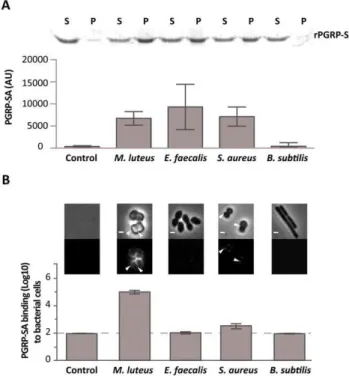
Initially, we used rPGRP-SA and mCherry-PGRP-SA in co-precipitation experiments in order to study binding to PG from different Gram-positive bacteria. Both bound with similar affinity to PG purified fromM. luteus,Enterococcus faecalis(E. faecalis), andS. aureus(data not shown and Figure 1A, respectively). For details of PG composition of these bacteria see Figure S2. Importantly, this indicated that the mCherry-tag appeared not to interfere with PGRP-SA binding, and thus, demonstrated that both proteins were able to bind Lys-type PG of different composition. We therefore assessedin vitro, the binding of mCherry-PGRP-SA to the surface of live bacteria harvested during exponential growth phase. Notably, the binding of the recombinant protein to live bacteria exhibited a range of different affinities in contrast to their respective purified PG. Binding to live E. faecalis and S. aureus was significantly reduced, when compared to binding toM. luteus
(Figure 1B). However, the binding levels of PGRP-SA to the purified PG from these bacteria were similar (Figure 1A). We also noticed that while mCherry-PGRP-SA was capable of binding the entire surface of M. luteus cells, it bound at specific sites at the surface of S. aureus cells, similar to what has been described recently for mammalian bactericidal PGRPs [20]. These results suggested that although the three types of bacterial PG were similarly recognized by PGRP-SA, the presence of other components found at the surface of live bacteria might have prevented PGRP-SA from finding its PG ligand.
The cell surface of a Gram-positive bacterium is a complex structure consisting of a thick layer of PG, surface proteins and glycopolymers such as capsular polysaccharides and WTA. As previous studies had shown that certain PG-binding proteins, such as bacterial autolysins, have a higher affinity for the surface of bacterial strains lacking WTA [21–23], it was decided to investigate whether presence of WTA could be preventing PGRP-SA from binding to the surface of live bacteria. Further support for the choice of WTA came from the fact that different Gram-positive bacteria can produce WTA with a variable composition [24–26]. M. luteus, for which mCherry-PGRP-SA
Author Summary
Gram-positive bacteria such as the opportunistic pathogen
Staphylococcus aureushave their cell wall exposed to the
displayed the highest affinity, does not produce WTA [24,27], (Figure S2C). To test whether WTA mediated the differential binding of PGRP-SA, we cultured bacteria in the presence of tunicamycin, thereby inhibiting their ability to synthesize WTA. At lower, sub-inhibitory concentrations as those used in this study, tunicamycin specifically inhibits TagO [28]: a glycosyltransferase that specifically localizes to the division septum ofS.aureus [29] and is required for the initial step of WTA biosynthesis, namely, the transfer of GlcNAc to the C55-P lipid anchor bactoprenol. We observed higher levels of mCherry-PGRP-SA binding to the newly
synthesized cell material, located at the division septum, when Gram-positive bacteria cells were treated with tunicamycin (Figure 2A). S. aureus and S. saprophyticus exhibited a similar increase in binding, 636and 846respectively, whilst E. faecalis
binding increased 86. It should be noted that the effect of
tunicamycin in these bacteria was not the same. While addition of the antibiotic resulted in binding of mCherry-PGRP-SA to the entire cell surface ofS.aureus, binding was observed predominantly at the division septum in S. saprophyticus and exclusively at this region in E. faecalis. We attribute these differences to how and where the new cell wall synthesis occurs in these bacteria.
Figure 1. Differential binding of PGRP-SA to the surface of live Gram-positive bacteria.(A) PGRP-SA and PG co-precipitation assay. Lys-type PG fromM. luteus,E. faecalis,S. aureus, and DAP-type PG from B. subtilis(this acts as a negative substrate control for PGRP-SA binding, which recognizes Lys-type PG), was incubated with rPGRP-SA for 30 minutes. Unbound rPGRP-SA remained in the supernatant fraction upon centrifugation (S). rPGRP-SA bound to the insoluble PG was co-precipitated and found in the pellet fraction (P). Quantified data (performed using ImageJ software) was plotted as mean values with 95% confidence limits: very little co-precipitation of rPGRP-SA occurred in the absence of PG (labelled Control) or in the presence ofB. subtilis DAP-type PG; however, PGRP-SA was co-precipitated similarly (One-way ANOVA,P.0.05) and at higher levels with the PG fromM. luteus, E. faecalis, or S. aureus. The data shown (mean with 95% confidence intervals) was obtained from 4 independent co-precipitation experi-ments. (B) mCherry-PGRP-SA was incubated with bacteria cells harvested in exponential phase, washed with PBS and visualized using fluorescence microscopy. Grey panels are phase-contrast images of bacterial cells (white scale bar represents 1mm), and black panels
mCherry-PGRP-SA binding: white arrowheads highlight binding to the lateral cell surface or the region of cell division. The total fluorescence of mCherry-PGRP-SA bound to a bacterium (covering all lateral and cell division regions, and including background) was quantified for each species (n= 50), and represented as the median (with 25% and 75% inter-quartile range). Dashed-line indicates the level of the background signal, control. Kruskal-Wallis analysis with Dunn’s multiple comparison post-test did not reveal significant differences (P.0.05) between mCherry-PGRP-SA binding to E. faecalis and B. subtilis, which were indistinguishable from the control. However, the protein bound more to S. aureus and M. luteus relative to the control, with the latter exhibiting highest binding (P,0.05 in all cases).
doi:10.1371/journal.ppat.1002421.g001
Figure 2. WTA reduce PGRP-SA binding at the bacterial cell surface.Grey panels are phase-contrast images of bacterial cells (white scale bar represents 1mm), and black panels mCherry-PGRP-SA binding;
white arrowheads highlight binding to the lateral cell surface or region of cell division. The binding of mCherry-PGRP-SA to individual bacterial cells (n= 50) was quantified, and represented as the median (with 25% and 75% inter-quartile range). (A) mCherry-PGRP-SA binding to Gram-positive bacteria grown with or without tunicamycin, an inhibitor of WTA synthesis. mCherry-PGRP-SA binding to the cell division region, rather than total binding, was measured because binding at the former was consistently enhanced for all treated bacteria species. Mann-Whitney U tests were used to compare differences for treated and untreated between each type of bacteria (P,0.05 in all cases). (B) RNDtagOmutant background rescued with variants of thetagOgene – expressed from a replicative pMAD vector – produce varying levels of WTA, given as a% relative to the wild type RN4220: pMAD vector (0%), ptagO(90%), ptagOD87A/D88A (0%), ptagOG152A (22%). Total binding of mCherry-PGRP-SA to the surface of live bacteria increases as the levels of WTA are reduced. Kruskal-Wallis analysis followed by Dunn’s multiple comparison post-test, revealed significant differences for all comparisons (P,0.05) except for that between PGRP-SA binding to pMAD and ptagOD87A/D88A.
Nevertheless, the results described above suggested that WTA in different bacteria might protect PG from exposure to host receptors.
To confirm that WTA were indeed required to reduce access of PGRP-SA at the cell surface, we quantified the binding of mCherry-PGRP-SA to S. aureus mutants that produced varying amounts of WTA due to mutations in the tagO gene [29]. We choseS.aureusbecause it is a major human pathogen with a well-characterised WTA synthetic pathway [30,31]. A complete absence of WTA, which occurs when tagO is entirely deleted (RNDtagO pMAD), or when two highly conserved residues have been mutated (RNDtagOptagOD87A/D88A), resulted in equivalently enhanced levels of mCherry-PGRP-SA binding, when compared to the wild type strain (,26103and,3.36103-fold respectively, Figure 2B). To verify that the observed result was indeed due to the loss of WTA, we expressed wild type tagO in the RNDtagO background (RNDtagOptagO): this rescued the loss of WTA (WTA levels restored to 90% of wild type levels) [29], and reduced mCherry-PGRP-SA binding to levels close to those observed for the wild type strain (Figure 2B). A tagO mutant that could only support production of a reduced amount of WTA (RNDtagO ptagOG152A; 24% levels of WTA compared to wild type) exhibited an intermediate level of mCherry-PGRP-SA binding relative to all strains (66102-fold increase relative to the wild type strain, Figure 2B). Overall, our data indicated that WTA found in the cell wall of different live Gram-positive bacteria restricted PGRP-SA from binding their PG, and in S. aureus this occurs in a dose dependent manner.
We next wanted to examine whether increased PGRP-SA binding – due to a lack of WTA – affected the ability of bacteria to survive in anin vivosystem. We choseD. melanogasterbecause it is a well-established model for dissecting pattern recognition in innate immunity [18]. We know for example thatin vitro, three PRRs – PGRP-SD/PGRP-SA/GNBP1 – form a ternary complex for binding to the PG ofS.aureus[14]. As a first approach wild type and mutantS. aureusstrains were injected into wild type flies and also into flies defective for PGRP-SD or PGRP-SA. We then determined the number of CFUs 6 and 17 hours post-infection; the latter time point being when the first flies succumb to infection (Figure 3 and S5). All flies were inoculated with low and statistically identical numbers of bacteria (,102CFUs per fly; Figure 3, Time 0). Our rationale was to induce infections that were comparable and that could evolve over time. For example, flies generally succumb to bacterial infection when their numbers increase beyond 106CFUs per fly [18,32], and therefore, high initial loads (e.g. 104–105CFUs per fly) may overwhelm the host and consequently may not be informative regarding the course of an infection. We observed that wild type S. aureus (NCTC8325-4) CFUs increased in all fly backgrounds over the period of infection to numbers that were statistically separable, with PGRP-SA deficient flies carrying the heaviest load (Figure 3). In contrast, the numbers of the S. aureus mutant, which lacked WTA (NCTCDtagO) [29], did not significantly increase in the wild type or PGRP-SD mutant background. However, the number of NCTCDtagO bacteria in the PGRP-SA mutant was significantly higher at both the 6 and 17 hours time points (Figure 3). Two-way ANOVA revealed a significant interaction between the bacteria and fly strains, which was due to the large increase of NCTCDtagO bacteria in the PGRP-SA mutant. Together, these data indicated that WTA were fundamental forS.aureusto counter recognition by PGRP-SA, and consequently, the bacteria were able to increase their number during the initial course of infection.
We have previously observed that PG produced by NCTCDtagO bacteria has reduced levels of cross-linking relative
to the wild type strain [29]. To evaluate whether this contributed to the inability of NCTCDtagObacteria to increase their number in wild type or PGRP-SD mutant flies, we assessed mCherry-PGRP-SA binding to NCTCDpbpDand determined CFUs at 6 and 17 hours. NCTCDpbpD is a derivative of NCTC8325-4 in which pbpD (the gene encoding to penicillin binding protein 4, PBP4) has been deleted. Deletion ofpbpDresults in a strain that produces PG with a similar level of cross-linking to that found in NCTCDtagO[29], but which still produces WTA. The inability of NCTCDpbpD and NCTCDtagO to produce a highly crosslinked PG did not interfere with bacteria growth in culture, as its duplication time at 30uC was very similar to the parental NCTC8325-4 strain (Figure S3B). In both experiments, NCTCDpbpDbehaved as the wild type bacteria. Firstly, binding mcherry-PGRP-SA similarly (Figure S3C) and secondly, for each fly background attaining numbers that were statistically insepara-ble from those for NCTC8325-4 (Figure 3, Time+17 hours).
To assess whether the developing trend in bacterial numbers at 17 hours post-infection resolved into differences in how flies survive, we monitored the number of flies alive at 24 hour intervals over 3 days. In addition, we infected GNBP1 mutant flies, because GNBP1 has been postulated to work as part of a complex with PGRP-SA [14,17]. Survival curves for a particular fly background when infected with either NCTC8325-4 or NCTCDpbpD were statistically inseparable, except for those obtained for the wild type background, where flies succumbed more to NCTCDpbpD (Figure 4; 62% and 38% survival at 72 hours post-infection, respectively). Nearly all PGRP-SA and GNBP1 mutant flies had died by 24 hours, whereas ,40% of PGRP-SD mutant flies survived beyond this time point, succumbing to infection around 48 hours (,5% of flies surviving). In contrast, ,95% of wild type and PGRP-SD mutant flies survived the NCTCDtagO infection up to 72 hours (furthermore, taking CFUs at this time-point revealed that NCTCDtagO had been eliminated from these flies, 0 CFUs per fly). The majority of PGPR-SA and GNBP1 flies had succumbed to infection by 48 hours (3% of flies surviving). A similar trend in survival outcome was observed with NCTC8325-4 after treatment with tunicamycin (Figure 4). These data confirmed that WTA were indeed required to counter host immunity, because without them, infection could be controlled in a PGRP-SA/GNBP1 dependent manner. Differences in CFUs were apparent 6 hours post-infection suggesting that recognition and reduction of propagation or killing of bacteria, occurs rapidly following infection. Interest-ingly, these results also showed that a requirement for PGRP-SD was bypassed when WTA are removed and PGRP-SA has far greater access to PG.
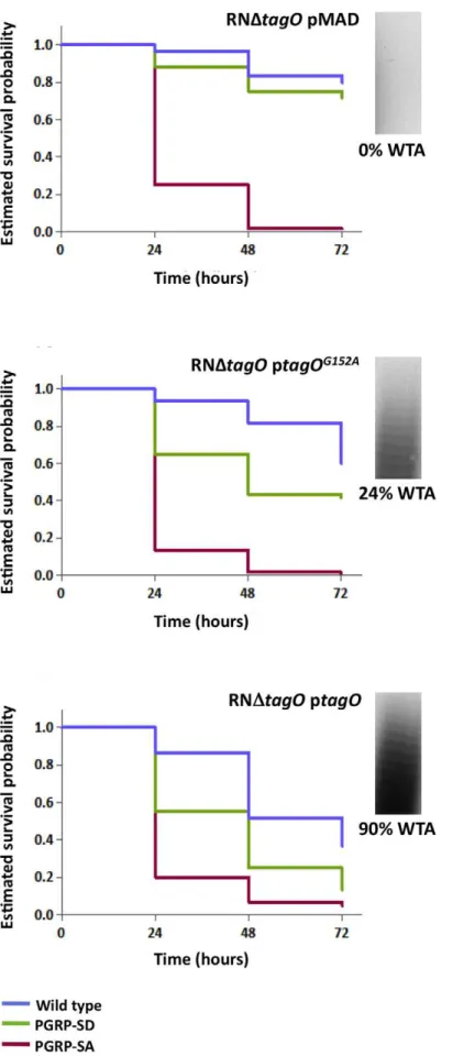
ptagOG152A (which produces ,24% WTA relative to RNDtagO ptagO but produces similar levels of the TagO protein) [29] survived to intermediary levels (Figure 5). Overall, survival of wild type flies decreased as WTA levels increased (with a concomitant decrease in PGRP-SA binding, Figure 2B), and likewise for the PGRP-SD mutant; with the difference between wild type and PGRP-SD mutant survival successively increasing. In contrast, survival of PGRP-SA mutant flies was independent of WTA levels,
with flies succumbing strongly for all infections in a statistically inseparable manner (Figure 5). These data confirmed that it was indeedin vivoprotection of PG by WTA against the consequences of PGRP-SA binding, and furthermore, suggested that a requirement for PGRP-SD gradually became redundant as WTA levels decreased.
It has been reported previously that D-alanylation of WTA is also required for the pathogenicity ofS.aureus[11]; D-alanylation Figure 3. PGRP-SA is fundamental for controlling bacterial numbers in flies infected with aS. aureusmutant that lacks WTA.Wild type flies, and those lacking PGRP-SD or PGRP-SA, were infected with differentS. aureus strains: NCTC8325-4 is the wild type; NCTCDpbpDis a mutant that produces WTA but has a PG similar to NCTCDtagO, both exhibiting reduced cross-linking; NCTCDtagOlacks WTA. The table gives the mean CFUs per fly (from 3 independent experiments). For each time point, the CFUs per fly data set was transformed via a Box-Cox transformation (which returns alnumber, where data-point = data-pointl
– 1/l) and represented as means with 95% confidence intervals. Flies were inoculated with a low
(,100 CFUs per fly) and comparable number of bacteria (Time 0; Two-way ANOVA did not reveal significant differences,P.0.05), and CFUs per fly
were determined at 6 and 17 hours post-infection. In contrast to NCTC8325-4 and NCTCDpbpD, the number of NCTCDtagO bacteria did not significantly increase in the wild type or PGRP-SD mutant background during the period of infection (Table); however, in the PGRP-SA mutant the number of bacteria increased significantly for all strains (P,0.05, Repeated Measures One-way ANOVA). Two-way ANOVA of the CFUs data at Time
+17 hours revealed a significant interaction (P,0.05) between the bacteria and fly strains, which was due to the large increase of NCTCDtagOCFUs in
the PGRP-SA mutant, whilst differences in CFUs were similar for NCTC8325-4 and NCTCDpbpD. One-way ANOVA and 95% Tukey’s HSD intervals were used to look for factor differences at this time. For each fly background NCTC8325-4 and NCTCDpbpDCFUs were equivalent (P.0.05). NCTCDtagO CFUs in the wild type and PGRP-SD backgrounds were similar (P.0.05), but separated from all other values (P,0.05). In the PGRP-SA mutant, NCTCDtagOCFUs reached levels seen with the other bacteria in wild type and PGRP-SD flies. The negative error bars for the NCTCDtagOinfection occur because of large variation of the biological repeats. This is consistent with the fact that NCTCDtagOoccasionally causes a lethal infection in both the wild type and PGRP-SD backgrounds.
is a process that incorporates D-alanine residues into the glycerol-/ ribitol-phosphate backbone of WTA, thereby reducing the negative charge of the polymer [33]. We examined therefore, whether aS.aureus mutant that lacks the D-alanylation pathway (RNDdltABCD) bound mCherry-PGRP-SA equivalently to RNDtagO. Binding of, mCherry-PGRP-SA to RNDdltABCD was similar to the binding to the wild type bacteria (Figure S3). This prompted us to assess how RNDdltABCDaffected survival of the wild type, PGRP-SD and PGRP-SA mutant flies. In contrast to RNDtagO, PGRP-SA mutant flies did not succumb strongly to RNDdltABCD infection, with 83% surviving at 72 hours post-infection (Figure S4); furthermore, survival was statistically inseparable for the different fly backgrounds (Figure S4). These data demonstrated that D-alanylation is not necessary for WTA to limit the access of SA, that neither SD nor PGRP-SA were required to control the RNDdltABCD infection and
consequently, the reduced killing effect of RNDdltABCD had nothing to do with recognition.
Discussion
The results shown here indicate that in respect to Gram-positive bacteria, where the cell wall is not concealed by outer membrane (e.g.staphylococci), pathogen recognition, via recognition of PG, is tightly linked to host survival. Our studies bring forward the notion that one of the strategies used by pathogens to reduce recognition is to restrict accessibility to inflammatory non-self components of the cell wall. Specifically, the results here show that presence of WTA in a range of Gram-positive bacteria impaired PGRP-SA binding. The use of tunicamycin to abolish WTA synthesis dramatically improved receptor recognition of bacteria as well as host survival of flies infected antibiotic treatedS. aureus. Genetically Figure 4. PGRP-SA and not PGRP-SD is required to control infection byS. aureusmutant lacking WTA.Flies assayed for survival were injected concurrently with those for determining CFUs. The survival of infected flies (n= 90) was monitored at 24-hour intervals for three days, and estimates of survival plotted (for clarity, 95% confidence intervals have been omitted). For each fly background – except wild type – survival curves were statistically inseparable for flies infected with NCTC8325-4 or NCTCDpbpD(log-rank test,P.0.05). PGRP-SD, PGRP-SA and GNBP1 mutant flies succumbed strongly to infection by 72 hours, whereas wild type survived up to,60%. In contrast, wild type and PGRP-SD mutant flies were barely
susceptible to infection with NCTCDtagO, however, PGRP-SA and GNBP1 flies succumbed strongly; a similar survival trend was seen when flies were
deleting a major component of the WTA synthesis (TagO) inS. aureusalso increased PGRP-SA binding leading to increased host survival. It should also be noted that, rPGRP-SA was capable of bindingin vitrosignificantly better to free PG than to WTA-linked PG that were purified from wild type S. aureus bacteria, treated with trypsin to remove any attached surface proteins and adjusted to the same concentration of PG (Figure S2). This observation confirmed the results obtained with live bacteria and allowed us to eliminate the notion that deletion oftagOgene may influence the amount of protein present at the cell surface and that this change in protein levels was influencing the binding of PGRP-SA. Effectively during the course of this work we have removed WTA from PG by treatment with antibiotic, by deletion of the
tagOgene and finally we have chemically removed them from PG. In all the cases binding of PGRP-SA to PG has increased.
S. aureus produces WTA composed of about 40 ribitol phosphate-repeating units modified with N-acetylglucosamine (GlcNAc) and D-alanine [7]. The latter modification is mediated by the D-Alanine ligase DltA and partially neutralizes the negative charge of the cell surface thus reducing attraction of cationic AMPs [33]. DdltA mutants are more susceptible to killing by cationic AMPs and neutrophilsin vitroand have markedly reduced virulence in several animal infection models including Drosophila [11,34]. In one of these studies [11], Tabuchi and colleagues showed thatS. aureusproducing WTA without D-alanylation were impaired in their ability to killDrosophila. Surprisingly, theDdltA mutant was more impaired in the ability to kill flies than an independently generatedtagOmutant [11]; the latter according to the authors had the same pathogenicity as wild typeS. aureus[11], contrary to our findings.
There is a crucial point to be made in reference to this however, which is at the heart of our experimental design and gives physiological relevance to our results. We propose that WTA are important to reduceS. aureusrecognition by the host and thus help the pathogen increase its numbers inside the fly. The host uses PGRP-SA to control bacterial numbers and the more PGRP-SA binds to the cell wall (see Figure 2B) the more the bacterial load is controlled (as seen by comparing CFUs between wild type NCTC8325-4 S. aureus and NCTCDtagO in Figure 3A). In PGRP-SA mutants the control mechanism is absent and NCTCDtagOwas able to proliferate and kill the host (Figure 3B). We were able to observe this because we started from a low bacterial load (102cells/initial infection/fly) and followed the progress of pathogen load inside the host. Tabuchiet al. injected 104–106cells per fly for all bacterial strains used [11]. In our hands this concentration overwhelmed the host from the beginning and it is not surprising that these authors were unable to resolve statistical differences in host survival.
In order to rule out possible pleiotropic effects produced by the inactivation of the tagO due to the insertion of non-replicative plasmids or reversion of the mutation by elimination of the plasmid from the chromosome, we have specifically deleted the
tagO gene in a manner that left no resistance marker in the bacterial chromosome and thus minimized possible alterations on the transcription of neighbouring genes. Finally, in order to increase the confidence of our results, we have complemented the
tagOnull mutant with plasmids that allowed the expression of a partially active (TagOG152A), TagO protein and have statistically analyzed the estimated host survival probability curves obtained. Finally we should emphasize that deletion of the tagO gene in NCTC8325-4 strain (an agr positive strain) and in RN4220 (an agr negative strains) resulted in similar outcomes (Figure S3) - a reduced pathogenicity in the Drosophila infection model and the production of a bacterial cell surface that was better recognized by mCherry-PGRP-SA.
In parallel experiments we have also generated aDdltAdeletion mutant (this study) as well as a deletion of the DdltA operon (DdltABCD) [29] and found that both were indeed less pathogenic than wild typeS. aureus(Figure S4), similar to what was previously reported [11]. However, this reduced pathogenicity was also observed in PGRP-SA and PGRP-SD single mutant flies (in contrast toDtagO). This indicated that the non-pathogenicity of DdltAwas not linked to recognition by PGRP-SA or PGRP-SD.
We propose that increased ‘‘visibility’’ of PG to PGRP-SA when WTA were removed, dramatically improved survival of the host. However, alternative interpretations of our results may exist. In the following section we will attempt to challenge and rule them out:
1. We have recently reported that removal of WTA has an impact on PG cross-linking and consequently on the susceptibility to host lysozyme [29]. The possibility that the increased host survival may be the result of decreased pathogen resistance to the lysozyme constitutively expressed in the fly (due to the reduced PG cross-linking in the S. aureus tagO null mutant) rather than removal of a physical entity (WTA), which blocked access to PG, was ruled out as follows. We generated an S. aureusmutant unable to produce high-level PG cross-linking but capable of producing regular levels of WTA, by deleting the
pbpD gene [29]. The pbpD gene encodes to PBP4 which is responsible for the final stages of PG maturation and results in highly cross-linked PG. As shown in Figure 4, bacteria that produce PG with a low level of cross-linking, but normal levels of WTA, were able to kill wild type flies similarly to the parental S. aureus strain. In addition, similar amounts of mCherry-PGRP-SA bound to the surface of both wild type bacteria and NCTCDpbpD(Figure S3C). These results indicate that inDtagOincreased recognition by mCherry-PGRP-SA and the inability to kill flies is due to the absence of the WTA and not due to modifications in PG cross-linking.
2. The hypothesis that the absence of teichoic acids could turnS. aureus bacteria more susceptible to enzymes present in the haemolymph of Drosophila, such as lysozyme-like enzymes, which would make the bacteria unable to kill flies, was also considered and ruled out. In accordance with previous reports [34] we have verified that theS. aureus tagOnull mutant is as resistant to lysozyme as the parental strain. The tagO null mutant only becomes susceptible to lysozyme when an additional mutation in the oat gene, encoding a protein responsible for PG O-acetylation, is introduced (data not shown). Most importantly, injection of S. aureus tagO null mutant into PGRP-SA mutant flies was lethal, indicating that
theS. aureus tagOnull mutant bacteria were able to multiply in the haemolymph of flies if undeterred by PGRP-SA.
3. The possibility that PGRP-SA is responsible for directly killing bacteria lacking WTA was also ruled-out as there was no alteration in the growth rate ofS. aureus tagOnull mutant when grown in the presence of recombinant PGRP-SA (data not shown). PGRP-SA is believed to be non-lytic [35]. Neverthe-less, this is a working hypothesis and has not been formally proven. In contrast, an unusual L,D-carboxypeptidase activity has been observed towards PG of some Gram-negative bacteria [36]. At the present moment, we cannot exclude that a protein existing in the haemolymph is capable of mediating killing ofS. aureus tagO in complex with PGRP-SA. In accordance to the latter hypothesis we have previously shown that PGRP-SA enhances the weak endomuramidase activity of GNBP1 for PG ofM luteus, the cell wall of which (liketagO), is devoid of WTA [37].
4. The possibility that the absence of WTA could turnS. aureus
bacteria more susceptible to AMPs (produced as a consequence of the recognition of an invading pathogen) was also tested. Injection ofS. aureus tagOnull mutant into mutant flies affected in the ability to produce AMPS, such asDif1-key1
,spzrm7
and
spz1was not lethal to the flies, indicating that theS. aureus tagO
null mutant bacteria were being eliminated in a way that was dependent on recognition by PGRP-SA but not dependent upon activation of the production of AMPs (Figure S6). At the moment we are unable to identify howDrosophilaflies are killing invadingS. aureus tagO null mutant bacteria. It is possible that bacteria, upon recognition by PGRP-SA, are more easily phagocytised or that, as in Tenebrio molitor [38], PGRP-SA binding recruits the local melanization cascade, triggering such a response.
Our results underline an important aspect of pathogen recognition by the host, which remains relatively unexplored. Namely, how does the host recognition machinery respond to changes in the surface of bacteria? Here we manipulated the amount of WTA on the cell surface ofS. aureus. Previously, two host PGRPs, PGRP-SA and PGRP-SD were found to be involved in recognition of wild typeS. aureus[14,15]. We found here that when WTA were genetically removed, the requirement for PGRP-SD was abolished. Flies deficient for PGRP-PGRP-SD had estimated survival probabilities comparable to wild type flies following infection byS. aureus DtagOorDtagOptagOD87/D88A. When a small amount of WTA was left on the surface through the residual activity of theS. aureus DtagOptagOG152A
then PGRP-SD mutants were less able to survive infection. However this sensitivity was not as pronounced as when infected with S. aureus DtagOptagO, the strain with reconstituted wild type levels of WTA. Previous studies have established that PGRP-SD does not bind Gram-positive Lys-type PG [14,39]. However, in its presence, PGRP-SA was able to bind substantially better to cell wall from S. aureus and S. saprophyticus [14]. Our results, combined with the latter observa-tion, support a role for PGRP-SD in neutralizing the effect of WTA obstructing access to PG. The alternate hypothesis that PGRP-SD may directly recognize WTA, and is therefore not necessary when flies are infected with bacteria that lack teichoic acids, is also a possibility.
The role of teichoic acids in concealing PG at the surface of Gram-positive bacteria may be also effective in preventing recognition by innate immune sensors of other organisms. It is now established that insect PGRPs have mammalian homologues and mice and humans express four genes encoding members of this family [35]. Our results correlate with data, which attributed a
significantly reduced virulence oftagOmutants in cotton rat nasal colonisation model [40] as well as a mouse endophthalmitis model [41] and suggest a mechanism for how this may happen: absence of teichoic acids may render PG at the bacteria surface more exposed to the host immune system.
Materials and Methods
Microbial and fly strains
Isogenic wild type flies (Bloomington#25174) were used as the wild type control. For the survival and bacterial Colony Forming Unit (CFU) experiments, and DD1 flies for assaying Drs levels visually or via qPCR; the latter carries aDrs-GFPand a Diptericin-lacz reporter [42]. The PGRP-SA and PGRP-SD mutant backgrounds are, respectively: flies with the semmelweis mutation inPGRP-SA[16] and a 1499 bp deletion inPGRP-SD(PGRP-SDD3) [15]. The spzrm7 [43] and spz1 [44] Toll pathway mutant backgrounds, and the Dif1-key1
[45] Toll-IMD pathways double mutant background, were used to assess survival of flies deficient for AMPs. All fly stocks were reared at 25uC. Bacterial strains are listed below. S. aureus strains were grown in tryptic soy broth medium (TSB; Difco) supplemented with antibiotic (erythromycin 10mg/ml; Sigma-Aldrich) when required.E. faecaliswas grown in
brain heart infusion medium (BHI; Fluka).M. luteuswas grown in LuriaBertani medium (LB; Difco). Bacteria were plated from -80uC stocks every 7 days Growth of all bacteria cultures were done at 30uC asS. aureusmutants impaired in the synthesis of teichoic acids are thermosensitive [46].
S. aureusstrains
NCTC8325-4 (S. aureus reference strain from R. Novick); NCTCDtagO (NCTC8325-4 tagO null mutant [29]); RN4220 (Restriction deficient derivative ofS. aureusNCTC8325-4 that can be electroporated); RNDtagO (RN4220 tagO null mutant [29]); RNDtagOpMAD (RNDtagOtransformed with pMAD [29]– shuttle vector with a thermosensitive origin of replication for Gram-positive bacteria); RNDtagO ptagO (RnDtagO transformed with ptagO [29]); RNDtagO ptagOD87A/D88A
(RNDtagO transformed ptagOD87A/D88A [29]); RNDtagO ptagOG152A
; RNDtagO trans-formed with ptagOG152A, [29]); RNDdltABCD (RN4220 dltABCD null mutant [29]); RNDdltABCD (RN4220 dltABCD null mutant [29]); RNDdltA(RN4220 dltA null mutant, this study).M. luteus strain: DMS20030 [47]; E. faecalis strain: JH2-2 [48]; B. subtilis
strain MB24 [49].
Construction of the RNDdltAnull mutant
To delete the dltA gene from the chromosome of S. aureus
RN4220 we started by amplifying two 0.55 Kb DNA fragments from the genome ofS. aureusNCTC 8325-4 strain, corresponding to the upstream (primers 59-AGATCTgaatgtatatatttgcgctgatg-39
Survival experiments and determination of CFUs Overnight 10 ml cultures of bacteria were washed and resuspended in an equal volume of sterile phosphate buffered saline (PBS), and further diluted 1/1000. Healthy looking adult flies from uncrowded bottles, 2–4 days old, were injected in the thorax with 32 nl of a bacterial cell suspension or PBS using a nanoinjector (Nanoject II, Drummond Scientific). For determina-tion of CFUs, injected flies (6 females) were crushed immediately in media appropriate for the bacteria injected and the homoge-nates were diluted and plated on tryptic soy agar-media (TSA). The plates were incubated at 30uC for 20–30 hours and the colony forming units (CFUs) per fly were measured by counting the number of colonies on each plate, the CFUs per fly were used to adjust the initial dose of bacteria injected to approximately 100 CFUs per fly. For the time course (0, 6, 17 hours) determination of CFUs, each value represents an arithmetic average derived from three biological repeat experiments (n= 3). Flies for survival and PGRP-SA mutant rescue assays were inoculated concurrently with those for determining CFUs, with ten or fifteen flies of each sex injected per bacteria-fly strain combination (or PBS-fly strain); each combination being repeated independently three times (n= 3). Following injection, flies were transferred to 30uC and survival assessed every 24 hours over a period of 3 days. Since the trends in survival were the same (i.e. survival curves were positioned similarly relative to one another) for each independent biological repeat, the data for each bacteria-fly strain combination was added (n= 60 orn= 90) and estimates of survival curves constructed. Flies injected with PBS were mostly unaffected for all fly backgrounds.
Purification of recombinant rPGRP-SA and mCherry-PGRP-SA fromE.coli
A truncated version of PGRP-SA (in which the N-terminal sorting sequence was replaced with a T7 tag, and a poly-histidine tag was added to the C-terminus) was expressed in E. coli and purified using cobalt affinity resin (Talon; BD Biosciences) under denaturing conditions. A mCherry tagged derivative, mCherry-PGRP-SA was produced using the same procedure. Proteins were stored in 20 mM Tris-HCl pH 8.0 and 150 mM NaCl.
Protein functionality assays
Functionality assays of the rPGRP-SA and mCherry-PGRP-SA proteins were performed as previously described [14]. Drs-GFP
expression was monitored after 24 hours of theM. luteusinfection through the production of fluorescent signal produced by the infected flies; and by qPCR using as template RNA extracted from 6 infected female flies, similar to what was previously described [50].
Purification of peptidoglycan
Peptidoglycan was prepared from exponentially growing cultures ofS. aureus,B. subtilis,M. luteus, andE. faecalisas previously described [13].
PGRP-SA-peptidoglycan co-precipitation assay
50mg of recombinant PGRP-SA was incubated with 0.2 mg of
peptidoglycan and 17mg of BSA (New England Biolabs) in
20 mM Tris-HCL pH 8.0 and 300 mM NaCl in a final volume of 300ml. Incubation was at 25uC with agitation for 30 minutes.
Peptidoglycan and co-precipitated proteins were harvested by centrifugation, washed twice with 20 mM Tris-HCl pH 8.0, 300 mM NaCl and then resuspended in 16SDS loading buffer,
boiled for 5 minutes and run on 12% SDS PAGE mini gels. An
aliquot of the supernatant, representing unbound protein, was also run. Gels were stained with Coommasie stain, destained and imaged using an ImageScanner (Amersham Biosciences/GE Healthcare). Quantifications of bands performed using ImageJ software [51]; each value represents an arithmetic average derived from three biological repeat experiments (n= 3).
mCherry-PGRP-SA binding to bacteria
Bacteria were grown to mid-exponential phase. Washed cell cultures in PBS (500ml) were incubated with 50ml of mCherry-PGRP-SA (2 mg/ml in 150 mM NaCl, 20 mM Tris pH 8.0) for 5 minutes on ice. The cells were washed twice with PBS and harvested at 4uC (3000 rpm, 10 minutes). Finally the bacteria were resuspended in 20ml PBS. A drop of this culture was placed on a PBS, 1% agarose slide and visualised. Images were obtained using a Zeiss Axio ObserverZ1 microscope equipped with a Photo-metrics CoolSNAP HQ2 camera (Roper Scientific using Meta-morph software, Meta Imaging series 7.5) and analyzed using ImageJ software.
WTA extraction
WTA were extracted by alkaline hydrolysis from overnight cultures were analyzed by native polyacrylamide gel electropho-resis and visualized by combined alcian blue silver staining, as previously described [52]. ImageJ software [51] was used to quantify the percentage of WTA produced by each strain as previously described [29]. The signal intensity of each lane was quantified and normalized against the corresponding value for the wild type (considered as 100%).
WTA inhibition
Tunicamycin minimum inhibitory concentration (MIC) assays were performed as previously described [28]. Overnight cultures of bacteria were grown in antibiotic free medium or in the presence of a subinhibitory concentration of tunicamycin (0.8 ug/ ml forE. faecalis– 176less than the MIC - and 0.4 ug/ml forS. aureus and S. saprophyticus – 326 less than MIC), that doesn’t interfere with the bacterial growth rate. For mCherry-PGRP-SA binding assays, overnight cultures were diluted 1:100 into fresh medium, with or without tunicamycin at the appropriate concentration, and were grown until mid-exponential phase. For survival experiments, we used S. aureus overnight culture grown with tunicamycin as above described.
Data analysis
As nonparametric tests lack statistical power with small samples, when required, data sets with three biological repeats (n= 3) were transformed to give a normal distribution (Lilliefors test,P.0.05) and then checked for equal variance (Levene’s test, P.0.05); subsequently, data was analysed using parametric tests.
Binding assays
Data for the PGRP-SA-peptidoglycan co-precipitation assay was normal with equal variance, thus not transformed; One-way ANOVA was applied to the data. For the mCherry-PGRP-SA binding to bacteria assays data (n= 50) was non-normal but with equal variance, therefore nonparametric Kruskal-Wallis test followed by Dunn’s multiple comparison was applied.
CFUs
were independently transformed via a Box-Cox transformation (Box-Cox returns alnumber, where a transformed data-point = data-pointl – 1/l) to give a normal distribution with equal variance, and statistical analysis performed as described. Firstly, for each bacterial strain (groups 1–3, graphical representations not shown), Repeated Measures Two-way ANOVA was used to look for differences over time and between the fly backgrounds. However, due to interactions between these two factors, Repeated Measures One-way ANOVA with 95% Tukey’s HSD Intervals was used to look for differences over time for each particular bacteria strain and fly background combination (i.e. 9 separate tests, data for each was normally distributed with equal variance). Secondly, at each time point (groups 4–6, Figure 3), Two-Way ANOVA was used to look for differences between the bacterial strains and between the fly backgrounds; where there was an interaction between these two factors, One-way ANOVA with 95% Tukey’s HSD Intervals was used to look for differences between the fly backgrounds for a particular bacterial strain.
Fly survival
Estimated survival curves were constructed from the raw data sets and the Log-rank (Mantel-Cox) test used to determine statistical significance between the curves. For clarity in display, 95% confidence intervals have been omitted from the graphs. All data was plotted and analyzed using GraphPad Prism 5 (GraphPad Software, Inc.) or MATLAB R2009a.
Supporting Information
Figure S1. Recombinant PGPR-SA proteins rescue Drs
expression in D. melanogaster.D. melanogasterrecombinant PGRP-SA proteins were produced inE. coli, except Sf9 rPGRP-SA, which was produced in an insect cell line. Flies carrying a Drs-GFP reporter were firstly injected with either 10 ng of a recombinant PGRP-SA (+), 10 ng of the fluorescent mCherry-tag (+), or an equivalent volume of sterile PBS when protein was not injected (PBS); after 2 hours the same flies were infected with
M. luteus. (A)Drs-GFPexpression was observed after 24 hours (Drs-GFP), and likewise mCherry fluorescence (mCherry). DD1 flies were used as a wild type control for Drs-GFP expression upon infection; all recombinant PGRP-SA proteins rescued Drs-GFP
expression in the PGRP-SA mutant background, whereas the mCherry-tag or sterile PBS did not. (B) The pooledDrs mRNA levels (normalised to the non-immune ribosomal geneRP49) from 12 female flies was determined 24 hours post-infection via qPCR. For each fly background, the Drs mRNA levels induced by M. luteuswere expressed as fold-change relative to the PBS injection (comparative CT method). Each column represents the mean value for three independent sets of injection (n= 3), and the error bars 95% confidence intervals. One-Way ANOVA and 95% Tukey HSD Intervals were used to analyse the data for PGRP-SA mutant flies: significant differences were not found between flies injected with PBS, M. luteus, or with only the recombinant proteins. However, the combination of a recombinant PGRP-SA withM. luteusgreatly enhanced the levels ofDrsmRNA (P,0.05). (TIF)
Figure S2. Substrates used in this study. (A) Substrates used in this study. A schematic representation of the different substrates used in binding reactions in this study. The surface of live cells is very complex and consists of Peptidoglycan (PG) with attached proteins (purple ovoids), large polymers (such as teichoic acids, red spheres) and other covalent modifications (including O-acetylation, blue triangles). The surface of live cells will also be influenced by the presence of other molecules that are not
covalently attached to the PG such as lipoteichoic acids (green spheres) which are anchored in the cell membrane and extra cellular proteins which are not covalently linked to the surface or are anchored in the cell membrane (purple stars and purple shapes in the membrane). It should also be noted that the surfaces of live cells are constantly undergoing remodelling processes and that the PG will be growing and dividing. Cell wall (CW) is produced from live cells by a treatment that subjects the cells to mechanical stress followed by boiling in detergent and treatment with proteases, DNases and RNases. CW consists of PG with covalently attached modifications such as teichoic acids and O-acetylation but free of protein, membrane and nucleic acids. PG is produced from CW by treatment with hydrofluoric acid that removes teichoic acids and O-acetylation, leaving just the naked PG mesh. CW and PG are metabolically inert, the structures should not change with time. (B) PGRP-SA co-precipitation assay in the presence of CW and PG. Binding of PGRP-SA to CW produced from NCTC8325-4 is very low (left panel, lane 4). On the other hand, binding of PGRP-SA to PG produced from NCTC8325-4 is high (right panel, lane 4). The difference between CW and PG is the presence or absence of O-acetyl groups and teichoic acids. Removal of these from CW makes the resulting PG a far better substrate for binding of PGRP-SA. (C) PG type and structure of the repeating unit of teichoic acids found in the strains used in this study. Note that the S. saphrophyticus strain used here (ATCC 15305) has a similar or identical teichoic acid composition to S.aureus. Most other strains ofS. saprophyticuscontain a teichoic acid based around a glycerol repeating unit. This glycerol repeating unit is modified by the addition of glucose.
(TIF)
negative) strains. (D) Estimated survival curves for wild type flies infected with S. aureus agr positive (NCTC8325-4 and NCTCDtagO) and negative strains (RN4220 and RNDtagO). Flies were infected with ,100 bacterial cells and fly survival was assessed every 24 hours over 3 days.S. aureusRN4220 strain with
agr negative phenotype is not affected in the ability to kill drosophila flies.
(TIF)
Figure S4. PGRP-SA mutant flies survive infection byS. aureus strains defective in the D-alanylation of WTA. The dltABCD operon encode proteins involved in the D-alanylation of WTA. Deletion ofdltA, or of thedltABCDoperon, result in bacteria that produce D-Alanine free WTA. With all backgrounds, more than 80% of flies survived infection by RNDdltABCDor RNDdltA; all curves being statistically inseparable (log-rank, P.0.05). Survival outcomes with the parental RN4220 strain are similar to those seen with NCTC8325-4.
(TIF)
Figure S5. Survival dynamics prior to 24 hours post-infection.As previously performed, the given fly strains (n= 90) were infected with eitherS. aureusNCTC8325-4 or NCTCDtagO strains, and survival monitored every 6 hours. This revealed that PGRP-SA and GNBP1 mutants succumb almost completely to NCTC8325-4 infection after approximately 18 hours, whereas for NCTCDtagO, this occurs after 24 hours.
(TIF)
Figure S6. Flies severely compromised in AMP produc-tion are able to survive upon infecproduc-tion with S. aureus
lacking WTA.To assess the contribution of AMPs with regards to determining how flies survive infection with NCTC8325-4 or NCTCDtagO, flies compromised in their ability to produce AMPs (PGRP-SA, Dif-key, spz1and spzrm7) were infected (,100 cells per fly) and survival recorded every 24 hours over 3 days. For each fly background – except wild type – survival curves were statistically inseparable for flies infected with NCTC8325-4 (log-rank test, P.0.05). Flies affected in the production of AMPs succumbed strongly to infection with wild type bacteria NCTC8325-4 by 72 hours, whereas wild type flies survived up to ,55%. When infected with NCTCDtagO, survival curves for each fly background were statistically different from the PGRP-SA mutant flies (log-rank test, P.0.05). PGRP-SA mutant flies succumbed to infection, whereas the rest of the mutants containing functional PGRP-SA but affected in the ability to produced AMPs survived up to more than 60% by 72 hours.
(TIF)
Acknowledgments
We would like to thank Hermı´nia de Lencastre, Adriano Henriques and Maria Fa´tima Lopes for providing bacterial strains; Bruno Lemaitre, Julien Royet, Dominique Ferrandon and the Bloomington Stock Centre for fly stocks and Bruno Lemaitre for fruitful discussions.
Author Contributions
Conceived and designed the experiments: SRF PL MLA JY MG. Performed the experiments: MLA JY MG. Analyzed the data: MLA MG JY SRF PL. Contributed reagents/materials/analysis tools: MLA JY SRF. Wrote the paper: PL SRF MLA MG.
References
1. Foster TJ (2005) Immune evasion by staphylococci. Nat Rev Microbiol 3: 948–958.
2. Chaput C, Boneca IG (2007) Peptidoglycan detection by mammals and flies. Microbes Infect 9: 637–647.
3. Vollmer W, Blanot D, de Pedro MA (2008) Peptidoglycan structure and architecture. FEMS Microbiol Rev 32: 149–167.
4. Schleifer KH, Kandler O (1972) Peptidoglycan types of bacterial cell walls and their taxonomic implications. Bacteriol Rev 36: 407–477.
5. Scott JR, Barnett TC (2006) Surface proteins of gram-positive bacteria and how they get there. Annu Rev Microbiol 60: 397–423.
6. Kadioglu A, Weiser JN, Paton JC, Andrew PW (2008) The role ofStreptococcus pneumoniaevirulence factors in host respiratory colonization and disease. Nat Rev Microbiol 6: 288–301.
7. Weidenmaier C, Peschel A (2008) Teichoic acids and related cell-wall glycopolymers in Gram-positive physiology and host interactions. Nat Rev Microbiol 6: 276–287.
8. Humann J, Lenz LL (2009) Bacterial peptidoglycan degrading enzymes and their impact on host muropeptide detection. J Innate Immun 1: 88–97. 9. Vollmer W (2008) Structural variation in the glycan strands of bacterial
peptidoglycan. FEMS Microbiol Rev 32: 287–306.
10. Boneca IG, Dussurget O, Cabanes D, Nahori MA, Sousa S, et al. (2007) A critical role for peptidoglycan N-deacetylation in Listeria evasion from the host innate immune system. Proc Natl Acad Sci U S A 104: 997–1002.
11. Tabuchi Y, Shiratsuchi A, Kurokawa K, Gong JH, Sekimizu K, et al. (2010) Inhibitory role for D-alanylation of wall teichoic acid in activation of insect Toll pathway by peptidoglycan ofStaphylococcus aureus. J Immunol 185: 2424–2431. 12. Kurokawa K, Gong JH, Ryu KH, Zheng L, Chae JH, et al. (2011) Biochemical
characterization of evasion from peptidoglycan recognition byStaphylococcus aureusD-alanylated wall teichoic acid in insect innate immunity. Dev Comp Immunol 35: 835–839.
13. Filipe SR, Tomasz A, Ligoxygakis P (2005) Requirements of peptidoglycan structure that allow detection by the Drosophila Toll pathway. EMBO Rep 6: 327–333. 14. Wang L, Gilbert RJ, Atilano ML, Filipe SR, Gay NJ, et al. (2008) Peptidoglycan
recognition protein-SD provides versatility of receptor formation in Drosophila immunity. Proc Natl Acad Sci U S A 105: 11881–11886.
15. Bischoff V, Vignal C, Boneca IG, Michel T, Hoffmann JA, et al. (2004) Function of the drosophila pattern-recognition receptor PGRP-SD in the detection of Gram-positive bacteria. Nat Immunol 5: 1175–1180.
16. Michel T, Reichhart JM, Hoffmann JA, Royet J (2001) Drosophila Toll is activated by Gram-positive bacteria through a circulating peptidoglycan recognition protein. Nature 414: 756–759.
17. Gobert V, Gottar M, Matskevich AA, Rutschmann S, Royet J, et al. (2003) Dual activation of the Drosophila toll pathway by two pattern recognition receptors. Science 302: 2126–2130.
18. Lemaitre B, Hoffmann J (2007) The host defense ofDrosophila melanogaster. Annu Rev Immunol 25: 697–743.
19. Wang L, Weber AN, Atilano ML, Filipe SR, Gay NJ, et al. (2006) Sensing of Gram-positive bacteria in Drosophila: GNBP1 is needed to process and present peptidoglycan to PGRP-SA. EMBO J 25: 5005–5014.
20. Kashyap DR, Wang M, Liu LH, Boons GJ, Gupta D, et al. (2011) Peptidoglycan recognition proteins kill bacteria by activating protein-sensing two-component systems. Nat Med 17: 676–683.
21. Schlag M, Biswas R, Krismer B, Kohler T, Zoll S, et al. (2010) Role of staphylococcal wall teichoic acid in targeting the major autolysin Atl. Mol Microbiol 75: 864–873.
22. Grundling A, Missiakas DM, Schneewind O (2006)Staphylococcus aureusmutants with increased lysostaphin resistance. J Bacteriol 188: 6286–6297.
23. Steen A, Buist G, Leenhouts KJ, El Khattabi M, Grijpstra F, et al. (2003) Cell wall attachment of a widely distributed peptidoglycan binding domain is hindered by cell wall constituents. J Biol Chem 278: 23874–23881.
24. Davison AL, Baddiley J (1963) The Distribution of Teichoic Acids in Staphylococci. J Gen Microbiol 32: 271–276.
25. Swoboda JG, Campbell J, Meredith TC, Walker S (2010) Wall teichoic acid function, biosynthesis, and inhibition. Chembiochem 11: 35–45.
26. Wang Y, Huebner J, Tzianabos AO, Martirosian G, Kasper DL, et al. (1999) Structure of an antigenic teichoic acid shared by clinical isolates of Enterococcus faecalis and vancomycin-resistant Enterococcus faecium. Carbohydr Res 316: 155–160.
27. Salton MRJ (1994) The bacterial cell envelope - a historical perspective. In: Ghuysen J-M, Hakenbeck R, eds. Bacterial Cell Wall Elsevier. pp 1–22. 28. Campbell J, Singh AK, Santa Maria JP, Jr., Kim Y, Brown S, et al. (2011)
Synthetic lethal compound combinations reveal a fundamental connection between wall teichoic acid and peptidoglycan biosyntheses inStaphylococcus aureus. ACS Chem Biol 6: 106–116.
29. Atilano ML, Pereira PM, Yates J, Reed P, Veiga H, et al. (2010) Teichoic acids are temporal and spatial regulators of peptidoglycan cross-linking inStaphylococcus aureus. Proc Natl Acad Sci U S A 107: 18991–18996.
30. Brown S, Zhang YH, Walker S (2008) A revised pathway proposed for Staphylococcus aureuswall teichoic acid biosynthesis based on in vitro reconstitution of the intracellular steps. Chem Biol 15: 12–21.
32. Galac MR, Lazzaro BP (2011) Comparative pathology of bacteria in the genus Providencia to a natural host, Drosophila melanogaster. Microbes Infect 13: 673–683.
33. Peschel A, Otto M, Jack RW, Kalbacher H, Jung G, et al. (1999) Inactivation of thedltoperon inStaphylococcus aureusconfers sensitivity to defensins, protegrins, and other antimicrobial peptides. J Biol Chem 274: 8405–8410.
34. Bera A, Biswas R, Herbert S, Kulauzovic E, Weidenmaier C, et al. (2007) Influence of wall teichoic acid on lysozyme resistance inStaphylococcus aureus. J Bacteriol 189: 280–283.
35. Dziarski R, Gupta D (2006) The peptidoglycan recognition proteins (PGRPs). Genome Biol 7: 232.
36. Chang CI, Pili-Floury S, Herve´ M, Parquet C, Chelliah Y, et al. (2004) A Drosophila pattern recognition receptor contains a peptidoglycan docking groove and unusual L,D-carboxypeptidase activity. PLoS Biol 2: e277. 37. Wang L, Weber AN, Atilano ML, Filipe SR, Gay NJ, et al. (2006) Sensing of
Gram-positive bacteria in Drosophila:GNBP1 is needed to process and present peptidoglycan to PGRP-SA. EMBO J 25: 5005–14.
38. Park JW, Je BR, Piao S, Inamura S, Fujimoto Y, et al. (2006) A synthetic peptidoglycan fragment as a competitive inhibitor of the melanization cascade. J Biol Chem 281: 7747–7755.
39. Leone P, Bischoff V, Kellenberger C, Hetru C, Royet J, et al. (2008) Crystal structure of Drosophila PGRP-SD suggests binding to DAP-type but not lysine-type peptidoglycan. Mol Immunol 45: 2521–2530.
40. Weidenmaier C, Kokai-Kun JF, Kulauzovic E, Kohler T, Thumm G, et al. (2008) Differential roles of sortase-anchored surface proteins and wall teichoic acid in Staphylococcus aureus nasal colonization. Int J Med Microbiol 298: 505–513.
41. Suzuki T, Campbell J, Swoboda JG, Walker S, Gilmore MS (2011) Role of wall teichoic acids in Staphylococcus aureus endophthalmitis. Invest Ophthalmol Vis Sci 52: 3187–3192.
42. Ferrandon D, Jung AC, Criqui M, Lemaitre B, Uttenweiler-Joseph S, et al. (1998) A drosomycin-GFP reporter transgene reveals a local immune response in Drosophila that is not dependent on the Toll pathway. EMBO J 17: 1217–1227. 43. Lemaitre B, Nicolas E, Michaut L, Reichhart JM, Hoffmann JA (1996) The dorsoventral regulatory gene cassette spatzle/Toll/cactus controls the potent antifungal response in Drosophila adults. Cell 86: 973–983.
44. Lindsley DL, Zimm GG (1992) The Genome ofDrosophila melanogaster. San Diego: Academic Press.
45. Rutschmann S, Kilinc A, Ferrandon D (2002) Cutting edge: the toll pathway is required for resistance to gram-positive bacterial infections in Drosophila. J Immunol 168: 1542–1546.
46. Vergara-Irigaray M, Maira-Litran T, Merino N, Pier GB, Penades JR, et al. (2008) Wall teichoic acids are dispensable for anchoring the PNAG exopolysaccharide to the Staphylococcus aureus cell surface. Microbiology 154: 865–877.
47. Wieser M, Denner EB, Kampfer P, Schumann P, Tindall B, et al. (2002) Emended descriptions of the genus Micrococcus, Micrococcus luteus (Cohn 1872) and Micrococcus lylae (Kloos, et al. 1974). Int J Syst Evol Microbiol 52: 629–637.
48. Jacob AE, Hobbs SJ (1974) Conjugal transfer of plasmid-borne multiple antibiotic resistance in Streptococcus faecalis var. zymogenes. J Bacteriol 117: 360–372.
49. Henriques AO, Glaser P, Piggot PJ, Moran CP, Jr. (1998) Control of cell shape and elongation by the rodA gene inBacillus subtilis. Mol Microbiol 28: 235–247. 50. Glittenberg MT, Silas S, MacCallum DM, Gow NA, Ligoxygakis P (2011) Wild-type Drosophila melanogaster as an alternative model system for investigating the pathogenicity of Candida albicans. Dis Model Mech 4: 504–514. 51. Abramoff M, Magalhaes P, Ram S (2004) Image processing with ImageJ.
Biophoton Int 11: 36–42.