Case report
Case 1
A 78-year-old female patient, without comor-bidities, presented with syncope, a drop in the level of consciousness and respiratory dysfunc-tion. The clinical hypothesis was cerebrovascular accident. The patient underwent emergency orotracheal intubation at home and was taken
Introduction
Tracheal injury due to orotracheal intubation is a rare and potentially fatal complication. It can be caused by a single-lumen or a double-lumen tube, typically in emergency orotracheal intuba-tion. It usually presents as a linear lesion in the membranous wall of the trachea, and it is more prevalent in women.(1) Diagnosis is confirmed by fiberoptic bronchoscopy, and the treatment can be either conservative or surgical.
Post-intubation tracheal injury: report of
three cases and literature review*
Laceração traqueal pós-intubação: análise de três casos e revisão de literatura
Carlos Remolina Medina, José de Jesus Camargo, José Carlos Felicetti, Tiago Noguchi Machuca, Bruno de Moraes Gomes, Iury Andrade Melo
Abstract
Post-intubation tracheal injury is a rare and potentially fatal complication. Among the most common causes, cuff overinflation and repetitive attempts of orotracheal intubation in emergency situations are paramount. Diagnosis is based on clinical and radiological suspicion, confirmed by fiberoptic bronchoscopy. Both conservative and surgical management apply, and the decision-making process depends on the patient profile (comorbidities, respiratory stability), characteristics of the lesion (size and location) and the time elapsed between the occurrence of the injury and the diagnosis. We report the cases of three patients presenting tracheal laceration due to traumatic orotracheal intubation, two submitted to surgical treatment and one submitted to conservative treatment.
Keywords: Tracheal diseases; Rupture; Intubation.
Resumo
A laceração traqueal pós-intubação é uma complicação rara e potencialmente fatal. Entre as principais causas, se destacam a hiperinsuflação do balonete e tentativas repetidas de intubação em situações de emergência. O diagnóstico depende da suspeita clínico-radiológica e da confirmação por fibrobroncoscopia. O manejo pode ser conservador ou cirúrgico, e essa opção depende de fatores do paciente (comorbidades, estabilidade ventilatória), das características da lesão (tamanho e topografia) e do tempo decorrido até o diagnóstico. O presente estudo relata três casos de laceração traqueal decorrente de trauma de intubação com dois pacientes submetidos a trata-mento operatório e um deles ao tratatrata-mento conservador.
Descritores: Doenças da traqueia; Ruptura; Intubação.
* Study carried out at the Pereira Filho Ward, Santa Casa Hospital Complex in Porto Alegre, Porto Alegre, Brazil. Correspondence to: Carlos Remolina Medina. Av. Independência, 75, CEP 90035-070, Porto Alegre, RS, Brasil. Tel 55 51 3214-8300. E-mail: [email protected]
Financial support: None.
difficulty in walking, dizziness and lip commis-sure displacement. The patient had a history of an ischemic cerebral event 18 months prior, type 2 diabetes, arterial hypertension and dysli-pidemia. Magnetic resonance imaging revealed cerebral bulb ischemia.
Nine days after admission, the patient presented neurological worsening associated with acute respiratory failure and required emer-gency orotracheal intubation. Five hours later, she presented significant cervicofacial subcuta-neous emphysema. A chest X-ray showed cervical subcutaneous emphysema without pneumoth-orax. A CT scan of the chest and cervical region showed a lesion measuring approximately 4.4 cm in the distal segment of the trachea, as to the emergency room. She was extubated due
to the fact that her condition improved, after which she did not respond well and required reintubation. Physical examination revealed crackling subcutaneous emphysema in the ante-rior chest wall, in the cervical region and on the face. Auscultation revealed breath sounds with bilateral rhonchi. A chest X-ray showed subcu-taneous emphysema.
Fiberoptic bronchoscopy revealed laceration of the membranous wall of the distal third of the trachea, near the level of the carina, with exposure of the esophagus. Antibiotic therapy against pathogens in the tracheobronchial tree was introduced.
A right posterolateral thoracotomy revealed a laceration (length, 80 mm) of the distal trachea at the junction of the membranous portion and the tracheal rings, extending to the right main bron-chus (Figure 1). Primary closure was performed using a continuous 4-0 polydioxanone suture interposed with a flap of parietal pleura. The patient was extubated 24 h later. The fiberoptic bronchoscopy performed on postoperative day 4 showed that the trachea remained patent and the suture line was healing well. Although the tracheal laceration was repaired, the patient suffered pulmonary sepsis, shock and multiple organ system failure, dying on postoperative day 8.
Case 2
An 82-year-old female patient presented with a cerebrovascular accident manifesting as
Tracheal carina Tracheal carina
Cartilaginous portion Cartilaginous portion
Right bronchus Right bronchus
Membranous portion Membranous portion
Figure 1 - Intraoperative photograph showing a lesion at the junction of the membranous and cartilaginous portions of the distal trachea on the right. Note the tracheal carina at the apex.
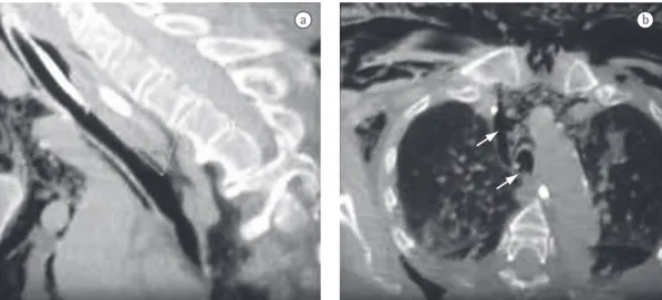
well as extensive subcutaneous emphysema and pneumomediastinum (Figure 2). Fiberoptic bronchoscopy confirmed the laceration (length, 4 cm) of the posterior wall of the trachea, located 0.5 cm from the carina. As a temporary measure, an orotracheal tube was selectively placed in the right main bronchus, distally to the lesion, and antibiotic therapy with coverage of lower airway pathogens was introduced.
Seven hours after the emergency orotracheal intubation, the patient underwent video-assisted right posterior thoracotomy. Primary closure of the lesion was performed using a contin-uous 4-0 polydioxanone suture. Postoperative control fiberoptic bronchoscopy showed that the suture line was healing well. Extubation was considered difficult in this patient due to her neurological condition, and she was therefore submitted to tracheostomy on postoperative day 8. Bronchoscopic examination 2 months later revealed a well-healed suture line and no evidence of stenosis.
Case 3
A 61-year-old female patient with a history of arterial hypertension and chronic renal failure was admitted with a drop in the level of consciousness and respiratory failure secondary to cerebrovascular accident. In the emergency room, the patient was sedated and, after multiple attempts at and great difficulty in accessing the airway, was intubated. The patient was admitted to the intensive care unit, where she remained sedated and on mechanical ventilation with low pressures. However, a chest X-ray revealed that the patient had developed hemoptysis and emphysema (subcutaneous and mediastinal). Fiberoptic bronchoscopy showed evidence of a laceration (length, approximately 3 cm) in the membranous wall, in the proximal third of the cervical trachea and 4 cm from the vocal cords. There was no apparent active bleeding, only clots, which were aspirated. Conservative treatment was chosen due to the acute neuro-logical condition of the patient. The orotracheal tube was inserted endoscopically with its cuff positioned distally to the lesion, and antibiotic therapy with coverage of tracheobronchial tree pathogens was introduced. The subcutaneous emphysema progressively decreased. The bron-choscopy performed on post-intubation day 8
showed that the lesion had healed completely Tabl
mechanical ventilation or a progressive increase in subcutaneous emphysema.(8) Regardless of the treatment proposed, the patient should receive antibiotics with coverage of the tracheobron-chial flora.(8)
In patients on mechanical ventilation, conservative treatment consists in positioning the tracheal tube cuff distal to the lesion. In patients who do not depend on mechanical ventilation, clinical observation is indicated, with surgical intervention at the first sign of ventilatory instability or mediastinitis.(7-9) When surgery is indicated, collar or transverse cervi-cotomy should be performed, depending on the side of the lesion and at the discretion of the surgeon; in cases of mediastinal tracheal injury, right thoracotomy should be performed in the fourth intercostal space.(3,8) Subsequently, the tracheal laceration is treated by primary closure using a single layer of absorbable suture.(3,8)
All of the case series of post-intubation tracheal injury have involved small numbers of patients, of varying ages, and there has been a predominance of females. In such studies, the time elapsed between the occurrence of the injury and the diagnosis has ranged from 0 to 124 h. The reported mortality is low, being slightly higher in patients submitted to surgical treatment. In patients who had poor general health status and multiple comorbidities, deaths were not related to the tracheal injury or to the surgery itself but rather to clinical complications (Table 1).
The data presented previously are corrobo-rated by our case series, in which all patients were female, required emergency orotracheal intuba-tion due to respiratory failure and were difficult to intubate. The diagnosis was made early, 1-5 h after the occurrence of the injury, based on the identification of clinical-radiological signs of subcutaneous emphysema, confirmed by fiberoptic bronchoscopy. Treatment was imme-diate. Two patients were submitted to surgical treatment, and one was submitted to conserva-tive treatment. In the first two cases reported here, the patients were treated surgically because they had an extensive lesion located in the distal trachea and the respiratory isolation of the lesion was difficult. One patient who had suffered a cerebrovascular accident died due to respiratory sepsis, despite evidence of adequate tracheal healing. In the second case, due to impaired without treatment. Due to the neurological
damage caused by the underlying disease, cervical tracheostomy was then performed.
Discussion
Iatrogenic tracheal injury due to orotra-cheal intubation is a rare entity. Its incidence is approximately 0.005% when a single-lumen tube is used and ranges from 0.05% to 0.19% when a double-lumen tube is used. Topographically, it occurs predominantly in the distal third of the trachea and in the main bronchi, at the junction of the membranous and cartilaginous portions. When it is due to cuff overinflation, it occurs predominantly in the proximal trachea.(2,3) It is most common in women, in patients with tracheal wall weakness due to inflammatory disease and in patients on corticosteroid therapy, also occurring in patients with congenital tracheal malformations.(3)
The major mechanisms of injury are the use of an inappropriate tube size, cuff overinflation and sudden movements in the tube. Direct injury caused by the tube usually occurs after multiple, vigorous attempts at orotracheal intubation in emergency situations.(4) Other mechanisms include inappropriate use of the guide and repo-sitioning of the tube without complete emptying of the cuff.(5) In orotracheal intubation using a double-lumen tube, there is a danger not only of laceration but also of bronchial rupture.(6)
The most common clinical manifestations are subcutaneous emphysema in the chest and neck, as well as pneumomediastinum, pneu-mothorax and respiratory failure. Radiological findings such as subcutaneous or mediastinal emphysema, extension of the tip of the endotra-cheal tube to the right and cuff overinflation, are usually indirect signs of injury.(2) The diag-nosis is confirmed by fiberoptic bronchoscopy.
5. Marty-Ané CH, Picard E, Jonquet O, Mary H. Membranous tracheal rupture after endotracheal intubation. Ann Thorac Surg. 1995;60(5):1367-71.
6. Bessa Junior RC, Jorge JC, Eisenberg AF, Duarte WL, Silva MS. Ruptura brônquica após intubação com tubo de duplo lúmen: Relato de caso. Rev Bras Anestesiol. 2005;55(6):660-4.
7. Denlinger CE, Veeramachaneni N, Krupnick AS, Patterson GA, Kreisel D. Nonoperative management of large tracheal injuries. J Thorac Cardiovasc Surg. 2008;136(3):782-3, 783.e1.
8. Schneider T, Storz K, Dienemann H, Hoffmann H. Management of iatrogenic tracheobronchial injuries: a retrospective analysis of 29 cases. Ann Thorac Surg. 2007;83(6):1960-4.
9. Conti M, Pougeoise M, Wurtz A, Porte H, Fourrier F, Ramon P, et al. Management of postintubation tracheobronchial ruptures. Chest. 2006;130(2):412-8. 10. Kaloud H, Smolle-Juettner FM, Prause G, List WF.
Iatrogenic ruptures of the tracheobronchial tree. Chest. 1997 Sep;112(3):774-8.
11. Lampl L. Tracheobronchial injuries. Conservative treatment. Interact Cardiovasc Thorac Surg. 2004;3(2):401-5.
neurological function, the patient required a tracheostomy and a prolonged hospital stay. In the third case, the patient was treated conserva-tively because she had a smaller, more proximal lesion, which facilitated respiratory isolation.
References
1. Grillo HC. Tracheal and Bronchial Trauma. In: Grillo HC, editor. Surgery of the Trachea and Bronchi. Lewiston: BC Decker; 2004. p. 274-5.
2. Borasio P, Ardissone F, Chiampo G. Post-intubation tracheal rupture. A report on ten cases. Eur J Cardiothorac Surg. 1997;12(1):98-100.
3. Massard G, Rougé C, Dabbagh A, Kessler R, Hentz JG, Roeslin N, et al. Tracheobronchial lacerations after intubation and tracheostomy. Ann Thorac Surg. 1996;61(5):1483-7.
4. Jougon J, Ballester M, Choukroun E, Dubrez J, Reboul G, Velly JF. Conservative treatment for postintubation tracheobronchial rupture. Ann Thorac Surg. 2000;69(1):216-20.
About the authors
Carlos Remolina Medina
Resident in Thoracic Surgery. Pereira Filho Ward, Santa Casa Hospital Complex in Porto Alegre, Porto Alegre, Brazil.
José de Jesus Camargo
Thoracic Surgeon. Pereira Filho Ward, Santa Casa Hospital Complex in Porto Alegre, Porto Alegre, Brazil.
José Carlos Felicetti
Thoracic Surgeon. Pereira Filho Ward, Santa Casa Hospital Complex in Porto Alegre, Porto Alegre, Brazil.
Tiago Noguchi Machuca
Resident in Thoracic Surgery. Pereira Filho Ward, Santa Casa Hospital Complex in Porto Alegre, Porto Alegre, Brazil.
Bruno de Moraes Gomes
Resident in Thoracic Surgery. Pereira Filho Ward, Santa Casa Hospital Complex in Porto Alegre, Porto Alegre, Brazil.
Iury Andrade Melo