2011;20:2610-2617. Published OnlineFirst September 29, 2011.
Cancer Epidemiol Biomarkers Prev
Bruno Marques Costa, Marta Viana-Pereira, Ricardo Fernandes, et al.
Outcome
Genetic Variants on Glioma Risk and Patient
EGFR
Impact of
Updated Version
10.1158/1055-9965.EPI-11-0340
doi:
Access the most recent version of this article at:
Material
Supplementary
http://cebp.aacrjournals.org/content/suppl/2011/09/27/1055-9965.EPI-11-0340.DC1.html
Access the most recent supplemental material at:
Cited Articles
http://cebp.aacrjournals.org/content/20/12/2610.full.html#ref-list-1
This article cites 43 articles, 23 of which you can access for free at:
E-mail alerts
Sign up to receive free email-alerts
related to this article or journal.
Subscriptions
Reprints and
.
[email protected]
Department at
To order reprints of this article or to subscribe to the journal, contact the AACR Publications
Permissions
.
[email protected]
Impact of
EGFR
Genetic Variants on Glioma Risk
and Patient Outcome
Bruno Marques Costa1,2, Marta Viana-Pereira1,2, Ricardo Fernandes1,2, Sandra Costa1,2, Paulo Linhares3, Rui Vaz3, Celine Pinheiro1,2, Jorge Lima4, Paula Soares4,5, Ana Silva6, Fernando Pardal6,
Julia Amorim7, Rui Nabi¸co
7
, Rui Almeida8, Carlos Alegria8, Manuel Melo Pires9, Celia Pinheiro10, Ernesto Carvalho10, Pedro Oliveira11, Jose M. Lopes4,5, and Rui M. Reis1,2,12
Abstract
Background:The epidermal growth factor receptor (EGFR) regulates important cellular processes and is frequently implicated in human tumors. Three EGFR polymorphisms have been described as having a transcriptional regulatory function: two single-nucleotide polymorphisms in the essential promoter region, 216G/T and 191C/A, and a polymorphic (CA)nmicrosatellite sequence in intron 1. We aimed to elucidate
the roles of theseEGFRpolymorphisms in glioma susceptibility and prognosis.
Methods:We conducted a case–control study with 196 patients with glioma and 168 cancer-free controls. Unconditional multivariate logistic regression models were used to calculate ORs and 95% confidence intervals. A Cox regression model was used to evaluate associations with patient survival. False-positive report probabilities were also assessed.
Results:None of theEGFR 216G/T variants was significantly associated with glioma risk. The 191C/A genotype was associated with higher risk for glioma when the (CA)nalleles were classified as short for16 or 17 repeats. Independently of the (CA)n repeat cutoff point used, shorter (CA)nrepeat variants were
significantly associated with increased risk for glioma, particularly glioblastoma and oligodendroglioma. In all tested models with different (CA)ncutoff points, only 191C/A genotype was consistently associated with
improved survival of patients with glioblastoma.
Conclusions:Our findings implicateEGFR 191C/A and the (CA)nrepeat polymorphisms as risk factors for
gliomas, and suggest 191C/A as a prognostic marker in glioblastoma.
Impact:Our data support a role of theseEGFRpolymorphisms in determining glioma susceptibility, with potential relevance for molecularly based stratification of patients with glioblastoma for individualized therapies.Cancer Epidemiol Biomarkers Prev; 20(12); 2610–7.2011 AACR.
Introduction
Glioma is a broad category of tumors divided into histologic subgroups based on the type of glial cell of origin or morphologic similarities between tumor and normal glial cells: astrocytomas (astrocytic lineage), oli-godendrogliomas (oligodendroglial lineage), and oligoas-trocytomas (mixed lineage) are the major subtypes, whereas ependymomas (ependymal lineage) are less common (1, 2). They are the most common primary central nervous system tumors and account for approximately 80% of those that are malignant (2), for which efficient therapies are not available (3–5). Despite recent advances in the field of neuro-oncology, the prognosis of patients with glioma remains very poor (6); in particular, patients with glioblastoma, the most common and aggressive (WHO grade IV) form of glioma, present median survival time ranging from 12 to 15 months and the majority die within 2 years (5). Their etiology remains largely unknown: so far, only exposure to high-dose therapeutic
Authors' Affiliations:1Life and Health Sciences Research Institute (ICVS), School of Health Sciences, University of Minho, Braga, Portugal;2ICVS/ 3B's - PT Government Associate Laboratory, Braga/Guimar~aes, Portugal; 3
Department of Neurosurgery, Hospital S. Jo~ao, Porto, Portugal; 4IPATIMUP
–Institute of Molecular Pathology and Immunology, University of Porto, Porto, Portugal;5Medical Faculty, Department of Pathology, University of Porto, Porto, Portugal; Departments of6Pathology,7 Oncol-ogy,8Neurosurgery, Hospital S. Marcos, Braga, Portugal;9Unit of Neuro-pathology,10Department of Neurosurgery, Hospital S. Antonio, Porto, Portugal;11Department of Population Studies, ICBAS, University of Porto, Porto, Portugal;12Molecular Oncology Research Center, Barretos Cancer Hospital, Barretos, S~ao Paulo, Brazil
Note:Supplementary data for this article are available at Cancer Epide-miology, Biomarkers & Prevention Online (http://cebp.aacrjournals.org/).
B.M. Costa and M. Viana-Pereira contributed equally to this work.
Corresponding Author:Rui M. Reis, Life and Health Sciences Research Institute (ICVS), School of Health Sciences, University of Minho, Campus de Gualtar, Braga 4710-057, Portugal. Phone: 351253604825; Fax: 351253604820; E-mail: [email protected]
doi:10.1158/1055-9965.EPI-11-0340
radiation has been firmly established as a risk factor, but other plausible causes include genetic syndromes, familial aggregation, and genetic polymorphisms (7, 8). Equally mysterious are truly clinically relevant prognostic fac-tors for patients with glioma; patient age and clinical performance status are clearly associated with patient outcome, but recent evidences suggest that molecular traits of tumor may also be important determinants of prognosis (9–11).
The epidermal growth factor receptor (EGFR) pathway plays prominent roles in regulating cell growth, apopto-sis, and differentiation, a process that is tightly regulated in normal epithelial cells (12). Deregulation of this path-way can occur, for example, via somaticEGFRmutations (e.g.,EGFRvIII), gene amplifications, and ligand-indepen-dent constitutive stimulation signaling loops, and has been implicated in several human tumors, including gli-omas (13). Aberrant EGFR signaling may ultimately affect major hallmarks of cancer including tumor growth, inva-sion, malignancy, and prognosis (13–16). In addition to these somatic molecular alterations, germ line polymor-phic variants in the EGF/EGFR pathway have also been implicated in tumor risk, therapy response, and patient outcome (17–20). We have previously shown that a func-tionalEGFpolymorphism (EGFþ61) is associated with glioma risk (19), which adds to the growing hypothesis that polymorphisms in the EGFR pathway may be rele-vant in the context of glioma.
Two single-nucleotide polymorphisms (SNP) were recently found in the essential promoter region of theEGFR
gene ( 216G/T and 191C/A), and the T variant in the 216 SNP was shown to increase the promoter activity in an Sp1-dependent manner, resulting in higher gene expres-sion bothin vitroandin vivo(21). A study by Carpentier and colleagues (22) implicated the 216T variant as a risk factor for glioblastoma. Likewise, a highly polymorphic micro-satellite sequence (CA)nrepeat was identified in the first
intron ofEGFR(23). It has been proposed that transcrip-tional activity ofEGFRdeclines with increasing number of
(CA)n repeats, suggesting that this polymorphism may
play a role in cancer susceptibility (24).
In the context of the widely acknowledged role of EGFR signaling in gliomas and the functional relevance of par-ticular EGFR polymorphisms, we investigated the rele-vance of EGFR 216G/T, 191C/A, and (CA)nrepeat
polymorphisms in glioma susceptibility and patient prog-nosis. To do so, we conducted a population-based case– control study of 196 patients with glioma and 168 cancer-free controls from Portugal.
Materials and Methods
Study population
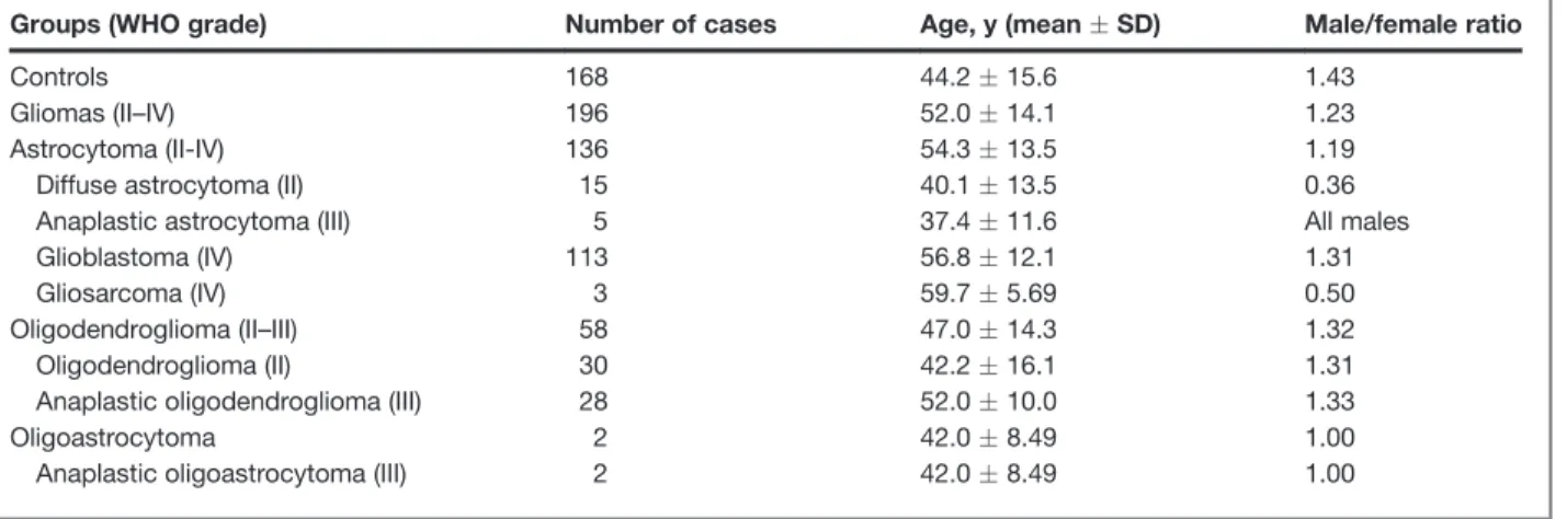
We studied a total of 196 patients with glioma recruited from Hospital S. Marcos, Braga, Hospital S. Jo~ao, and Hospital S. Antonio. Tumors were classified by experienced neuropathologists according to WHO standards (1). The control group is composed of 168 cancer-free individuals randomly selected from blood donors’ bank. All patients and controls were from Northwest Portugal and of Caucasian ethnicity. Table 1 summarizes the clinicopathologic features of patients and controls. This study was conducted in accordance with institutional ethical committees. Blood samples were obtained following informed signed consent. All the samples enrolled were unlinked and unidentified from their donors.
Genotyping
Genomic DNA from patients with glioma was obtained from blood (n¼70) or formalin-fixed, paraffin-embedded tumor samples (n ¼ 126) as previously reported (19).
EGFR 216G/T and 191C/A genotyping was carried out by PCR amplification of the promoter region from nucleotides 352 to 155 (Fw primer: 50
-CTCCTCC-TCCTCTGCTCCTC-30; Rv primer: 50
-GGGGCTAGC-TCGGGACTC-30
), followed by RFLP withBseRI andSacII respectively, as previously described (21). To evaluate
Table 1.Clinicopathologic features of gliomas and controls
Groups (WHO grade) Number of cases Age, y (meanSD) Male/female ratio
Controls 168 44.215.6 1.43
Gliomas (II–IV) 196 52.014.1 1.23
Astrocytoma (II-IV) 136 54.313.5 1.19
Diffuse astrocytoma (II) 15 40.113.5 0.36
Anaplastic astrocytoma (III) 5 37.411.6 All males
Glioblastoma (IV) 113 56.812.1 1.31
Gliosarcoma (IV) 3 59.75.69 0.50
Oligodendroglioma (II–III) 58 47.014.3 1.32
Oligodendroglioma (II) 30 42.216.1 1.31
Anaplastic oligodendroglioma (III) 28 52.010.0 1.33
Oligoastrocytoma 2 42.08.49 1.00
Anaplastic oligoastrocytoma (III) 2 42.08.49 1.00
EGFR/EGFPolymorphisms and Glioma
EGFR(CA)nrepeats in intron 1, a 5-carboxyfluorescein
(FAM)-labeled PCR reaction was carried out (Fw primer: 5-FAM–labeled 50-GGCTCACAGCAAACTTCTCC-30; Rv
primer: 50
-AAGCCAGACTCGCTCATGTT-30
) and the products subsequently analyzed by single capillary genet-ic analysis, as previously described (25). Briefly, PCR cycles were 95C for 7 minutes; followed by 35 cycles of
95C for 45 seconds, 60C for 30 seconds, and 72C for 30
seconds; and a final step at 72C for 7 minutes. PCR
fragments ranging from 194 to 212 bp containing the polymorphic region were obtained. One microliter of PCR product and 0.6mL of GeneScan 500 TAMRA size standard (Applied Biosystems) were mixed with 14.4mL of formamide. PCR products were then separated using an ABI Prism 310 (Applied Biosystems), and the frag-ment lengths were determined with GeneScan Analysis software version 3.7 (Applied Biosystems).
Data analyses and statistical methods
EGFR 216G/T and 191C/A groups were catego-rized as previously described (21). Because EGFR
(CA)n repeats vary between n ¼ 14 and n ¼ 23 and
considering different approaches used in other studies (26–30), we evaluatedEGFR(CA)nrepeat length on the
basis of 2 different methods. First, by the genotype-based method (26, 30), the genotypes ofEGFR (CA)n
repeat were categorized into 3 groups considering a cutoff point of 17 (CA) repeats: homozygous short (SS) contained 2 short alleles (both alleles17 CA repeats), homozygous long (LL) contained 2 long alleles (both alleles>17 CA repeats), and heterozygous short/long (SL) contained one short (17) and one long (>17) allele. Because the cutoff value to consider short and long alleles has been somewhat controversial in the literature (30–33), we also tested 2 additional cutoff values to define short alleles with 16 and 18 CA repeats, and long alleles with>16 and>18 CA repeats, respectively (33). Second, we investigated the EGFR
(CA)n repeat polymorphism by the summed allele
lengths method (28), where individuals were catego-rized into short (sum of alleles length<36) or long (sum of alleles length36).
Thex2test was used to assess whether the observed
allele distributions of all polymorphisms were in Hardy– Weinberg equilibrium in the control group. ORs and 95% confidence intervals (95% CI) for the effect of EGFR
variants on the risk for each glioma type were estimated by multivariate logistic regression analyses, adjusted for
EGFþ61A/G genotype, which was previously reported to affect glioma susceptibility (19), and patient age (as a continuous variable) and sex. The false-positive report probability (FPRP) was calculated for significant associa-tions observed in multivariate tests according to the study of Wacholder and colleagues (34). Associations between
EGFRvariants [ 216G/T, 191C/A, and (CA)nrepeat]
and patient survival were assessed using a multivariate Cox regression model adjusted forEGFþ61A/G, patient age, and sex (35).
Results
EGFR 216G/T, 191C/A, and (CA)nrepeat
polymorphisms and risk of glioma
We studied a total of 196 patients with glioma and 168 cancer-free control individuals. Successful determination of EGFR genotypes was achieved for all samples. The distributions ofEGFþ61A/G,EGFR 216G/T, and (CA)n
allele frequencies in the control group were in Hardy– Weinberg equilibrium (P¼0.121,P¼0.899, andP¼0.150, respectively); however,EGFR 191C/A alleles in controls were not in equilibrium (P ¼ 0.034). A summary of clinicopathologic features of the controls and cases is presented in Table 1.
The frequencies of eachEGFRgenotype in controls and cases are presented in Table 2. TheEGFRallele frequencies in our cancer-free control population are similar to those found in other control populations (21, 31, 33). The most frequent genotypes for each polymorphism in the control group (homozygous 216G/G, homozygous 191C/C, and heterozygous (CA)nrepeat SL) were considered as
references. To assess associations between each variant of the studiedEGFRpolymorphisms and risk for glioma, we used an unconditional multivariate logistic regression analysis adjusted for potential confounding variables (EGFþ61A/G genotype, patient age, and sex). The control group was compared with all glioma cases (WHO grades II–IV) and also with glioblastoma (WHO grade IV) and oligodendroglioma (WHO grades II and III; Table 2), as these were the most frequent subtypes in our series (Table 1).
No statistically significant associations were found betweenEGFR 216G/T genotype variants and risk for glioma, glioblastoma, or oligodendroglioma (Table 2; all values ofP>0.05). In contrast, when compared with the 191C/C reference genotype, the heterozygous 191C/ A genotype was significantly associated with increased risk for glioma (OR¼1.81; 95% CI, 1.01–3.24; Table 2). No significant associations between the 191A/A genotype and risk for glioma, glioblastoma, or oligodendroglioma were found (Table 2; all values ofP>0.05). In addition, the SS genotype of (CA)nrepeat (considering short alleles17
CA repeats) was significantly associated with increased risk for glioma (OR¼2.38; 95% CI, 1.42–3.98), glioblas-toma (OR ¼ 2.25; 95% CI, 1.20–4.25), and oligodendro-glioma (OR¼2.45; 95% CI, 1.17–5.12), as well as the LL genotype was significantly associated with increased risk for glioma (OR ¼1.95; 95% CI, 1.02–3.73). As expected (19),EGFþ61A/G and G/G genotypes were significantly associated with increased risk for glioma (OR¼1.75; 95% CI, 1.03–2.96 and OR ¼ 1.90; 95% CI, 1.03–3.52, respectively; Table 2), particularly oligodendroglioma (OR ¼ 2.80; 95% CI, 1.18–6.67 and OR¼ 2.75; 95% CI 1.06–7.09, respectively; Table 2) and nearly statistically significant for glioblastoma (OR¼1.72; 95% CI, 0.91–3.26 and OR¼1.92; 95% CI, 0.91–4.03, respectively; Table 2).
specificallyn¼16 orn¼18 (i.e., S16 and L>16, or S18 and L>18, respectively), similar results were obtained; particularly, the (CA) repeat homozygous SS genotype was again significantly associated with increased risks for glioma, glioblastoma, and oligodendroglioma both for a cutoff value of S16 repeats (Supplementary Table S1) and S 18 repeats (Supplementary Table S2). The LL
genotype was also associated with increased risk for glioma and glioblastoma for the cutoff value of S 16 (Supplementary Table S1) and for glioblastoma only for the cutoff value of S18 (Supplementary Table S2). Of note, the heterozygous 191C/A genotype was also associated with increased risk for glioma when the cutoff value for (CA)n short and long repeats was 16
(Supplementary Table S1) but not in the case of a cutoff value of 18 repeats (Supplementary Table S2). We also analyzed the associations between EGFRvariants and risk considering the sum of alleles for the (CA)nrepeat
polymorphism (28), which in our series varied from 29 to 41 repeats. Because the median value for the sum of alleles in the control group was 36, we categorized the sum of alleles as short<36 and long36 but none of the
EGFRvariants were significantly associated with risk in this analysis (Supplementary Table S3).
The calculation of FPRP showed that all of the above-mentioned EGFRassociations with risk remained note-worthy (FPRP0.5) when a prior probability of
associ-ation of 10% or greater was considered (Table 3). This was also the case forEGFþ61A/G associations, except in the case of the G/G genotype in oligodendroglioma risk (FPRP¼0.562; Table 3). For a prior probability of 5% or more, only the associations between (CA)nSS genotype
and risk for glioma and glioblastoma remained notewor-thy, which remained significant even for a prior proba-bility of 1% in the case of glioma risk (Table 3).
Taken together, these data strongly suggest that shorter variants of theEGFRintron 1 (CA)nrepeat polymorphism
increase the risk for gliomas, particularly glioblastoma and oligodendroglioma.
EGFR 216G/T, 191C/A, and (CA)nrepeat
polymorphisms and survival of patients with glioblastoma
In a subset of patients with glioblastoma, we also had available follow-up data (n¼63). Thus, we investigated the associations between eachEGFRvariant ( 216G/T, 191C/A, and (CA)nrepeat) and overall survival by a
multivariate Cox proportional hazard model, adjusted for
EGFþ61A/G, patient age, and sex.
None of theEGFR 216G/T variants was associated with survival of patients with glioblastoma (Table 4; Supplementary Tables S4–S6 when the (CA)nrepeat
var-iants were classified considering a cutoff value of S17, S16, S18, or sum of alleles<36, respectively).
Table 2.Multivariate logistic regression analysis of associations betweenEGFR/EGFpolymorphisms and
risk for glioma groups
Polymorphism Control Glioma (grades II–IV)
OR (95% CI) Glioblastoma (grade IV)
OR (95% CI) Oligodendroglioma (grades II–III)
OR (95% CI)
EGFR 216G/T
G/G 77 91 — 55 — 28 —
G/T 74 85 1.12 (0.68–1.84) 45 1.09 (0.58–2.03) 25 1.01 (0.49–2.07)
T/T 17 20 1.15 (0.52–2.59) 13 1.30 (0.50–3.40) 5 1.02 (0.31–3.40) EGFR 191C/A
C/C 130 140 — 77 — 41 —
C/A 32 49 1.81 (1.01–3.24) 30 1.97 (0.96–4.03) 16 2.08 (0.93–4.63)
A/A 6 7 0.68 (0.19–2.45) 6 0.88 (0.21–3.70) 1 0.47 (0.05–4.84) EGFR(CA)nrepeata
SL 92 75 — 42 — 23 —
LL 28 40 1.95 (1.02–3.73) 25 2.14 (0.96–4.76) 10 1.83 (0.68–4.94)
SS 48 81 2.38 (1.42–3.98) 46 2.25 (1.20–4.25) 25 2.45 (1.17–5.12)
EGFþ61A/G
A/A 57 47 — 30 — 9 —
A/G 73 96 1.75 (1.03–2.96) 54 1.72 (0.91–3.26) 32 2.80 (1.18–6.67)
G/G 38 53 1.90 (1.03–3.51) 29 1.92 (0.91–4.03) 17 2.75 (1.06–7.09)
Age 1.04 (1.02–1.06) 1.07 (1.04–1.09) 1.02 (1.00-1.04)
Sex
Female 69 88 — 49 — 25 —
Male 99 108 0.95 (0.60–1.49) 64 1.02 (0.59–1.78) 33 0.98 (0.51–1.88)
NOTE: Bold-faced values indicate significant difference at 5% level.
a(CA)
nrepeat considered short17 and long>17.
EGFR/EGFPolymorphisms and Glioma
In contrast, the heterozygous 191C/A genotype was significantly associated with improved overall survival of patients with glioblastoma, as compared with homozy-gous 191C/C, which was consistent across all analyses, that is, when (CA)nalleles were classified as S17 (OR¼
0.37; 95% CI, 0.16–0.88; Table 4), S16 (OR¼0.35; 95% CI, 0.15–0.82; Supplementary Table S4), S18 (OR¼0.37; 95% CI, 0.16–0.82; Supplementary Table S5), or sum of alleles<36 (OR¼0.39; 95% CI, 0.17–0.86; Supplementary Table S6).
The homozygous SS and LL (CA)nrepeat genotypes
were also significantly associated with a longer survival of patients with glioblastoma when the (CA)nrepeat variants
were classified as S17 (OR¼0.33; 95% CI, 0.11–0.95 for LL genotype and OR ¼ 0.41; 95% CI, 0.18–0.93 for SS genotype; Table 4). In the case of the SS genotype, this association with longer survival was maintained when a cutoff value of S16 was considered (OR¼0.42; 95% CI, 0.18–0.95; Supplementary Table S4).
FPRP calculations showed that the association between 191C/A and (CA)n repeat SS genotypes and longer
survival of patients with glioblastoma remained notewor-thy for a prior probability of association of 10% or more (FPRP<0.5 for all associations), which was not the case for LL genotypes (FPRP>0.5, data not shown).
Taken together, these data strongly suggest thatEGFR
191C/A and intron 1 (CA)nrepeat polymorphisms are
prognostic markers in patients with glioblastoma, where-as 216G/T variants do not seem to predict the outcome of patients with glioblastoma.
Discussion
Gliomas are the most frequent and malignant primary central nervous system tumors. These result in more years
of life lost than do any other tumors (36) and are a significant source of cancer-related death (37). It is gen-erally assumed that genetic and environmental factors contribute to gliomagenesis, but the etiology of gliomas remains very poorly understood. Presently, one of the several lines of brain tumor research focuses on the rel-evance of germ line genetic polymorphisms in glioma risk, grade, prognosis, and response to specific therapies. We have recently shown that an SNP in theEGFgene (which encodes one of the main EGFR ligands),EGFþ61A/G, has
functional consequences and associated the G allele with increased risk for glioma, particularly glioblastoma and oligodendroglioma (19).
The EGFR pathway is commonly altered in gliomas. Approximately 50% of glioblastomas showEGFR ampli-fication and overexpression, 40% of which express the mutant formEGFRvIII, resulting in constitutive activation of the EGFR pathway (13, 38–40). Previously, Carpentier and colleagues (22) showed an association between the 216T allele and increased risk for glioblastoma. We attempted to replicate these findings, and examined, for the first time, the implication of 216G/T and 191C/A SNPs in other types of glioma. In opposition to the results of the study of Carpentier and colleagues, we have not seen association betweenEGFR 216T allele and increased risk for glioblastomas or any other glioma subtype. This dis-crepancy may be partially explained by distinct popula-tion sampling and the fact that we conducted a logistic regression adjusted for 2 other EGFR polymorphisms, together withEGFþ61A/G, patient age, and sex. Impor-tantly, the allele distribution of the 216G/T polymor-phism in the control cancer-free population, we report here (67.9% G, 32.1% T,n¼168) is very similar to that
reported by other studies [68.3% G, 31.7% T,n¼60 (ref. 21); 68.2% G, 31.8% T,n¼22 (ref. 41)]. Inversely, the study of
Table 3.FPRP for significant associations with risk
Polymorphism OR (95% CI) Powera ReportedP Prior probability
0.1 0.05 0.01 0.001
Glioma risk
EGFR 191C/A (C/C ref.) 1.81 (1.01–3.24) 0.632 0.046 0.395 0.579 0.878 0.980 EGFR(CA)nLL (SL ref.) 1.95 (1.02–3.73) 0.530 0.044 0.425 0.609 0.890 0.988 EGFR(CA)nSS (SL ref.) 2.38 (1.42–3.98) 0.254 0.001 0.033 0.066 0.270 0.789 EGFþ61A/G (A/A ref.) 1.75 (1.03–2.96) 0.691 0.037 0.325 0.504 0.841 0.982 EGFþ61G/G (A/A ref.) 1.90 (1.03–3.51) 0.565 0.040 0.391 0.576 0.876 0.986
Glioblastoma risk
EGFR(CA)nSS (SL ref.) 2.25 (1.20–4.25) 0.358 0.012 0.238 0.398 0.775 0.972
Oligodendroglioma risk
EGFR(CA)nSS (SL ref.) 2.45 (1.17–5.12) 0.295 0.017 0.344 0.525 0.852 0.998 EGFþ61A/G (A/A ref.) 2.80 (1.18–6.67) 0.224 0.020 0.447 0.630 0.899 0.989 EGFþ61G/G (A/A ref.) 2.75 (1.06–7.09) 0.255 0.036 0.562 0.730 0.934 0.993
NOTE: Bold-faced values indicate the FPRP0.5 for the most likely prior probability.
aEstimation of statistical power to detect an OR of 2.0 with an
Carpentier and colleagues shows a significantly different distribution of 216G/T alleles in the control group (53.2% G, 46.8% T,n¼176) when compared with our and others data [x2(3)¼19.4,P<0.001] (21, 41). Even though the allele
frequencies of this SNP vary greatly based on the ethnic background (21), this feature cannot explain the observed differences because all patients in our and others studies (21, 22, 41) were Caucasians. Concerning the 191C/A SNP, we observed that the heterozygous 191C/A genotype was associated with increased risk for glioma. A recent study by Schwartzbaum and colleagues (42) identified 3EGFRSNPs consistently associated with glio-blastoma risk across 4 independent data sets. In addition, these SNPs were highly correlated with 4 other
EGFRSNPs previously found to be significantly associated with risk for glioma (43). Collectively, these and our data seem to supportEGFRSNPs as potential risk factors for glioma.
Several studies suggest thatEGFRexpression is depen-dent on the number of the intron 1 (CA)n repeats
(28, 30, 44, 45). In addition, this polymorphism has been associated with risk of breast, lung, and colorectal cancers
(31–33) but was never studied in gliomas. In this study, we provide the first evidence on the relevance of theEGFR
(CA)n repeat length polymorphism in glioma risk and
patient survival. Because different criteria have been published for the analysis of this polymorphism and no consensual cutoff point exists to distinguish short and longEGFR(CA)nrepeat alleles (30–33), we used 3
differ-ent cutoff points to cover most of the previously published analysis (considering short alleles16,17, or18 CA repeats) and evaluated the (CA)nrepeat by the genotype,
and the sum of the alleles length (considering the cutoff point for the sum of alleles as the median value in our control group). Our data show that the homozygous SS genotype of the (CA)nrepeat polymorphism was
asso-ciated with increased risk for glioma, glioblastoma, or oligodendroglioma, regardless of the selected cutoff point. The homozygous LL genotype was also found to be associated with increased risk for glioma and glioblastoma but only in some of the tested cutoff values. Thus, caution must be taken in the interpretation of the results and validation in an independent series is required.
Investigating the prognostic value of these 3 EGFR
polymorphisms, we found a significant association between the heterozygous 191C/A genotype and improved survival of patients with glioblastoma, regard-less of the criteria used for the cutoff value of the (CA)n
repeat genotypes in the multivariate Cox model (Table 4, Supplementary Tables S4–S6). The SS and LL genotypes for the (CA)nrepeat polymorphism were also associated
with a better survival of patients with glioblastoma but only for specific cutoff values (SS when S17 or S16; LL when S17); thus, the clinical relevance of this polymor-phism warrants further confirmation.
In our study, the genotype assays of most patients with glioma were carried out in tumor tissue, raising the possibility that somatic alterations in the EGFR locus (7p12) could lead to misgenotyping. However, we believe that this is not the case because of the following reasons: (i) the overall distribution ofEGFR 216 and 191 and (CA)nrepeat genotypes and alleles was not
statistically different for the group of patients whose genotyping was done in DNA from blood or tumor tissue (P ¼0.241 for EGFR 216; P ¼0.176 for EGFR
191; P ¼ 0.155 for (CA)n repeats; data not shown);
(ii) genotyping of all 3 polymorphisms was done with 100% concordance in 20 glioma cases from whom DNA was available from both peripheral blood and tumor tissue; (iii) we found no statistically significant associa-tions between EGFRpolymorphic variants and partic-ular EGFR molecular alterations (EGFR amplification and EGFRvIII mutation; Supplementary Table S7) we had previously analyzed in a subset of tumors (13). Accordingly, in lung cancer, it was previously shown for the (CA)nrepeats polymorphism that
regard-less the amplification status there was 100% concor-dance in the genotyping of tumor and nontumor tissues of 450 cases (15).
Table 4.Multivariate Cox proportional hazard model analysis of associations betweenEGFR/ EGFpolymorphisms and survival of patients
with glioblastoma (adjusted for patient age and sex)
Polymorphism Glioblastoma (grade IV)
OR (95% CI)
EGFR 216G/T
G/G 33 —
G/T 23 0.49 (0.23–1.01)
T/T 7 0.61 (0.21–1.73)
EGFR 191C/A
C/C 41 —
C/A 18 0.37 (0.16–0.88)
A/A 4 0.56 (0.12-2.69)
EGFR(CA)nrepeat a
SL 17 —
LL 17 0.33 (0.11–0.95)
SS 29 0.41 (0.18–0.93)
EGFþ61A/G
A/A 16 —
A/G 28 1.06 (0.47–2.38)
G/G 19 2.10 (0.89–4.99)
Age 1.00 (0.97–1.03)
Sex
Female 25 —
Male 38 1.02 (0.54–1.92)
NOTE: Bold-faced values indicate significant difference at 5% level.
a(CA)
nrepeat considered short17 and long>17.
EGFR/EGFPolymorphisms and Glioma
In summary, our data consistently indicate thatEGFR
intron 1 homozygous (CA)nrepeat short genotypes confer
higher susceptibility to develop different histologic enti-ties of glioma, and implicate the heterozygous 191C/A genotype as a predictive marker of worse survival in patients with glioblastoma. Because EGFR is one of the most frequently altered molecules in high-grade glioma, it is natural to think of it as an attractive therapeutic target. Therefore, further studies are warranted to investigate how theseEGFRpolymorphisms may affect response of patients with glioma to EGFR-targeting therapies.
Disclosure of Potential Conflicts of interest
R.M. Reis has commercial research grant from Schering-Plough, Portugal. No potential conflicts of interest were disclosed by other authors.
Acknowledgments
The authors thank the Immunochemotherapy Department of Hos-pital S. Marcos, and Clnica Laboratorial Dr. Edgar Botelho Moniz, S. Tirso, Portugal, for their helpful assistance in the management of controls.
Grant Support
The study was supported by Funda¸c~ao para a Ci^encia e Tecnologia,
Portugal (SFRH/BPD/33612/2009; SFRH/BD/29145/2006). Schering-Plough Farma, Portugal.
The costs of publication of this article were defrayed in part by the payment of page charges. This article must therefore be hereby marked
advertisementin accordance with 18 U.S.C. Section 1734 solely to indicate this fact.
Received April 11, 2011; revised August 17, 2011; accepted September 21, 2011; published OnlineFirst September 28, 2011.
References
1. Louis DN, Ohgaki H, Wiestler OD, Cavenee WK. WHO classification of tumours of the central nervous system. Lyon, France: IARC; 2007.
2. CBTRUS. Statistical report: primary brain tumors in the United States, 1998–2002. Hinsdale (IL): CBTRUS; 2005.
3. Reardon DA, Rich JN, Friedman HS, Bigner DD. Recent advances in the treatment of malignant astrocytoma. J Clin Oncol 2006;24: 1253–65.
4. Jaeckle KA, Ballman KV, Rao RD, Jenkins RB, Buckner JC. Current strategies in treatment of oligodendroglioma: evolution of molecular signatures of response. J Clin Oncol 2006;24:1246–52.
5. Clarke J, Butowski N, Chang S. Recent advances in therapy for glioblastoma. Arch Neurol 2010;67:279–83.
6. Butowski NA, Sneed PK, Chang SM. Diagnosis and treatment of recurrent high-grade astrocytoma. J Clin Oncol 2006;24:1273–80.
7. Ohgaki H, Kleihues P. Epidemiology and etiology of gliomas. Acta Neuropathol (Berl) 2005;109:93–108.
8. Bondy ML, Scheurer ME, Malmer B, Barnholtz-Sloan JS, Davis FG, Il'Yasova D, et al. Brain tumor epidemiology: consensus from the Brain Tumor Epidemiology Consortium. Cancer 2008;113:1953–68.
9. Costa BM, Smith JS, Chen Y, Chen J, Phillips HS, Aldape KD, et al. Reversing HOXA9 oncogene activation by PI3K inhibition: epigenetic mechanism and prognostic significance in human glioblastoma. Can-cer Res 2010;70:453–62.
10. Hegi ME, Diserens AC, Gorlia T, Hamou MF, de TN, Weller M, et al. MGMT gene silencing and benefit from temozolomide in glioblastoma. N Engl J Med 2005;352:997–1003.
11. Phillips HS, Kharbanda S, Chen R, Forrest WF, Soriano RH, Wu TD, et al. Molecular subclasses of high-grade glioma predict prognosis, delineate a pattern of disease progression, and resemble stages in neurogenesis. Cancer Cell 2006;9:157–73.
12. Yarden Y, Sliwkowski MX. Untangling the ErbB signalling network. Nat Rev Mol Cell Biol 2001;2:127–37.
13. Viana-Pereira M, Lopes JM, Little S, Milanezi F, Basto D, Pardal F, et al. Analysis of EGFR overexpression, EGFR gene amplification and the EGFRvIII mutation in Portuguese high-grade gliomas. Anticancer Res 2008;28:913–20.
14. Zhu Y, Parada LF. The molecular and genetic basis of neurological tumours. Nat Rev Cancer 2002;2:616–26.
15. Nomura M, Shigematsu H, Li L, Suzuki M, Takahashi T, Estess P, et al. Polymorphisms, mutations, and amplification of the EGFR gene in non-small cell lung cancers. PLoS Med 2007;4:e125.
16. Weiss WA, Burns MJ, Hackett C, Aldape K, Hill JR, Kuriyama H, et al. Genetic determinants of malignancy in a mouse model for oligoden-droglioma. Cancer Res 2003;63:1589–95.
17. Araujo A, Ribeiro R, Azevedo I, Coelho A, Soares M, Sousa B, et al. Genetic polymorphisms of the epidermal growth factor and related
receptor in non-small cell lung cancer–a review of the literature. Oncologist 2007;12:201–10.
18. Zhang YM, Cao C, Liang K. Genetic polymorphism of epidermal growth factor 61A>G and cancer risk: a meta-analysis. Cancer Epi-demiol 2010;34:150–6.
19. Costa BM, Ferreira P, Costa S, Canedo P, Oliveira P, Silva A, et al. Association between functional EGFþ61 polymorphism and glioma risk. Clin Cancer Res 2007;13:2621–6.
20. Liu W, He L, Ramirez J, Krishnaswamy S, Kanteti R, Wang YC, et al. Functional EGFR germline polymorphisms may confer risk for EGFR somatic mutations in non-small cell lung cancer, with a predominant effect on exon 19 microdeletions. Cancer Res 2011;7:2423–7
21. Liu W, Innocenti F, Wu MH, Desai AA, Dolan ME, Cook EH Jr, et al. A functional common polymorphism in a Sp1 recognition site of the epidermal growth factor receptor gene promoter. Cancer Res 2005;65: 46–53.
22. Carpentier C, Laigle-Donadey F, Marie Y, Auger N, Benouaich-Amiel A, Lejeune J, et al. Polymorphism in Sp1 recognition site of the EGF receptor gene promoter and risk of glioblastoma. Neurology 2006;67:872–4.
23. Chi DD, Hing AV, Helms C, Steinbrueck T, Mishra SK, Donis-Keller H. Two chromosome 7 dinucleotide repeat polymorphisms at gene loci epidermal growth factor receptor (EGFR) and pro alpha 2 (I) collagen (COL1A2). Hum Mol Genet 1992;1:135.
24. Gebhardt F, Zanker KS, Brandt B. Modulation of epidermal growth factor receptor gene transcription by a polymorphic dinucleotide repeat in intron 1. J Biol Chem 1999;274:13176–80.
25. Butler JM, Buel E, Crivellente F, McCord BR. Forensic DNA typing by capillary electrophoresis using the ABI Prism 310 and 3100 genetic analyzers for STR analysis. Electrophoresis 2004;25:1397–412.
26. Kang D, Gridley G, Huang WY, Engel LS, Winn DM, Brown LM, et al. Microsatellite polymorphisms in the epidermal growth factor receptor (EGFR) gene and the transforming growth factor-alpha (TGFA) gene and risk of oral cancer in Puerto Rico. Pharmacogenet Genomics 2005;15:343–7.
27. McKay JA, Murray LJ, Curran S, Ross VG, Clark C, Murray GI, et al. Evaluation of the epidermal growth factor receptor (EGFR) in colorectal tumours and lymph node metastases. Eur J Cancer 2002;38:2258–64.
28. Amador ML, Oppenheimer D, Perea S, Maitra A, Cusatis G, Iacobuzio-Donahue C, et al. An epidermal growth factor receptor intron 1 polymorphism mediates response to epidermal growth factor receptor inhibitors. Cancer Res 2004;64:9139–43.
30. Etienne-Grimaldi MC, Pereira S, Magne N, Formento JL, Francoual M, Fontana X, et al. Analysis of the dinucleotide repeat polymorphism in the epidermal growth factor receptor (EGFR) gene in head and neck cancer patients. Ann Oncol 2005;16:934–41.
31. Zhang W, Weissfeld JL, Romkes M, Land SR, Grandis JR, Siegfried JM. Association of the EGFR intron 1 CA repeat length with lung cancer risk. Mol Carcinog 2007;46:372–80.
32. Zhang W, Park DJ, Lu B, Yang DY, Gordon M, Groshen S, et al. Epidermal growth factor receptor gene polymorphisms predict pelvic recurrence in patients with rectal cancer treated with chemoradiation. Clin Cancer Res 2005;11:600–5.
33. Brandt B, Hermann S, Straif K, Tidow N, Buerger H, Chang-Claude J. Modification of breast cancer risk in young women by a polymorphic sequence in the egfr gene. Cancer Res 2004;64:7–12.
34. Wacholder S, Chanock S, Garcia-Closas M, El GL, Rothman N. Assessing the probability that a positive report is false: an approach for molecular epidemiology studies. J Natl Cancer Inst 2004;96: 434–42.
35. Costa BM, Caeiro C, Guimaraes I, Martinho O, Jaraquemada T, Augusto I, et al. Prognostic value of MGMT promoter methylation in glioblastoma patients treated with temozolomide-based chemoradia-tion: a Portuguese multicentre study. Oncol Rep 2010;23:1655–62.
36. Burnet NG, Jefferies SJ, Benson RJ, Hunt DP, Treasure FP. Years of life lost (YLL) from cancer is an important measure of population burden–and should be considered when allocating research funds. Br J Cancer 2005;92:241–5.
37. Jemal A, Siegel R, Ward E, Hao YP, Xu JQ, Thun MJ. Cancer statistics, 2009. CA Cancer J Clin 2009;59:225–49.
38. Humphrey PA, Wong AJ, Vogelstein B, Zalutsky MR, Fuller GN, Archer GE, et al. Anti-synthetic peptide antibody reacting at the fusion junction of deletion-mutant epidermal growth factor receptors in human glioblastoma. Proc Natl Acad Sci U S A 1990;87:4207–11.
39. Wong AJ, Ruppert JM, Bigner SH, Grzeschik CH, Humphrey PA, Bigner DS, et al. Structural alterations of the epidermal growth factor receptor gene in human gliomas. Proc Natl Acad Sci U S A 1992; 89:2965–9.
40. Heimberger AB, Suki D, Yang D, Shi W, Aldape K. The natural history of EGFR and EGFRvIII in glioblastoma patients. J Transl Med 2005;3:38.
41. Liu W, Wu X, Zhang W, Montenegro RC, Fackenthal DL, Spitz JA, et al. Relationship of EGFR mutations, expression, amplification, and poly-morphisms to epidermal growth factor receptor inhibitors in the NCI60 cell lines. Clin Cancer Res 2007;13:6788–95.
42. Schwartzbaum JA, Xiao Y, Liu Y, Tsavachidis S, Berger MS, Bondy ML, et al. Inherited variation in immune genes and pathways and glioblas-toma risk. Carcinogenesis 2010;31:1770–7.
43. Andersson U, Schwartzbaum J, Wiklund F, Sj€ostr€om S, Liu Y, Tsava-chidis S, et al. A comprehensive study of the association between the EGFR and ERBB2 genes and glioma risk. Acta Oncol 2010;49:767–75.
44. Buerger H, Packeisen J, Boecker A, Tidow N, Kersting C, Bielawski K, et al. Allelic length of a CA dinucleotide repeat in the egfr gene correlates with the frequency of amplifications of this sequence–first results of an inter-ethnic breast cancer study. J Pathol 2004;203:545–50.
45. Buerger H, Gebhardt F, Schmidt H, Beckmann A, Hutmacher K, Simon R, et al. Length and loss of heterozygosity of an intron 1 polymorphic sequence of egfr is related to cytogenetic alterations and epithelial growth factor receptor expression. Cancer Res 2000;60:854–7.
EGFR/EGFPolymorphisms and Glioma