Introduction
Simvastatin (SMT), chemically known as (1S, 2S, 8S, 8aR)-1,2,6,8,8a-hexahydro-1-(2-((2R, 4R)-terahyro-4-hydroxy-6-oxo-2H-pyran-2-yl)-2,6-dimethylnapthalen-8-yl 2,2-dimethylbutanoate (Fig.1) is a cholesterol lowering lactone,and is iden-tified as being among the most widely prescribed drugs in the world in 2007. It is found to lower cho-lesterol by inhibiting the synthesis of movalinic
acid, which is a key precursor in cholesterol synthe-sis. SMT is administered as a prodrug, and in liver, it is hydrolysed to the β-hydroxy acid form [1].
The therapeutic importance of SMT justifies research to develop analytical methods for its deter-mination in body fluids and pharmaceuticals. The most widely used technique for the assay of SMT has been the high performance liquid chromatogra-phy (HPLC) with UV, ms-ms or fluorescence detec-tion. The drug in plasma has been assayed by LC www.scielo.br/eq
www.ecletica.iq.unesp.br
Volume 33, número 2, 2008
Cerimetric determination of simvastatin in pharmaceuticals
based on redox and complex formation reactions
K. Basavaiah*, and O. Z. Devi
Department of Chemistry, University of Mysore, Manasagangotri, Mysore-570006, India *[email protected]
Abstract:Two sensitive spectrophotometric methods are described for the determination of simvastatin (SMT) in bulk drug and in tablets. The methods are based on the oxidation of SMT by a measured excess of cerium (IV) in acid medium followed by determination of unreacted oxidant by two different reaction schemes. In one procedure (method A), the residual cerium (IV) is reacted with a fixed concentration of fer-roin and the increase in absorbance is measured at 510 nm. The second approach (method B) involves the reduction of the unreacted cerium (IV) with a fixed quantity of iron (II), and the resulting iron (III) is com-plexed with thiocyanate and the absorbance measured at 470 nm. In both methods, the amount of cerium (IV) reacted corresponds to SMT concentration. The experimental conditions for both methods were opti-mized. In method A, the absorbance is found to increase linearly with SMT concentration (r = 0.9995) whereas in method B, the same decreased (r = -0.9943). The systems obey Beer’s law for 0.6-7.5 and 0.5-5.0 µg mL-1for method A and method B, respectively. The calculated molar absorptivity values are 2.7 X 10⁴and 1.06 X 10⁵Lmol-1 cm-1, respectively; and the corresponding sandel sensitivity values are 0.0153 and 0.0039µg cm-2, respectively. The limit of detection (LOD) and quantification (LOQ) are reported for both methods. Intra-day and inter-day precision, and accuracy of the methods were established as per the current ICH guidelines. The methods were successfully applied to the determination of SMT in tablets and the results were statistically compared with those of the reference method by applying the Student’s t-test and F-test. No interference was observed from the common excipients added to tablets. The accuracy and validity of the methods were further ascertained by performing recovery experiments viastandard addition procedure.
with UV-detection [2-6], mass spectrometric detec-tion [7-13] and with fluorescence detecdetec-tion [14]; and in serum, the assay of drug has been accom-plished by derivative UV-spectrophotometry [15] and voltammetry [16]. Not many methods have been reported for the determination of SMT in phar-maceutical substances. Here also, HPLC with UV detection [17-22] is the widely used technique although High-Performance Thin Layer Chromatography (HPTLC) [23], UV-spectropho-tometry [15,24-28], voltammetry [16] and Micellar Electro kinetic Chromatography (MEKC) [29] have sparsely been used. The US pharmacopoeia [30] describes a HPLC-UV detection procedure for the assay of SMT in pharmaceuticals.
Figure 1. Structure of simvastatin
Although visible spectrophotometry is a rapid, fairly sensitive, accurate and precise, and cost-effective technique, it has not been applied yet to the assay of SMT in pharmaceuticals. The present study includes two new and indirect spectrophoto-metric methods for the sensitive determination of SMT. The study represents the use of cerium (IV) sulphate as the oxidimetric reagent, and ferroin, iron (II) and thiocyanate as subsidiary reagents.
Experimental details Apparatus
A Systronics model 106 digital spectropho-tometer provided with 1-cm matched quartz cells was used for all absorbance measurements.
Reagents and materials
All chemicals used were of analytical reagent grade and distilled water was used to pre-pare solutions.
Cerium (IV) sulphate (300 and 400 µg mL-1).
A stock standard solution equivalent to 0.01M ceri-um (IV) sulphate was prepared by dissolving 1.011 g of the chemical (LOBA Chemie, Mumbai,India, assay 99.9%) in 0.5 mol L-1H
2SO4and diluting to 250 ml with the same acid in a 250 ml standard flask and standardised [31]. The stock solution was then diluted appropriately with 0.5mol L-1H
2SO4 to obtain a working concentrations of 300 µg mL-1 and 400 µg mL-1 in cerium (IV) sulphate for method A and method B, respectively.
Ferroin [36 µg ml-1with respect to iron (II)].
Prepared by dissolving 44.8 mg of FeSO4.7H2O (assay 99%) and 96 mg of 1,10-phenanthroline monohydrate (assay 100%) in water, and diluted to volume in a 250 ml calibrated flask.
Ferrous ammonium sulphate, FAS (400 µg ml-1). Accurately weighed 100 mg of the
chemical (LOBA Chemie, Mumbai, India, assay 99-101%) and dissolved in water containing 5 ml of dilute H2SO4and diluted to volume in a 250 ml calibrated flask.
Potassium thiocyanate (3 mol L-1).
Prepared by dissolving 29 g of the chemical (S.d Fine Chem, Mumbai, India, assay 98%) in water and diluting to 100 ml.
Hydrochloric acid (5 mol L-1).
Concentrated hydrochloric acid (S.d. Fine Chem, Mumbai, India;sp.gr.1.18) was diluted appropri-ately with water.
Acetic acid (3:2). Prepared by diluting appropriately glacial acetic acid (S.d. Fine Chem, Mumbai, India; sp.gr. 1.05) with water.
Dosage forms. The following dosage forms were purchased from local commercial sources and subjected to analysis: Simlip (10 and 20 mg), Simvas (5,10 and 20 mg) and Zosta (5,10 and 20 mg).
Procedures
Method A. Different aliquots (0.0,0.5,1.0…3.0 ml) of standard 25 µg ml-1SMT solution were measured accurately and transferred into a series of 10 ml calibrated flasks .To each flask 1.5 ml of 5 mol L-1HCl was added and the total vol-ume was adjusted to 5 ml by adding water. One ml of cerium (IV) sulphate (300 µg ml-1) was added to each flask, content mixed, and after 10 minutes, 1ml of ferroin was added, diluted to the mark with water and mixed well. The absorbance of the solution was measured at 510 nm against the reagent blank.
Method B. Varying aliquots (0.0,0.2,0.4…1.0 ml) of 50 µg ml-1 SMT solution were transferred into a series of 10 ml calibrated flasks by means of a micro burette and the total ume was adjusted to 3 ml by adding requisite vol-ume of water. To each flask, it was added 3 ml of 5 mol L-1HCl followed by 1 ml of 400 µg ml-1 ceri-um (IV) sulphate the last being added from a micro burette. The content was mixed and the flasks were let stand for 10 min before adding 1 ml of accurate-ly measured 400 µg ml-1FAS. After 5 min, 1ml of 3 mol L-1 thiocyanate solution was added to each flask, diluted to the mark with water and the absorbance measured at 470 nm against water blank. In either method, analytical curve was pre-pared by plotting the increasing absorbance values
in method A or decreasing absorbance in method B as a function of SMT concentration. The concen-tration of the unknown was read from the analyti-cal curve or computed from the respective regres-sion equation derived using Beer’s law data.
Procedure for tablets. Twenty tablets were weighed accurately and finely powdered. A quan-tity of the powdered tablet containing 20 mg of SMT was weighed accurately and transferred into a 100 ml calibrated flask, 60 ml of 3:2 acetic acid and the content shaken thoroughly for 15-20 min to extract the drug into the liquid phase; the vol-ume was finally diluted to the mark with the same acetic acid, mixed well and filtered using a Whatman No 42 filterpaper. First 10 ml of the fil-trate was discarded, and suitable aliquots of the subsequent portion were diluted with 3:2 acetic acid to get tablet extracts containing 25 and 50 µg ml-1SMT. A convenient aliquot was then subject-ed to analysis by either method describsubject-ed above.
Results and discussion
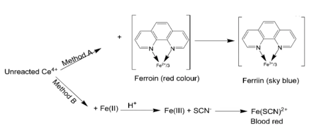
A number of oxidisable pharmaceutical substances have earlier been assayed using ceri-um (IV) sulphate by several workers [32-45] based on several reaction schemes. The present methods entail adding a known excess of cerium (IV) sulphate to SMT in acid medium followed by the determination of unreacted oxidant by two reaction schemes involving the use of ferroin, and iron (II) and thiocyanate as reagents. The possible reaction scheme is given in Fig.2.
Method A
One of the popular methods for cerium (IV) uses its oxidizing property and is based on the bleaching of the ferroin [46,47] by cerium (IV). In the present method, a known excess of cerium (IV) sulphate was reacted with SMT and after the oxidation of the drug was ensured to be complete, the unreacted oxidant was reacted with a fixed quantity of ferroin, and the resulting change in absorbance was measured at 510 nm, and related to the drug concentration. When a fixed concen-tration of cerium (IV) sulphate was made to react with increasing concentration of SMT, there occurred a concomitant fall in the oxidant concen-tration. The decreasing concentrations of cerium (IV) sulphate upon reacting with a fixed concen-tration of ferroin resulted in increasing absorbance at 510 nm due to the bleaching of ferroin colour by the oxidant, the bleaching being caused by the oxi-dation of ferroin to ferriin by cerium (IV) sulphate. This is observed as a proportional increase in the absorbance ferroin with increase in the SMT con-centration which formed the basis for the assay.
One ml of ferroin (36 µgml-1in Fe2+) in a total volume of 10 ml was found to give a conven-ient maximum absorbance at 510 nm and this was bleached to a minimum and constant absorbance by 1 ml of 300 µg ml-1cerium (IV) sulphate in acid medium. Hence, different concentrations of SMT were reacted with 1ml of 300 µgml-1cerium (IV) sulphate in acid medium and the unreacted oxidant was determined by reacting with 1 ml of ferroin equivalent to 36 µg ml-1iron (II). One and a-half ml of 5 mol L-1HCl in a total volume of 6 ml was found optimum for the oxidation of SMT by ceri-um (IV) sulphate which was complete in 10 min. Hydrochloric acid volume upto 2.5 ml and reaction time upto 60 min had no adverse effect on the absorbance. The same acid concentration was maintained for the reaction between the cerium (IV) and ferroin, which was instantaneous. The colour was found to be stable for several hours in the presence of the reaction product/s.
Method B
Cerium (IV) in the past has been deter-mined by reducing it by known excess of iron (II) and complexing the iron (III) with thiocyanate [48]. This method is based on the oxidation of
SMT by a measured excess of cerium (IV) sul-phate in HCl medium, reduction of the residual oxidant by a fixed amount of iron (II) and subse-quent formation of iron (III)-thiocyanate complex, which is measured at 470 nm. When a fixed con-centration of cerium (IV) sulphate is reacted with increasing concentrations of SMT, there will be a proportional decrease in the concentration of the oxidant. The unreacted oxidant, when treated with a fixed concentration of iron (II) accounts for a proportional decrease in the iron (III) concentra-tion. This is observed as a proportional decrease in the absorbance of iron (III)-thiocyanate complex with the drug concentration, which formed the basis for the assay of drug.
The conditions for the determination of iron (III) by thiocyanate are well established [49]. Hence, experimental variables for the oxi-dation of SMT by cerium (IV) sulphate and its reduction by iron (II) were optimized. Three ml of 5 molL-1HCl in a total volume of 7 was deter-mined to be optimum for the oxidation step which was complete in 10 min and the same acidic condition was used for the reduction of residual cerium (IV) by iron (II) and subsequent formation of iron (III)-thiocyanate complex. The last two reaction steps were instantaneous. The colour was stable for at least 60 min in the pres-ence of reaction product/s.
Taking 5.5 µg ml-1iron (III) as the upper limit that could be determined by thiocyanate method, stoichiometrically this could be generated from 386.2 µg ml-1FAS by 400 µg ml-1 cerium (IV) sulphate in a total volume of 10 ml. However, a slightly higher concentration (400 µg ml-1FAS) was used to ensure the complete reduction of resid-ual oxidant. Hence, different concentrations of SMT were reacted with 1 ml of 400 µg ml-1cerium (IV) sulphate followed by the reduction of residual oxidant by 1 ml of 400 µg ml-1FAS to determine the concentration range within which the drug could be determined by the present method.
Method validation
Y=a + bX
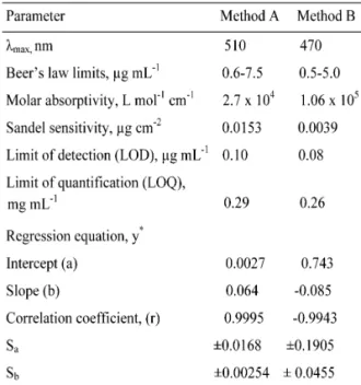
where Y=absorbance, a=intercept, b=slope and X=concentration in µg ml-1 obtained by the method of least squares. Correlation coefficients, intercepts and slopes for the calibration data are also presented in Table 1.
Table 1. Analytical and regression parameters.
*Y=a+bX, where Y is the absorbance and X con-centration in µg mL-1
Sa.standard deviation of intercept. Sb.standard deviation of slope.
Table 2. Evaluation of accuracy and precision Method
*SMT taken/found, range, SD and SEM are in µg mL-1.
RE. Relative error; SD. Standard deviation; SEM. Standard error of mean; RSD. Relative standard deviation; a. Intra-day precision, b. Inter-day precision.
Sensitivity. Sensitivity parameters such as molar absorptivity and Sandel sensitivity, and lim-its of detection and quantification calculated as per the current ICH guidelines [50] are compiled in Table1. The limits of detection (LOD) and quan-tification (LOQ) were calculated according to the same guidelines using the formulae:
LOD=3.3σ/s and LOQ=10σ/s
where σ is the standard deviation of seven reagent blank determinations and s is the slope of the calibration curve.
Robustness and ruggedness. To evaluate the method robustness, two experimental vari-ables such as HCl volume and reaction time were slightly altered, and the same were found to have no significant effect on the accuracy and precision of the methods when the studies were made on a single concentration of SMT. The ruggedness of the methods was assessed by cal-culating the RSD for results obtained by per-forming the analysis using three different instru-ments and by three different persons. The inter-instrumental RSD values ranged from 2.5 to 4.5% whereas the inter-personal RSD values varied from 2.5-3.5% for three concentrations of SMT employed for accuracy and precision studies.
Effect of interferences. To assess the use-fulness of the methods, synthetic mixtures con-taining SMT and inactive ingredients with the following composition were prepared and analysed for SMT content: SMT (20 mg), talc (40 mg), starch (50 mg), sodium alginate (50 mg), magnesium stearate (20 mg), lactose (20 mg), calcium gluconate (30 mg), calcium dihy-drogen orthophosphate (50mg) and titanium dioxide (10mg). The percent recovery of SMT by method A was 96.58±1.6 (n=5) and 97.62±2.1 (n=5) by method B indicating that there is no interference from the common excip-ients added to tablet preparations.
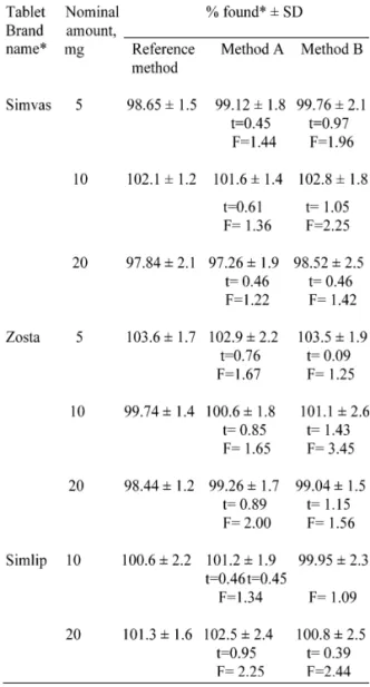
Application to tablet assay. Of the seven-teen brands of tablets available in the Indian market, three representative brands were sub-jected to analysis by the proposed methods. Results presented in Table 3 reveals that there is a close agreement between the results obtained by the proposed methods and the label claim. The results were also compared statistically with those obtained by a reference method [30] by applying Student’s t-test for accuracy and F-test for precision. At the 95% confidence level, the calculated t- and F-values did not exceed the tabulated values suggesting that the proposed methods are as accurate and precise as the ref-erence method.
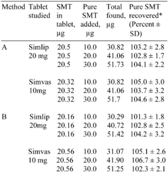
Recovery study. Accuracy and validity of the methods were further ascertained by per-forming recovery studies via standard-addition procedure. Pre-analysed tablet powder was
Table 3. Results of assay of tablets and statistical comparison.
*Mean value of five determinations
#Marketed by; a.Cipla Ltd ; b. Morepen Labs. Ltd; c. Glenmark Pharm. Ltd.,
Tabulated t-value at 95% confidence level is 2.77 Tabulated F-value at 95% confidence level is 6.39.
Table 4. Results of recovery study.
*Mean value of three determinations.
Conclusions
This is a first report on the application of visible spectrophotometry for the assay of sim-vastatin. The methods are based on well-estab-lished and characterised chemical reactions and use cheaper and readily available chemicals. The procedures do not involve any critical experi-mental condition or tedious sample preparation. The methods are highly sensitive as shown by the molar absorptivity values besides being fairly accurate and precise, the last two parameters being unaffected by slight variations in experi-mental conditions such as acid concentration and reaction time. The methods have been demon-strated to be free from interference from common tablet excipients and additives.
Received 26 February 2008 Accepted 06 May 2008
References
[1] G.K. Mc Evoy, American Society of Health System Pharmasists, (2002) 779-782.
[2] The United state Pharmacopoeia 25, the National Formulary 20, United States Pharmacopocial Convention Inc. (2002) 9571-572.
[3] K.Bac-Chan, K. Chong-kook, B. Eunmi , Jeong-sook, P.S. Yun-Kyoung, J. Liq.Chromotogra. Rel.Technol., 27 (2004) 3089-3102.
[4] S.Jianzhong , W. Lihua , S Meifu , Z Xia, C Zhigen, Yaown Fenei Zahzi, 22(2002) 18.Sci Finder, CAN 137:27751, AN 2002:269841.
[5] SS. Ling, L. Tan, LL.Yang , YS Yuan, X. Zhang , Chinese Journal of chromatography.18(2002) 232-4; Su.Finder,CA N 133:83802,AN 2000:509394.
[6] L. Biordi , M. Bologna , G. Carlucci , R. Iannarelli, P. Mazzeo, Int.J. Immunopathol.Pharmacol. 7 (1994) 79-85. [7] L.Biordi , M. Bologua , G. Calucci, P. Mazzeo, J.Pharm.Biomed.Anal. 10 (1992) 693-697.
[8] B. Barrett , V. Borek-Dokalshy, J. Huclova , I. Jelinek, B. Nemec, J.Pharm.Biomed.Anal.41(2006) 517-526.
[9] DG. Musson, L. Sun, A.Y. Yang, JJ. Zhao, Pharm. Biomed.Anal. 38(2005) 521-527.
[10] Y. Feng, Y. Luan and H. Yang, J.Chromatogr. B. Anal.Technol.Biomed.Life Sci., 785(2003) 369-375. [11] Y. Feng, L. Sheng, L. Zhan, Zhongguo Yaoke Daxue Xuebao, 32(2001) 282-285; Sci Finder, CAN 137:72493,AN 2001:825947.
[12] BA. Roadeap, JD. Rogers, IH. Xie, AY. Yang, JJ. Zhao, J.Mass Spectrom. 35 (2000) 1133-1143.
[13] JD. Rogers, A. Yang, N. Zhang, JJ. Zhao, J.Pharm.Biomed.Anal. 34(2004) 175-187.
[14] O. Hisao, I. Kazuhide, U. Naotaka, H. Shunsuke, K. Toshio, J.Chromatography, B. 694(1994) 211-217.
[15] N. Erk, Pharmazie. 57(2002) 817-819.
[16] O. Coruh, SA. Ozkan, Die Pharmazie, 61(2006) 285-290. [17] X. Jianwei, L. Ying, Yaowu Fenxi Zazhi.25(2005) 523-525.
[18] A. Ali, ESM. Nameh, RA. Shawabkeh, J. Anal. Chem. 61(2006) 63-66.
[19] G. Carolina, GC. Gloria, DD. Marta, G. Ricardo, J.Chilean Chem. Soc., 49(2004) 289-290.
[20] X.Yan, G. Cao, X. He, X. Hu, D. Gu, Huaxi Yaoxue Zazhi, 15(2000) 205-206.
[21] J. Wang, Thongguo Yiyao Gongye ZaZhi.31 (2000) 121-122; Sci Finder, CAN 132:284323; AN 2000:293986. [22] B.G. Chandhari, N.M. Patel, P.B. Shah, J.AOAC Int. 90(2007) 1242-9.
[23] B.G. Chandhari, N.M. Patel, P.B. Shah, Indian J. Pharm. sci. 69(2007) 130-132.
[24] L. Xu, Thongguo Yiyao Gongye Zazhi.32 (2001) 271-272;Sci. Finder CAN 135:362691;AN 2001: 606960. [25] Z. Li, S. Jang, Thongguo Yaoxue Zazhi, 35(2000) 554-556.Sci Finder,CAN 133:301278;AN 2000: 675973. [26] L.Wang, M. Asgharnejad, J.Pharm.Boimed.Anal.21 (2000) 1243-1248.
[27] G. Carlcucci, P. Mazzeo, Farmaco.47(1992) 817-823. [28] M.S. Arayne, N. Sultana, F. Hussain, S.A. Ali, J.Anal. Chem. 62(2007) 536-541.
[29] M.K. Srinivasu, A.N. Raju, G.O. Reddy, J.Pharm Biomed Anal.29(2002) 715-721.
[30] K.Basavaiah, U.R. Anil Kumar, J. Mexican. Chem. Society, 51 (2007) 106-112.
[31] J. Basset,R.C. Denney,G.H. Jeffery,J. Mendham,“Vogel’s Quantitative Inorganic Analysis”,English Language Book Society and Longman, Essex,4th Edn,1978,pp.360.
[32] H. Raber,Sci.Pharm.,35 (1967) 220. [33] I.P. Koka, Farm. Zh (Kiev), 3 (1983) 55. [34] I.P. Koka, Farm. Zh (Kiev), 3(1983) 74.
[37] M. Kuchavski, T. Szumilo, Chem. Anal(Warsaw), 28 (1983) 727.
[38] C.A.P. Sastry, M. Aruna, A.R.M. Rao, Talanta, 35 (1988) 113
[39] C.S.P. Sastry, T.N.V. Prasad, B.S. Sastry, E.V. Rao, Analyst(London), 113 (1988) 255.
[40] K. Basavaiah, U. Chandrashekar, H.C. Prameela, Turkish J. Chem., 27 (2003) 591.
[41] K. Basavaiah, B.C. Somashekar, Curr. Pharm. Res.J., 2 (2006) 69.
[42] K. Basavaiah, V. Ramakrishna, U.R. Anilkumar, Ecletica Quimica, 31 (2006) 67.
[43] K. Basavaiah, V. Ramakrishna, U.R. Anilkumar, Acta Pharm. 57 (2007) 211.
[44] K. Basavaiah, B.C. Somashekar, Proc. Natt. Acad. Sci.
India, 76 (A) (2006) 29
[45] K. Basavaiah, U. Chandrashekar, Nagegowda, Bulg. Chem. Commun., 35 (2003) 174.
[46] F. Culkin, J.P. Riley, Anal. Chim. Acta, 24 (1961) 167. [47] F. Culkin.J.P. Riley, Anal. Chim. Acta, 32 (1965) 197. [48] F. Verbeck, Bull. Soc. Chem. Belg. 70 (1961) 415. [49] E.B. Sandel, “Colorimetric Determination of Traces of Metals” , Interscience Publishers Inc, New York., 3rd Edn,
1959, pp.524.