Co llate ral m e tho tre xate re sistance in
cisplatin-se le cte d m urine le uke m ia ce lls
1Department of Pharmaceutical Sciences, College of Pharmacy, Idaho State University,
Pocatello, ID, USA
2Department of Health Sciences, Grand Valley State University, Allendale, MI, USA 3Haverford College, Haverford, PA, USA
A. Bhushan1,
M.P. Hacker2 and
T.R. Tritton3
Abstract
Resistance to anticancer drugs is a major cause of failure of many therapeutic protocols. A variety of mechanisms have been proposed to explain this phenomenon. The exact mechanism depends upon the drug of interest as well as the tumor type treated. While studying a cell line selected for its resistance to cisplatin we noted that the cells expressed a >25,000-fold collateral resistance to methotrexate. Given the magnitude of this resistance we elected to investigate this intri-guing collateral resistance. From a series of investigations we have identified an alteration in a membrane protein of the resistant cell as compared to the sensitive cells that could be the primary mechanism of resistance. Our studies reviewed here indicate decreased tyrosine phosphorylation of a protein (molecular mass = 66) in the resistant cells, which results in little or no transfer of methotrexate from the medium into the cell. Since this is a relatively novel function for tyrosine phosphorylation, this information may provide insight into possible pharmacological approaches to modify therapeutic regimens by analyzing the status of this protein in tumor samples for a better survival of the cancer patients.
Co rre spo nde nce
A. Bhushan
Department of Pharmaceutical Sciences
College of Pharmacy
Idaho State University Pocatello, ID 83209-8334 USA
Presented at the I International Symposium on “Signal Transduction and Gene Expression in Cell
Proliferation and Differentiation”, São Paulo, SP, Brasil,
August 31-September 2, 1998.
Research supported by the American Cancer Society (No. DHP-170).
Received November 4, 1998 Accepted November 19, 1998
Ke y wo rds
·Methotrexate transport ·Resistance
·Cisplatin ·Leukemia ·Phosphorylation
Intro ductio n
Cisplatin [cis-diammine dichloroplatinum (II)] (DDP) is among the most active chemo-therapeutic agents available for treatment of patients with ovarian, testicular, bladder, head and neck, and lung cancers (1-3). Despite the extensive use of DDP, its clinical value has been limited, in part due to the development of resistant cancer cells. Therefore, a great deal of research has been directed towards overcoming resistance by understanding the interactions between cisplatin and DNA.
Acquisition of resistance to the primary drug, DDP, and cross resistance to unrelated drugs has plagued the treatment of ovarian cancer and most solid tumors (4-7). Studies by several investigators have implicated an
interaction between DDP and folate metabo-lism in the acquisition of clinical resistance (8,9). DDP resistance is sometimes associ-ated with cross resistance to the folate an-tagonist methotrexate (MTX) as seen in P388 murine leukemia (8), SSC-25 human squa-mous cell carcinoma (9) and A2780 human ovarian cancer cell lines (10). The link be-tween DDP resistance and the expression of collateral resistance to MTX is not under-stood.
has been explained by a number of mecha-nisms: enhanced repair of DDP-DNA le-sions, altered DDP uptake, and altered intra-cellular sequestration of DDP by such en-dogenous compounds as glutathione and me-tallothionein.
Me tho tre xate actio n and re sistance
The cytotoxicity of methotrexate occurs through competitive inhibition of dihydrofo-late reductase (DHFR) (14). MTX binds very tightly, in a nearly stoichiometric relation-ship to DHFR and prevents DHFR from maintaining a cellular pool of the reduced folate, tetrahydrofolate. Tetrahydrofolates serve as cofactors in the de novo synthesis of
purine nucleotides and thymidylate (15). The most frequent explanation of MTX-induced cell death is through unbalanced growth re-sulting from a depletion of thymidine pools. Resistance to MTX has been reported to be mediated through increased DHFR gene copy number, altered DHFR binding to MTX, impaired polyglutamation of MTX and de-creased MTX uptake. The latter mechanism will be the focus of this review.
Me tho tre xate transpo rt and tyro sine pho spho rylatio n
Unlike DDP, MTX utilizes a specific active folate transport system to gain access to the cellular interior. L1210 mouse leuke-mia cells contain a complex high-affinity transport system for accumulation of MTX and reduced folate compounds. MTX trans-port in L1210 cells is mediated by a 48-kDa integral membrane protein and a 38-kDa cytosolic or peripheral protein which are also involved in intracellular transport of reduced folates (16). Using a photoaffinity probe, Freisheim and his co-workers (17) observed that MTX was first associated with this larger molecular weight protein (66 kDa) and was subsequently transported to the 48-kDa protein. Given the close similarity in
weight to albumin this 66-68-kDa protein was assumed to be albumin. We demon-strate that this protein is not albumin and is intimately involved in MTX transport.
Others have observed that resistance to MTX is often manifested by a decrease in the transport of drug from the extracellular space to the interior of the cell (18-21). It has been demonstrated that folate-binding pro-teins in human leukemia CEM cells selected for MTX resistance have relatively poor af-finity for MTX (22,23).
Me chanism o f m e tho tre xate
re sistance in cisplatin-se le cte d ce lls
Below we provide a stepwise analysis of the two cell lines which led to our first reports depicting the association of tyrosine phosphorylation with methotrexate transport in cisplatin-selected cells (24,25). The re-sults reviewed here demonstrate that L1210 cells made 40-60-fold resistant to DDP ex-press very pronounced collateral resistance to MTX and that the resistance to MTX appears to be directly related to changes in membrane-associated folate transport pro-teins. In these resistant cells, MTX associa-tion to the membrane is only 2-fold less, but no transport to the inside of these cells is detected.
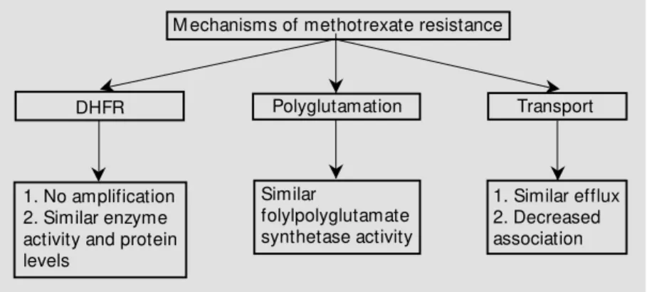
While defining the profile of resistance to antimetabolites in L1210/DDP cells we found that the L1210/DDP cells were >25,000-fold resistant to MTX (24). Many cell lines have been described that are resistant to MTX through DHFR gene amplification. To deter-mine whether a similar situation existed in our L1210/DDP cells we investigated DHFR gene amplification, expression, protein lev-els, enzyme activity and inhibition of enzyme activity by MTX (24). None of these param-eters were altered in L1210/DDP cells as compared to L1210 cells. It is thus clear that MTX resistance seen in the L1210/DDP cells is not due to changes in DHFR.
mediates formation of polyglutamates of MTX and other biologically important folates (26,27). This polyglutamation enhances their cellular retention. Further, it has been shown that impaired polyglutamation alone can cause resistance to MTX (26). In order to adequately test this possibility, we measured FPGS activity in both cell lines (24). FPGS activity in L1210/DDP cells is not signifi-cantly different than in sensitive cells.
Given that other potential mechanisms of resistance, such as altered drug uptake and efflux, can also contribute to resistance, we compared drug transport in the two cell lines. If less MTX enters the cell, DHFR inhibition decreases and therefore less MTX cytotoxic-ity is observed. L1210/0 and L1210/DDP cells were incubated with tritiated MTX and at selected times aliquots were removed to measure the amount of cell-associated drug. A two-fold difference between the two cell lines was seen. While this represents a sig-nificant difference, it cannot by itself ex-plain the remarkable resistance expressed by L1210/DDP cells. Additionally, we tested whether MTX efflux was greater in L1210/ DDP cells than in L1210/0 cells. Our results show MTX efflux to be similar in the two cell lines. All of the above results are sum-marized in Figure 1.
Me tho tre xate transpo rt in L1210 and L1210/D D P ce lls
The studies described above can only ascertain differences in MTX association but not actual MTX uptake and intracellular distribution. Hence, it is possible that L1210/ DDP cells have a vastly different intracellu-lar MTX concentration compared to L1210/ 0 cells.
To address this possibility, we used an [125
I]-labeled photoaffinity probe for MTX. L1210/0 and L1210/DDP cells were incu-bated with the photo probe for 5 min at 4o
C, washed and resuspended in prewarmed buf-fer. Aliquots were removed at selected time
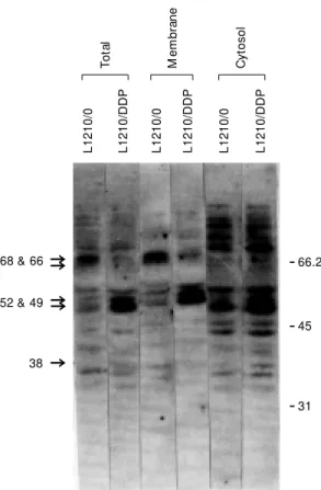
intervals and exposed to UV light to co-valently link the photo probe to its associ-ated protein. The cells were then washed, lysed and the cell lysates were subjected to SDS-PAGE. Protein bands linked to the MTX photoaffinity probe, including 66, 48, 38, and 21 kDa, were identified by autoradiogra-phy (Figure 2). In contrast, the MTX analog was only associated with a 68-kDa protein in L1210/DDP cells with no transfer to addi-tional folate transport proteins.
Figure 1 - Summary of mechanisms of methotrexate resistance in cisplatin-selected L1210/ 0 cells. DHFR, Dihydrofolate reductase.
196
-106
-71
-66 & 6844
-38 U
28
-21U U U
0 1 2 5 10 20 0 1 2 5 10 20
L1210/0 L1210/DDP
Time (min)
Figure 2 - Photoaffinity labeling of L1210/0 and L1210/DDP cells. Equal numbers of cells w ere treated w ith a photoaffinity analog of methotrexate, exposed to UV after various time intervals. Cells w ere w ashed, and lysates w ere analyzed by SDS-PAGE and autoradio-graphed. (Reprinted from Ref. 25, w ith permission from Elsevier Science).
M echanisms of methotrexate resistance
DHFR
1. No amplification 2. Similar enzyme activity and protein levels
Polyglutamation
Similar
folylpolyglutamate synthetase activity
1. Similar efflux 2. Decreased association
Transport
S
S
S S S
It is important to realize that while most drugs enter cells through passive diffusion, methotrexate utilizes the reduced folate up-take system. If the upup-take system is inopera-tive in L1210/DDP cells, as we hypothesize, then [3
H]-MTX will not measure true drug uptake. Instead, it demonstrates association with membrane-bound proteins. Changes in MTX transport are measured by the covalent binding of the photoaffinity analog (Figure 2). [3
H]-MTX shows only the association with the cells.
The temporal relationship in MTX trans-fer seen in the L1210/0 cells supports an orderly transfer of MTX from 66- to 48- to 38- to 21-kDa proteins. These results pro-vide valuable information with respect to possible components of MTX resistance. No affinity label was associated with any of the putative folate transport proteins in the L1210/DDP cells, indicating that MTX is not delivered to DHFR. This strengthens the argument that the mechanism of resistance
in L1210/DDP cells rests in blockade of MTX transport and not in altered DHFR. That the MTX analog was not transported beyond the 68-kDa membrane protein impli-cates the 68- and 48-kDa proteins in faulty MTX transport. While MTX association does occur in the L1210/DDP cells, transport stops at the membrane. This could account for the rather small differences in MTX association (2-fold) but large differences in drug sensi-tivity (25,000-fold) between L1210/0 and L1210/DDP cells.
Binding studies done by other investiga-tors (17) using L1210 cells showed the 66-kDa band, but they suggested it to be un-washed bovine serum albumin (BSA). We have addressed this concern by Western blot and amino acid analyses (25).
Me chanism o f im paire d transpo rt
The results discussed above implicate a faulty protein shuttle system in the type of MTX resistance expressed by the L1210/ DDP cells. Why MTX is not transported in the L1210/DDP cells was the focus of the next series of experiments.
Protein phosphorylation is required for activation of a number of functional proteins related to growth regulation and signal trans-duction. To date, however, tyrosine phos-phorylation has not generally been associ-ated with membrane transport proteins. Our work indicates that the tyrosine phosphory-lation of the folate-binding proteins may be involved in the altered transport of MTX into the cell. Figure 3 shows a Western blot of total, membrane and cytosolic proteins of L1210/0 and L1210/DDP cells probed with an antibody to phosphotyrosine. Differences are seen in cytosolic proteins with molecular weight 74- and 38-kDa, and also membrane-bound 68-, 66-kDa proteins and proteins in the range of 48-52 kDa. Of specific interest are the membrane proteins, especially the 66- and 68-kDa proteins, since we have dem-onstrated, using the photoaffinity analog of
68 & 66 Z
52 & 49 ZZ
Z
38
-
66.2-
45-
31L
1
2
1
0
/0
L
1
2
1
0
/D
D
P
L
1
2
1
0
/0
L
1
2
1
0
/D
D
P
L
1
2
1
0
/0
L
1
2
1
0
/D
D
P
T
o
ta
l
M
e
m
b
ra
n
e
C
y
to
s
o
l
Figure 3 - Western blot anal-ysis of cell lysates, mem-branes and cytosolic frac-t ions f rom L1210/0 and L1210/DDP cells using an antibody to phosphotyrosine. (Reprint ed f rom Ref . 25, w ith permission from Else-vier Science).
MTX (17), that the transfer of the MTX analog to DHFR is blocked at this stage in L1210/DDP cells. Altered tyrosine phospho-rylation at the 68-kDa protein in the resistant cells may explain the decrease in photoaffin-ity analog binding in resistant cells at 0o
C. The increase in phosphorylation of the 48-kDa protein in the L1210/DDP cells as com-pared to L1210/0 cells may block MTX bind-ing or MTX transfer from the 68-kDa protein to the 48-kDa protein.
To further confirm that the tyrosine phos-phorylated 66-68-kDa band was a metho-trexate-binding protein, the cells were la-beled with the [125
I]-labeled photoaffinity analog in the presence and absence of meth-otrexate. Cell lysates were prepared and im-munoprecipitated with an antiphosphotyro-sine antibody. The 66-68-kDa band was found to be labeled and MTX competed with the labeling. That the labeling competed with methotrexate shows that the methotrexate-binding protein as seen by the photoaffinity labeling (25) is tyrosine phosphorylated and that the photoaffinity analog competitively binds the methotrexate-binding proteins.
To determine if the cellular membrane (with protein 66-68 kDa) was the barrier to methotrexate transport, the sensitive and re-sistant cells were disrupted by sonication, treated with the methotrexate photoaffinity analog, and exposed to UV light. The pro-teins were separated by SDS-PAGE, the gel dried and autoradiographed (Figure 4). The methotrexate analog was bound to dihydro-folate reductase (21 kDa) in both cell lines equally, showing that it is a defect in the membrane transport proteins that results in impaired transport to DHFR. A strong band at the bottom is the free unbound [125
I]-labeled photoaffinity analog.
Although we have shown by different studies that impaired MTX uptake is respon-sible for this resistance, the sonication ex-periment substantiates the importance of the membrane in MTX sensitivity. For DHFR to be inhibited by MTX, it has to bind the drug.
These studies emphasize that it can do so equally in the sonicated cell lysates of both cell lines. The high resistance in L1210/DDP cells can only be explained due to reduced uptake of MTX. Without a functional trans-port protein, the membrane barrier stops MTX from entering the cell and results in high drug resistance. Once the membrane barrier is eliminated, MTX goes to its target enzyme. The issue is the lack of transport, as shown in photoaffinity experiments using intact L1210/DDP cells (Figure 2).
Mo dulatio n o f tyro sine pho spho rylatio n re sults in m e tho tre xate se nsitivity
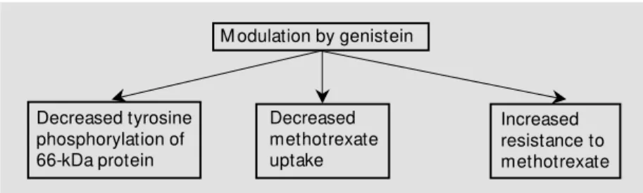
To determine if the transport of MTX requires tyrosine phosphorylation, we used the tyrosine kinase-inhibiting drug, genistein. We studied three effects of genistein treat-ment (28) on L1210/0 cells: 1) MTX
sensi-1 2 kDa
-
45-
29-
18-
15tivity, 2) MTX association and 3) tyrosine phosphorylation. Genistein pretreatment par-tially protected the L1210/0 cells from MTX cytotoxicity and decreased both MTX asso-ciation and 68-kDa protein tyrosine phos-phorylation (Figure 5). This information pro-vides further links for tyrosine phosphoryla-tion with MTX transport.
Co nclusio n
This observation of different tyrosine phosphorylation patterns was somewhat sur-prising. While tyrosine phosphorylation has been commonly associated with cell growth and differentiation (29-31), we now propose a role in membrane transport. Approximately one-third of all oncogenes encode tyrosine kinases which transfer the terminal phos-phate of ATP to the hydroxyl group of ty-rosine residues in a protein. In addition, many growth factor receptors function as tyrosine kinases. These proteins, as well as class I oncogenes, are located on the plasma membrane.
In normal cells the growth factor recep-tors function as tyrosine kinases only after the growth factor binds to its receptor. Spe-cific protein targets within the cell are then phosphorylated as part of the signal
trans-M odulation by genistein
Decreased tyrosine phosphorylation of 66-kDa protein
Decreased methotrexate uptake
Increased resistance to methotrexate
S S
S
Figure 5 - Summary of results obtained after treatment of L1210/0 cells w ith genistein (28).
duction pathway. As a result, the cell under-goes growth and division. This signal input lasts for only a prescribed period of time after which the signal is lost. Tyrosine kinase activity is then down-regulated and cell growth and division cease.
In malignant cells a different scenario exists. In many situations, tyrosine kinases are coded for by activated oncogenes and phosphorylate their substrates in a constitu-tive manner. In such cells the oncogene ex-pression is deregulated, hence tyrosine phos-phorylation continues and growth signals remain unabated. Inhibition of tyrosine ki-nase by such compounds as genistein results in growth arrest and differentiation (32).
Although the preponderance of data ac-cumulated to date relates to the role of ty-rosine kinases in cell growth and differentia-tion, proteins known to be crucially involved in cell-substratum adhesion have also been identified as substrates for this enzyme sys-tem. Our studies provide yet another poten-tial role for tyrosine phosphorylation.
In summary, as we pursued the observa-tion that L12010/DDP cells demonstrate col-lateral resistance to MTX, it became increas-ingly more relevant to observe that the resis-tance appears not to be related to well-char-acterized mechanisms of resistance such as increased DHFR and polyglutamation. The mechanism seems to involve control of drug transport across the cell membrane by a 66-kDa protein.
Ackno wle dgm e nts
Re fe re nce s
1. Hakes T, M arkm an M , Reichm an B, Hoskins W, Jones W & Lew is J (1989). Pilot trial of high intensity intravenous cyt oxan/cisplat in and int raperit oneal cisplatin for advanced ovarian cancer. Pro-ceedings of the American Society of Clini-cal Oncology, 8: 152 (Abstract).
2. Ozols RF & Young RC (1984). Chemo-therapy of ovarian cancer. Seminars in Oncology, 11: 251-263.
3. Ozols RF (1989). Cisplatin dose intensity. Seminars in Oncology, 16: 22-30. 4. Fujii R, M utoh M , Niw a K, Yamada K,
Aikou T, Nakagaw a M , Kuw ano M & Akiyama S (1994). Active efflux system for cisplatin in cisplatin-resistant human KB cells. Japanese Journal of Cancer Re-search, 85: 426-433.
5. Bier H (1993). Chemotherapeutic drug re-sistance in the management of head and neck cancer. European Archives of Oto-Rhino-Laryngology, 250: 200-208. 6. Osmak M , Beketic-Reskovic L, M atulic M
& Soric J (1993). Resistance of human larynx carcinoma cells to cisplatin, gamma irradiation and methotrexate does not in-volve over expression of c-myc or c-Ki-ras oncogenes. M utation Research, 303: 113-120.
7. Hill BT, Shellard SA, Hosking LK, Dempke WC, Fichtinger-Schepman AM , Tone T, Scanlon KJ & Whelan RD (1992). Charac-terization of cisplatin-resistant human o-varian carcinom a cell line expressing cross-resistance to 5-FU but collateral sensitivity to methotrexate. Cancer Re-search, 52: 3110-3118.
8. Schabel Jr FM , Skipper HE, Trader M W, Laster WR, Grisw old DP & Corbett TH (1983). Establishment of cross resistance profiles for new agents. Cancer Treat-ment Reports, 67: 905-922.
9. Teicher BA, Cucchi CA, Lee JB, Flatow JL, Rosow sky A & Frei 3rd E (1986). Alky-lating agents: in vitro studies of cross re-sistance patterns in human cell lines. Can-cer Research, 46: 4379-4383.
10. Scanlon KJ, New man EM , Lu Y & Priest DG (1986). Biochemical basis for cisplatin and 5-fluorouracil synergism in human ovarian carcinoma cells. Proceedings of the National Academy of Sciences, USA, 83: 8923-8925.
11. Rosenberg B (1979). Anticancer activity of cisplatin and some relevant chemistry. Cancer Treatment Reports, 63: 1433-1438.
12. Erickson LC, Zw elling LA, Durcoe JM ,
Sharkey NA & Kohn KW (1981). Differen-tial cytotoxicity and DNA cross-linking in normal and transformed human fibro-blasts treated w ith cis -diamminedichloro-platinum (II). Cancer Research, 41: 2791-2794.
13. Roberts JJ, Knox RJ, Perna M F, Friedlos F & Lydall DA (1988). The role of platinum drug-DNA interactions in cellular toxicity and anti-tumor effects. In: Nicolini M (Edi-tor), Platinum and Other M etal Coordina-t ion Com pounds in Cancer Chem o-therapy. M artinus-Nijhoff Publishing, Bos-ton, 69-70.
14. Futterman S & Silverman M (1957). The inactivation of folic acid by liver. Journal of Biological Chemistry, 224: 31-43. 15. Taylor IW & Tattersall M H (1981). M
eth-otrexate cytotoxicity in cultured human leukemic cells studied by flow cytometry. Cancer Research, 41: 1549-1558. 16. Price EM , Ratnam M , Rodeman KM &
Freisheim JH (1988). Characterization of the methotrexate transport pathw ay in murine L1210 leukemia cells: involve-ment of a membrane receptor and cyto-solic protein. Biochemistry, 27: 7853-7858.
17. Price EM & Freisheim JH (1987). Photoaf-finity analogues of methotrexate as folate antagonist binding probes. 2. Transport studies, photoaffinity labeling and identifi-cation of the membrane carrier protein for methotrexate from murine L1210 cells. Biochemistry, 26: 4757-4763.
18. Sirotnak FM , M occio DM , Kelleher LE & Guotas LJ (1981). Relative frequency and kinetic properties of the transport defec-tive phenotypes among methotrexate re-sistant L1210 clonal cell lines derived in vivo. Cancer Research, 41: 4447-4452. 19. Rosow sky A, W right JE, Cucchi CA,
Lippke JA, Tantravahi R, Ervin TJ & Frei 3rd E (1985). Phenotypic heterogeneity in cultured human head and neck squamous cell carcinoma lines w ith low level meth-otrexate resistance. Cancer Research, 45: 6205-6212.
20. Dembo M & Sirotnak FM (1984). M em-brane transport of folate compounds. In: Folat e Ant agonist s as Therapeut ic Agents. 1: 173-217.
21. M cCormick JI, Susten SS & Freisheim JH (1981). Characterization of the methotrex-ate transport defect in a resistant L1210 lymphoma cell line. Archives of Biochem-istry and Biophysics, 212: 311-318. 22. Jansen G, Westerhof R, Kathmann I,
Rademaker BC, Rijksen G & Schornagel JH (1989). Identification of a membrane associated folate binding protein in hu-m an leukehu-m ic CCRF-CEM cells w it h transport related methotrexate. Cancer Research, 49: 2455-2459.
23. Freisheim JH, Price EM & Ratnam M (1989). Folate coenzyme and antifolate transport proteins in normal and neoplas-tic cells. Advances in Enzyme Regulation, 29: 13-26.
24. Wroblew ski DH, Bhushan A, Xuan Y, Tritton TR & Hacker M P (1996). Investiga-tions on the mechanisms of methotrex-ate resistance in a cisplatin resistant L1210 urine leukemia cell subline. Cancer Chemotherapy and Pharmacology, 37: 337-342.
25. Bhushan A, Wroblew ski DH, Tritton TR, Xuan Y & Hacker M P (1996). Altered ty-rosine phosphorylation in cisplatin resis-tance. Biochemical Pharmacology, 51: 477-482.
26. M cGuire JJ & Bertino JR (1981). Enzy-m at ic synt hesis and f unct ion of folypolyglutamates. M olecular and Cellu-lar Biochemistry, 38: 19-48.
27. M cClosky DE, M cGuire JJ, Russell CA, Row an BG, Bertino JR, Pizzorno G & M ini E (1991). Decreased folypolyglutamate synthetase activity as a mechanism of methotrexate resistance in CCRF-CEM human leukemia sublines. Journal of Bio-logical Chemistry, 266: 6181-6187. 28. Xuan Y, Hacker M P, Tritton TR & Bhushan
A (1998). M odulation of methotrexate re-sistance by genistein in murine leukemia L1210 cells. Oncology Reports, 5: 419-421.
29. Bishop JM (1983). Cellular oncogenes and retroviruses. Annual Review of Biochem-istry, 52: 301-354.
30. Hanks SK, Quinn AM & Hunter T (1988). The protein kinase family: conserved fea-tures and deduced phylogeny of the cata-lytic domains. Science, 241: 42-52. 31. Glenney Jr JG, Zokas L & Kamps M P
(1988). M onoclonal antibodies to phos-photyrosine. Journal of Immunological M ethods, 109: 277-285.