of gestational trophoblastic neoplasia
Diagnóstico, classificação e tratamento
da neoplasia trofoblástica gestacional
Abstract
Gestational trophoblastic neoplasia (GTN) is the term to describe a set of malignant placental diseases, including invasive mole, choriocarcinoma, placental site trophoblastic tumor and epithelioid trophoblastic tumor. Both invasive mole and choriocarcinoma respond well to chemotherapy, and cure rates are greater than 90%. Since the advent of chemotherapy, low-risk GTN has been treated with a single agent, usually methotrexate or actinomycin D. Cases of high-risk GTN, however, should be treated with multiagent chemotherapy, and the regimen usually selected is EMA-CO, which combines etoposide, methotrexate, actinomycin D, cyclophosphamide and vincristine. This study reviews the literature about GTN to discuss current knowledge about its diagnosis and treatment.
Resumo
Neoplasia trofoblástica gestacional (NTG) é o termo que descreve o conjunto de anomalias malignas da placenta, incluindo a mola invasora, coriocarcinoma, tumor trofoblástico do sítio placentário e tumor trofoblástico epitelióide. Ambos a mola invasora e o coriocarcinoma respondem bem à quimioterapia, com taxas de cura superiores a 90%. Desde o advento da quimioterapia, NTG de baixo risco tem sido tratada com monoquimioterapia, pelo geral methotrexate ou actinomicina-D. Casos de NTG de alto risco, contudo, devem ser tratados com poliquimioterapia, e o regime usualmente escolhido é o EMA-CO que combina etoposide, methotrexate, actinomicina-D, ciclofosfamida e vincristina. Esse estudo revê a literatura sobre NTG a im de discutir os conhecimentos atuais sobre seu diagnóstico e tratamento.
School of Medicine, Universidade Federal Fluminense – UFF – Rio de Janeiro (RJ), Brazil; School of Medicine, Universidade Federal do Rio de Janeiro – UFRJ – Rio de Janeiro (RJ), Brazil; New England Trophoblastic Disease Center, Brigham and Women’s Hospital – Harvard Medical School – Boston, United States.
1Professional Masters Degree Program in Maternal and Child Health, Universidade Federal Fluminense – UFF – Rio de Janeiro (RJ), Brazil. 2Universidade Federal do Rio de Janeiro – UFRJ – Rio de Janeiro (RJ), Brazil; Universidade Federal Fluminense – UFF – Rio de Janeiro (RJ), Brazil. 3Department of Obstetrics, Gynecology and Reproductive Biology, Harvard Medical School – Boston, United States. Brigham and
Women’s Hospital – Harvard Medical School – Boston, United States. Conlict of interests: none.
Keywords Gestational trophoblastic disease/diagnosis Gestational trophoblastic disease/classiication Gestational trophoblastic disease/drug therapy Choriocarcinoma/drug therapy
Palavras-chave Doença trofoblástica gestacional/diagnóstico Doença trofoblástica gestacional/classiicação Doença trofoblástica gestacional/quimioterapia Coriocarcinoma/quimioterapia
Correspondence Antonio Braga Maternidade Escola da UFRJ Rua das Laranjeiras, 180, Laranjeiras Zip Code: 22240-003 Rio de Janeiro (RJ), Brazil
Received 10/28/2014
Accepted with modiications 11/19/2014
Introduction
Gestational trophoblastic neoplasia (GTN) is the term used to describe malignant lesions that originate in the chorionic villi and the extravillous trophoblast. This designation includes four different clinical entities, each with different degrees of proliferation, invasion and dissemination: invasive mole (IM), choriocarcinoma (CCA), placental site trophoblastic tumor (PSTT) and epithelioid trophoblastic tumor (ETT)1-5.
About 50% of all cases of GTN occur postmolar gestations, 25% after abortions or ectopic pregnancies and 25%, after term or preterm deliveries4. PSTT and
ETT, however, develop after term deliveries or non-molar abortions in 95% of the cases6.
The largest epidemiological study conducted in Brazil about trophoblastic disease found that GTN developed in 24.6% of the patients that had complete hydatidiform moles and in 7.6% of the cases of partial hydatidiform moles7.
Most cases of GTN are IM and CCA. These dis-eases, characterized by high levels of human chorionic gonadotropin (hCG), are highly responsive to chemo-therapy (ChT) and have cure rates greater than 90%4.
In contrast, the rarer PSTT and ETT have low hCG levels8 and are relatively resistant to ChT. Therefore, the
irst-line treatment in these cases is surgery, particularly in no metastatic cases4.
The objective of this paper is to present an update of clinical aspects, diagnosis and treatment of GTN in order to guide the Brazilian gynecologists and obstetricians on the major advances in the management of patients affected by this neoplasm of pregnancy.
Methods
A search was conducted in the PubMed and the Cochrane Library databases from January 2004 to June 2014, using the term “gestational trophoblastic neo-plasia”. The search retrieved 1,950 publications, 370 abstracts were read in detail, and 52 studies, prefer-ably those published more recently and that focused on GTN diagnosis and treatment, were included in this review.
Although the priority was to select meta-analyses and methodologically well-designed studies, reviews conducted by organizations and institutions, as well as expert opinions, were also included because of the low prevalence of this disease. Although not published within the period deined for the search, the original Federation of Gynecology and Obstetrics (FIGO) 2002 study about GTN staging was also included because of its relevance,
as its publication marked the beginning of the use of a universal GTN classiication.
Clinical presentation
The clinical presentation of GTN is variable and depends on the gestational event that originated the disease, as well as on its extension and histological diagnosis1,4.
Enlarged uterus, vaginal bleeding and persistence of theca lutein cysts in the ovaries are suggestive of post-molar GTN4. However, over 50% of patients with GTN
do not have any clinical indings, and the diagnosis is made only according to plateaued or rising serum hCG concentrations measured during postmolar follow-up after uterine evacuation9.
When CCA is associated with a previous non-molar pregnancy, signs and symptoms are most often associated with tumor invasion in the uterus or sites of metastases, especially the lungs and the pelvis1,4.
GTN metastases occur by hematogenous spread to the lungs (80%), vagina (30%), brain (10%) and liver (10%)10. Lung metastases are usually
symptom-less, but may be extensive and cause dyspnea, cough, hemoptysis and thoracic pain1,4. Vaginal metastatic
nodules are more frequent in the fornices and subu-rethral region. They may cause purulent leucorrhea and difficult-to-control bleeding, as they are richly vascularized1,11.
Bleeding due to uterine perforation or metastases is associated with abdominal pain, hemoptysis, melena and signs and symptoms of increased intracranial pressure, such as headaches, seizures, impaired speech, impaired vision and hemiplegia1,4. GTN perfusion involves
anoma-lous and aberrant circulation in fragile vessels that tend to bleed. Metastatic site biopsies are not recommended because of the high risk of bleeding11.
Almost all patients with PSTT and ETT have ab-normal uterine bleeding a long time after the previous gestational event8. Virilization and nephrotic syndrome
have also been rarely described1,4,12.
As symptoms may be minimal or even absent and the antecedent gestational may be remote, a GTN diagnosis should be suspected in all reproductive age women who present with pulmonary symptoms or unexplained systemic symptoms, particularly in the case of metastases of a primary neoplasia whose site is unknown11.
Evaluation and management
In addition to a detailed patient history and careful clinical examination, a complete blood count, blood type, Rh factor, coagulation tests, liver and renal function evaluation and serum hCG levels should be requested2,4,13.
The concentration of serum hCG is the diagnostic pillar of postmolar GTN. The other diagnostic criteria for postmolar GTN are as follows14: 4 or more plateaued
hCG concentrations over three weeks (that is, on days 1, 7, 14 and 21); increase of hCG concentrations for three or more consecutive measurements for at least two weeks (that is, on days 1, 7 and 14); histological diagnosis of choriocarcinoma and elevated hCG concentrations for six months or longer.
Agarwal et al.15 evaluated the rigorous clinical and
laboratory follow-up of patients with elevated hCG concentrations for six months or longer after molar evacuation. They found that a prolonged follow-up was acceptable and commonly precluded the use of chemo-therapeutic drugs15.
The clinical events listed as indications for treat-ment by the Charing Cross Trophoblastic Disease Center should also be included as GTN diagnostic criteria: abundant vaginal bleeding, signs of gastrointestinal or intraperitoneal hemorrhage, evidence of brain, liver or gastrointestinal metastasis and radiological opacities >2 cm on chest X-ray5.
Transvaginal ultrasound (TVUS) imaging is a fundamental tool for the diagnosis of GTN, but differ-ent diseases may have similar appearances on imaging studies16. The most common inding is a focal
myome-trial mass. The image may be hypo- or hyperechoic, or complex and even multicystic. Intramyometrial anechoic spaces result from hemorrhage and tissue necrosis or vascular spaces16,17.
In extensive disease, imaging may reveal a large, heterogenous and lobulated uterus or an undifferenti-ated pelvic mass16,17. Color Doppler ultrasound may
demonstrate intense chaotic vascularity and loss of vessel discreteness. It demonstrates high-velocity, low-resistance low in contrast with the low usually detected in normal myometrial arteries. However, PSTT may be hypo- or hypervascular16.
A chest X-ray is the imaging method recommended by FIGO to evaluate lung metastases14. However, up
to 41% of the patients with lung metastases on com-puted tomography (CT) have a normal chest X-ray. Pulmonary micrometastases are better evaluated us-ing chest CT, but its use is questionable because the presence of micrometastases does not seem to affect long-term survival17.
Other imaging studies, such as magnetic resonance imaging (MRI) and CT, are not part of the routine
assessment of GTN and are used only in equivocal cases or when metastatic disease is suspected5,16.
CT is the most suitable method to evaluate the common sites of metastases, except for vaginal and brain lesions, which are accurately detected using MRI16.Although few studies have thoroughly
inves-tigated it, positron emission CT seems to be capable of detecting sites of metabolically active disease not found in other imaging studies. Moreover, it may be useful in differentiating uterine scars of ibrosis from active disease16.
Classification and staging
Several staging systems, classiications and prognostic systems have been used globally in the past for GTN, which has made it dificult to compare results of studies conducted in different reference centers.
Because of the need for a universal language, common treatment criteria and a staging system accepted world-wide, the FIGO published a new classiication system for GTN in 2002 (Table 1). The system combined its former anatomic staging system with a modiied scoring system with risk factors deined by the World Health Organization (WHO)4,14.
This classiication excluded blood type from the list of risk factors, assigned a score of four instead of three to liver metastases, and excluded the group of moderate risk neoplasia. According to this system, tumors may be classiied into two groups: low-risk GTN, if the score ≤6; and high-risk, if the score ≥713,14. Staging notation
uses a Roman numeral followed by an Arabic numeral that indicate FIGO anatomic staging and the WHO modiied score, respectively.PSTT and ETT are classi-ied separately4.
Treatment is deined according to the total score of risk factors, which represents the chance that the patient may develop resistance to irst-line single-agent treatment4,5.
Treatment
Half a century ago, before the introduction of ChT in the management of GTN, mortality rates associated with invasive moles reached 15%, more frequently due to hemorrhage, sepsis, embolic phenomena or surgical complications. In the presence of metastases, CCA had a mortality rate of 100%, and of about 60% when hysterectomy was performed to treat apparent nonmetastatic disease1.
A multicenter study conducted in Brazil found that 21.8% of 5,250 patients with molar pregnancy developed GTN, classified as low risk in 81.3%, high risk in 17.5% and PSTT in 1.2%. The overall cure rate was 96.4%, with a mortality of 0.4% for low-risk disease, 9.5% for high-risk and 21.4% for PSTT7.
Methotrexate (MTX), actinomycin-D (ActD), cy-clophosphamide, vincristine, etoposide, cisplatin and paclitaxel are some of the drugs used to treat GTN effectively19.
After hCG returns to normal levels, additional ChT courses, called consolidation ChT, are repeated three to four times, especially in cases of high-risk disease, to avoid recurrence20. A recent study
con-ducted by Lybol et al.21 found a greater recurrence rate
among patients with low-risk GTN treated with two instead of three ChT consolidation courses21.However,
their data were retrospective, and further prospective randomized studies should be conducted to confirm their findings.
Low-risk disease
Low-risk GTN includes nonmetastatic disease (stage I) and metastatic disease with FIGO/WHO scores <711,14. These patients should be first treated
with a single agent, either MTX or ActD4,20,22.
A retrospective study found that 9,4% of women undergoing a second uterine evacuation did not need ChT to attain remission23.Benefits seem to be greater
when hCG concentration is below 1500 IU/L at the time of evacuation24. However, this recommendation
is controversial, and prospective randomized studies should be conducted to confirm the benefits of a repeat uterine evacuation.
In the group of patients with low-risk disease, the choice of the irst line treatment depends on their wish to preserve fertility. Hysterectomy and an adjuvant single-agent ChT course may be recommended for patients that completed childbearing, because this choice treats any occult metastases11.
Extensive experience has been accumulated in treating low-risk GTN over time, and over 14 differ-ent types of ChT regimens have been described, but no consensus has been reached about the preferred irst-line treatment. As there is no strong evidence to conirm the superiority of any one method, several treatments have been arbitrarily used in different centers22,25. However,
a consensus has been reached about the use of a single agent, such as MTX or ActD, for patients with low-risk disease3. These drugs have induction remission rates of
50 to 90%5.
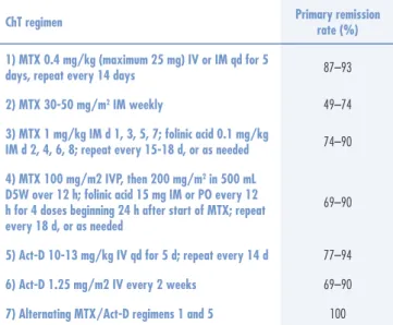
The three most common regimens are: (1) weekly low-dose intramuscular (IM) MTX; (2) pulsed doses of ActD every two weeks; and (3) several other dosing regimens of MTX with or without folinic acid (FA) res-cue25. Table 2 shows primary remission rates according
to ChT regimen.
Primary response variability results from differences in drug doses, times and administration modes, as well as patient selection. In general, weekly IM or intermittent IV infusion of MTX and biweekly ActD are less effective than MTX and ActD for ive days or MTX/FA for eight days. However, almost all patients are cured and have their fertility preserved despite the differences in initial remission after primary ChT3,4.
The weekly MTX 30–50 mg/m2 regimen has the
advantage of convenience and low cost and toxicity, but has the lowest complete response rate among all other regimens, and is not appropriate for the treatment of metastatic disease or CCA3.
ActD has been used as primary therapy in case of renal or hepatic compromise or MTX contraindication and as secondary therapy when the patient develops resistance Table 1. International Federation of Gynecology and Obstetrics and World Health Organization
staging and classiication of gestational trophoblastic neoplasia14
GTN: FIGO staging and classiication (Washington, 2000)
FIGO anatomic staging:
Stage I: Disease conined to the uterus
Stage II: GTN extends outside of the uterus, but is limited to the genital structures (adnexa, vagina, broad ligament)
Stage III: GTN extends to the lungs, with or without known genital tract involvement
Stage IV: All other metastatic sites
Modiied WHO prognostic scoring system as adapted by FIGO
Prognostic factors Score
0 1 2 4
Age <40 ≥40 – –
Antecedent gestation mole abortion term –
Interval (months) <4 4–6 7–12 >12
Pretreatment serum hCG (mIU/mL) <103 103 to
<104 104 to <105 >105
Largest tumor size (including uterus) <3 3 to 4 ≥5 –
Site of metastases lung spleen,
kidney GIT brain, liver
Number of metastases – 1–4 5–8 >8
Previous failed chemotherapy – – single drug ≥2
to MTX. It has more side effects (nausea, alopecia) than MTX and has the risk of local tissue injury in case of extravasation during IV infusion. The most effective regimens are ActD 10–12 mg/kg IV daily for ive days every two weeks, or a single 1.25 mg/m2 IV dose every
two weeks3,26.
Several studies, most retrospective and not random-ized, have investigated the eficacy of MTX in comparison with ActD in the treatment of low-risk GTN.
A recent prospective randomized study conducted by the Gynecologic Oncology Group found that ActD 1.25 mg/m2 IV every two weeks was signiicantly
supe-rior to MTX 30 mg/m2 IM every week, with complete
response rates of 70 and 53% (p=0.01). However, both regimens were less effective when the GTN score was ive or six, or when there was a histologic diagnosis of choriocarcinoma27.
Other studies also found greater primary remission rates for pulsed ActD than weekly MTX28,29, MTX for
ive days30 and MTX/FA for eight days31.
A study about GTN treatment at the John I. Brewer Trophoblastic Disease Center in Chicago included 359 pa-tients treated from 1979 to 2006 and found a rate of complete remission after treatment with a single agent of 79% (78% for MTX and 86% for ActD), and 92% complete response after treatment with a single sequential agent. The other 8% reached remission after treatment with multiple agents or adjuvant surgery32.
A Brazilian study compared three ChT regimens for low-risk GTN: MTX for ive days, ActD for ive days and combined MTX and ActD (MACT). Primary
remission rates were 69, 71.4 and 79.1% respectively, and the differences were not signiicant. Side effects were signiicantly more frequent in the MACT group than in the single-agent groups. The authors found that single-agent regimens are as effective as the combination of drugs under study and suggested that ActD is a less toxic drug with a better cost-effectiveness for the treat-ment of low-risk GTN. However, because of its ease of administration, MTX may be the irst choice in areas where resources are limited33.
Although the FIGO/WHO staging system is use-ful to deine the type of ChT to be used, several authors have suggested that a few points should be clariied, such as the scores assigned according to hCG concentration before treatment34.
A recent study found that resistance to first line ChT may develop when the FIGO/WHO score is six or when hCG is higher than 100,000 IU/L. Based on those findings, the authors suggested a change in the cut-off point for low-risk disease from six to five, or the assignment of a score of six, and not of four, to patients with hCG higher than 100,000 IU/L before treatment35.
Evidence suggests that patients with hCG above 400,000 IU/L should begin ChT with multiple agents because of the signiicantly greater resistance to single-agent ChT34.
A study conducted by the Charing Cross Trophoblastic Disease Center also found that the ef-ficacy of the MTX/FA regimen decreased as the prog-nostic score increased. The rate of complete response in the group of patients with scores of 0 and 1 was 75%, but this rate fell to less than 50% when the score was 3 to 5 and to 31% when the score was 636.
Regardless of which single-agent ChT regimen is used, ChT should continue until hCG returns to normal values, and at least three more ChT cycles should be ad-ministered after the irst normal hCG result. The drug in use should be replaced by another when hCG plateaus or when toxicity precludes the use of appropriate doses or treatment frequency. Multiagent ChT should be used when there is signiicant elevation of hCG concentra-tions, appearance of metastases or sequential resistance to single-agent ChT3.
Findings suggest that any regimen with ActD has primary remission rates higher than those achieved with MTX regimens, but most studies have compared pulsed ActD with weekly MTX, a regimen found to be less ef-fective than those in which treatment is administered along ive to eight days22.
The comparison of side effects between studies is dif-icult because of the differences between patients. The most common side effects for both agents are nausea, anemia Table 2. Primary remission rates of low-risk gestational trophoblastic neoplasia according
to regimen used3
ChT regimen Primary remission rate (%)
1) MTX 0.4 mg/kg (maximum 25 mg) IV or IM qd for 5
days, repeat every 14 days 87–93
2) MTX 30-50 mg/m2 IM weekly 49–74
3) MTX 1 mg/kg IM d 1, 3, 5, 7; folinic acid 0.1 mg/kg
IM d 2, 4, 6, 8; repeat every 15-18 d, or as needed 74–90
4) MTX 100 mg/m2 IVP, then 200 mg/m2 in 500 mL
D5W over 12 h; folinic acid 15 mg IM or PO every 12 h for 4 doses beginning 24 h after start of MTX; repeat every 18 d, or as needed
69–90
5) Act-D 10-13 mg/kg IV qd for 5 d; repeat every 14 d 77–94
6) Act-D 1.25 mg/m2 IV every 2 weeks 69–90
7) Alternating MTX/Act-D regimens 1 and 5 100
and fatigue22, similar in both pulsed ActD and low-dose
MTX regimens. However, Lertkhachonsuk et al.31 found
more severe side effects, such as alopecia and mucositis, in the group treated with ActD.
A prospective randomized study comparing 8-day MTX/FA and 5-day MTX with pulsed ActD is currently being conducted by the Gynecology Oncology Group. Its results, to be published in 2016, may help to deine which drug should be used as irst-line treatment22.
High-risk disease
Patients with high-risk GTN (FIGO stages II-III with score >7 and stage IV) should be treated with mul-tiagent chemotherapy with or without adjuvant surgery and radiotherapy3,4,11 .
The multiagent therapy of choice has changed over the years. In the 1970s and 1980s, MTX, ActD and cy-clophosphamide or chlorambucil (MAC) were the irst line treatment, and cure rates reached 63 to 71%. In the early 1980s, studies found that the regimen with cyclo-phosphamide, hydroxyurea, ActD, MTX/FA, vincristine and doxorubicin (CHAMOCA) increased primary remis-sion to 82%. However, CHAMOCA had lower sustained primary remission rates, as well as greater toxicity, than the MAC regimen3,4.
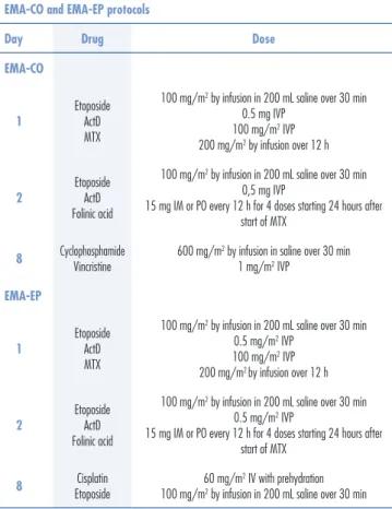
In 1980, etoposide was found to be a very effec-tive agent in the treatment of GTN. Regimens using this drug in combination with high doses of MTX, FA ActD, cyclophosphamide and vincristine (EMA-CO) resulted in higher remission and survival rates3.
The EMA-CO regimen (Table 3) became the first choice for the treatment of high-risk GTN because of its low toxicity and high complete response and survival rates3-5.
Primary or secondary hysterectomy is not effective in reducing the need of ChT or improving remission rates in women with metastatic high-risk GTN, probably because of the higher load of extra uterine disease found in these patients37.
Primary remission rates for the EMA-CO regimen range from 54 to 91%. Although EMA-CO is the most commonly used regimen in the treatment of patients with high-risk GTN, current evidence is not conclusive, as there are no high-quality studies in the literature to support the superiority of this regimen in comparison with other multiagent ChT regimens38.
Induction ChT with EP (etoposide 100 mg/m2
and cisplatin 20 mg/m2) for one or two courses before
the start of EMA-CO for selected high-risk patients (hCG>100,000 IU/L and FIGO/WHO>12) seems to increase overall survival and decrease early deaths39.
To deine which treatment is more effective and less toxic for high-risk patients, carefully conducted multi-site studies should control variables that may affect remission and survival rates, such as risk score, liver and brain metastases and use of adjuvant treat-ments (surgery, radiotherapy, granulocyte colony-stimulating factor)38.
Placental site trophoblastic tumor and epithelioid trophoblastic tumor
Because of the rarity of placental site trophoblastic tumors and epithelioid trophoblastic tumors, their treat-ment is based on indings of small case series described retrospectively12. These tumors are relatively resistant
to ChT and have a propensity for lymphatic spread. Therefore, hysterectomy with or without lymph node dissection and bilateral salpingo-oophorectomy plays a major role in their treatment when the disease is con-ined to the uterus3,11,12 and is curative in two thirds of
the cases37.
Table 3. EMA-CO and EMA-EP protocols4
EMA-CO and EMA-EP protocols
Day Drug Dose
EMA-CO
1
Etoposide ActD MTX
100 mg/m2 by infusion in 200 mL saline over 30 min
0.5 mg IVP 100 mg/m2 IVP
200 mg/m2 by infusion over 12 h
2
Etoposide ActD Folinic acid
100 mg/m2 by infusion in 200 mL saline over 30 min
0,5 mg IVP
15 mg IM or PO every 12 h for 4 doses starting 24 hours after start of MTX
8 CyclophosphamideVincristine 600 mg/m2 by infusion in saline over 30 min1 mg/m2 IVP
EMA-EP
1
Etoposide ActD MTX
100 mg/m2 by infusion in 200 mL saline over 30 min
0.5 mg/m2 IVP
100 mg/m2 IVP
200 mg/m2 by infusion over 12 h
2
Etoposide ActD Folinic acid
100 mg/m2 by infusion in 200 mL saline over 30 min
0.5 mg/m2 IVP
15 mg IM or PO every 12 h for 4 doses starting 24 hours after start of MTX
8 Cisplatin
Etoposide
60 mg/m2 IV with prehydration
100 mg/m2 by infusion in 200 mL saline over 30 min
There may be a response to the EP-EMA (Table 3) or the paclitaxel/cisplatin-paclitaxel/etoposide (TE-TP) regimen, protocols indicated for patients with unfavorable prognostic factors or metastatic disease3,10.
Resistant or recurrent disease
GTN is resistant to ChT when hCG levels plateau or increase, regardless of whether new metastases develop, while the patient is undergoing treatment. In contrast, a diagnosis of recurrence is made when there are two elevations of hCG concentrations in the absence of preg-nancy after a normal hCG result40. Both conditions are
challenging in the treatment of GTN.
Recent data indicate that the number of consolidation ChT administered, a clinical and histologic diagnosis of choriocarcinoma, a high pretreatment hCG concentra-tion, disease spread (brain, liver and gastrointestinal metastases) and a high WHO score are associated with increased rates of resistant disease26,41.
About 5% of the patients with low-risk GTN without metastases and 10% to 15% of those that have metastases develop resistance to primary ChT42.
For low-risk disease, a different single-agent regimen (e.g., ActD after ChT with MTX) is usually enough when hCG levels plateau4,43. When there is no response
to sequential single-agent therapy, multiagent ChT should be initiated, and the EMA-CO regimen is the most commonly used multiagent ChT4.
Recent studies suggest that a uterine artery pulsatil-ity index equal to or lower than one predicts an increase in the risk of resistance to MTX/FA in women with low-risk GTN and may be useful in patient staging for irst line therapy44,45. Prospective studies are underway
evaluate this inding.
Resistance to ChT and recurrent disease are more frequent in patients with high-risk GTN25.About 20
to 30% of the high-risk patients have an incomplete response to irst line ChT or recurrence after remis-sion and eventually need salvage ChT. Treatment with alternative agents, particularly those containing cisplatin, are usually necessary after failure of initial combined ChT43.
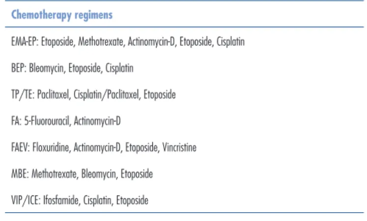
Because of the few cases of resistance to ChT, most studies with this type of patients are retrospective and based on case series. Several salvage regimens (Table 4) are used all over the world, and it is unclear which regimens are more effective and less toxic38, but
the EP-EMA regimen is preferred and recommended by the FIGO. The rate of complete response to this regimen is higher among patients that develop resistance (81.8%) than in those with disease recurrence (42.9%), and the most common side effects are myelosuppression, nausea,
Chemotherapy regimens
EMA-EP: Etoposide, Methotrexate, Actinomycin-D, Etoposide, Cisplatin
BEP: Bleomycin, Etoposide, Cisplatin
TP/TE: Paclitaxel, Cisplatin/Paclitaxel, Etoposide
FA: 5-Fluorouracil, Actinomycin-D
FAEV: Floxuridine, Actinomycin-D, Etoposide, Vincristine
MBE: Methotrexate, Bleomycin, Etoposide
VIP/ICE: Ifosfamide, Cisplatin, Etoposide
Table 4. Salvage chemotherapy for resistant or relapsed gestational trophoblastic neoplasia40
vomiting and hepatotoxicity46. With divergent results,
Powles et al.47 found an overall 5-year survival of 93% for
patients with recurrent disease and 43% for those with chemoresistant disease.
Recent studies have suggested the use of hCG regression normograms to predict resistance to ChT with EMA-CO and the initiation of ChT with a platinum-containing drug instead of EMA-CO when pretreatment hCG concentrations are above the 90th
percentile41.
In addition to salvage ChT, associated procedures, such as hysterectomy, surgical resection of sites of resistant disease, radiotherapy and chemoemboliza-tion techniques, are part of the adjuvant treatment for these patients43,48.
Follow-up after gestational trophoblastic neoplasia treatment
After three consecutive normal weekly hCG mea-surements and ChT completion, serum hCG should be measured monthly for 12 months20. Some centers
recommend additional follow-up procedures for this period. In the New England Trophoblastic Disease Center (Harvard Medical School), specialists recommend two years of follow-up for high-risk disease, and in the Charing Cross Trophoblastic Disease Center (United Kingdom) follow-up continues for the rest of life with urine hCG measurements every six months after ive years of follow-up4,49. Overall risk of recurrence is 3 to
9% in the irst year after treatment and is unusual after 12 months of normal hCG concentrations4.
Contraception is obligatory during follow-up, preferably using combined oral contraceptives. Intrauterine devices should not be used before hCG levels become undetectable4.
simultaneously. Patients with active metastatic disease run greater risks of developing severe psychosocial problems and should receive counseling, psychological support and interventions that may mitigate these disturbances50.
Most patients have a normal ovarian function after ChT, and numerous successful pregnancies have been reported, without any increase in abortions, stillbirths, congenital anomalies, prematurity or other serious ob-stetric complications3.
A Brazilian study about the irst pregnancy after single-agent and multiagent ChT found 68.2% normal term pregnancies and births of healthy newborns. Maternal adverse effects and abortions were signiicantly greater when conception occurred in up to six months after the end of ChT51.
As there is a risk of 1 to 2% of a new molar pregnancy in subsequent pregnancies, the use of TVUS is recom-mended in the beginning of the irst trimester to conirm that the pregnancy is normal. Moreover, the products of contraception or placentas of future pregnancies should be examined histologically. Serum hCG level should be measured six weeks after the end of any future pregnancy to detect occult GTN3,4.
The introduction of regimens with etoposide in the treatment of GTN has been associated with an increase in the risk of secondary neoplasia, such as acute myeloid
leukemia (1%), colon and breast cancer and melanoma. This increase of susceptibility seems to be dose depen-dent and to affect primarily patients that received doses that exceeded 2 g/m2, who should be strictly followed4.
Conclusion
Early adequate treatment ensures an excellent prog-nosis for patients with GTN. The FIGO/WHO staging system (2002)14 has provided a universal language for the
discussion of this neoplasia and has been used to deine prognostic factors more accurately and to compare dif-ferent treatment protocols for low- and high-risk GTN in several parts of the world.
Because of the low frequency of this disease, current treatments are based on retrospective studies and small case series. Well-designed prospective and multi-site studies should be conducted to answer issues that remain controversial, such as the role of a second uterine evacu-ation in the progression of the disease, irst-line ChT for low- and high-risk disease, when to discontinue ChT, when to end hCG monitoring and allow the patient to become pregnant again. It is essential that these patients be followed in Reference Centers where they will receive specialized care which directs impacts survival and quality of life52.
1. Lurain JR. Gestational trophoblastic disease I: epidemiology, pathology, clinical presentation and diagnosis of gestational trophoblastic disease, and management of hydatidiform mole. Am J Obstet Gynecol. 2010;203(6):531-9.
2. Royal College of Obstetricians and Gynaecologists [Internet]. The management of gestational trophoblastic disease. London: RCOG; 2010. (Green-top Guideline No. 38) [cited 2014 Oct 7]. Available from: <https://www.rcog.org.uk/globalassets/ documents/guidelines/gt38managementgestational0210.pdf> 3. Lurain JR. Gestational trophoblastic disease II: classiication and
management of gestational trophoblastic neoplasia. Am J Obstet Gynecol. 2011;204(1):11-8.
4. Goldstein DP, Berkowitz RS. Current management of gestational trophoblastic neoplasia. Hematol Oncol Clin North Am. 2012;26(1):111-31.
5. Seckl MJ, Sebire NJ, Fisher RA, Golier F, Massuger L, Sessa C, et al. Gestational trophoblastic disease: ESMO clinical practice guidelines for diagnosis, treatment and follow-up. Ann Oncol. 2013;24 Suppl 6:vi39-50.
6. Osborne R, Dodge J. Gestational trophoblastic neoplasia. Obstet Gynecol Clin North Am. 2012;39(2):195-212.
7. Braga A, Uberti EMH, Fajardo MC, Viggiano M, Sun SY, Grillo BM, et al. Epidemiological report on the treatment of patients with gestational trophoblastic disease in 10 Brazilian referral centers. Results after 12 years since International FIGO 2000 consensus. J Reprod Med. 2014;59(5-6):241-7.
8. Sung WJ, Shin HC, Kim MK, Kim MJ. Epithelioid trophoblastic tumor: clinicopathologic and immunohistochemical analysis of three cases. Korean J Pathol. 2013;47(1):67-73.
9. Khoo SK, Sidhu M, Baartz D, Yip WL, Tripcony L. Persistence and malignant sequelae of gestational trophoblastic disease: clinical presentation, diagnosis, treatment and outcome. Aust N Z J Obstet Gynaecol. 2010;50(1):81-6.
10. Hui P, Martel M, Parkash V. Gestational trophoblastic diseases: recent advances in histopathologic diagnosis and related genetic aspects. Adv Anat Pathol. 2005;12(3):116-25.
11. Berkowitz RS, Goldstein DP. Current management of gestational trophoblastic diseases. Gynecol Oncol. 2009;112(3):654-62.
12. Hyman DM, Bakios L, Gualtiere G, Carr C, Grisham RN, Makker V, et al. Placental site trophoblastic tumor: analysis of presentation, treatment and outcome. Gynecol Oncol. 2013;129(1):58-62.
13. Soper JT, Mutch DG, Schink JC; American College of Obstetricians and Gynecologists. Diagnosis and treatment of gestational trophoblastic disease: ACOG Practice Bulletin No. 53. Gynecol Oncol. 2004;93(3):575-85.
14. FIGO Oncology Committee. FIGO staging for gestational trophoblastic neoplasia 2000. Int J Gynaecol Obstet. 2002;77(3):285-7.
15. Agarwal R, Teoh S, Short D, Harvey R, Savage PM, Seckl MJ. Chemotherapy and human chorionic gonadotropin concentrations 6 months after uterine evacuation of molar pregnancy: a retrospective cohort study. Lancet. 2012;379(9811):130-5.
16. Kani KK, Lee JH, Dighe M, Moshiri M, Kolokythas O, Dubinsky T. Gestational trophoblastic disease: multimodality imaging assessment with special emphasis on spectrum of abnormalities and value of imaging in staging and management of disease. Curr Probl Diagn Radiol. 2012;41(1):1-10.
17. Allen SD, Lim AK, Seckl MJ, Blunt DM, Mitchell AW. Radiology of gestational trophoblastic neoplasia. Clin Radiol. 2006;61(4):301-13.
18. Kohorn EI. Worldwide survey of the results of treating gestational trophoblastic disease. J Reprod Med. 2014;59(3-4):145-53. 19. Cyriac S, Rajendranath R, Sridevi V, Sagar TG. Etoposide,
cisplatin-etoposide, methotrexate, actinomycin-D as primary treatment for management of very-high-risk gestational trophoblastic neoplasia. Int J Gynaecol Obstet. 2011;115(1):37-9.
20. Maestá I, Braga A. [Challenges of the treatment of patients with gestational trophoblastic disease]. Rev Bras Ginecol Obstet. 2012;34(4):143-6. Portuguese.
21. Lybol C, Westerdijk K, Sweep FC, Ottevanger PB, Massuger LF, Thomas CM. Human chorionic gonadotropin (hCG) regression normograms for patients with high-risk gestational trophoblastic neoplasia treated with EMA/CO (etoposide, methotrexate, actinomycin D, cyclophosphamide and vincristine) chemotherapy. Ann Oncol. 2012;23(11):2903-6.
22. Alazzam M, Tidy J, Hancock BW, Osborne R, Lawrie TA. First-line chemotherapy in low-risk gestational trophoblastic neoplasia. Cochrane Database Syst Rev. 2012;7:CD007102.
23. van Trommel NE, Massuger LF, Verheijen RH, Sweep FC, Thomas CM. The curative effect of a second curettage in persistent trophoblastic disease: a retrospective cohort survey. Gynecol Oncol. 2005;99(1):6-13.
24. Pezeshki M, Hancock BW, Silcocks P, Everard JE, Coleman J, Gillespie AM, et al. The role of repeat uterine evacuation in the management of persistent gestational trophoblastic disease. Gynecol Oncol. 2004;95(3):423-9.
25. Berkowitz RS, Goldstein DP. Current advances in the management of gestational trophoblastic disease. Gynecol Oncol. 2013;128(1):3-5. 26. Chapman-Davis E, Hoekstra AV, Rademaker AW, Schink JC,
Lurain JR. Treatment of nonmetastatic and metastatic low-risk gestational trophoblastic neoplasia: factors associated with resistance to single-agent methotrexate chemotherapy. Gynecol Oncol. 2012;125(3):572-5.
27. Osborne RJ, Filiaci V, Schink JC, Mannel RS, Alvarez Secord A, Kelley JL, et al. Phase III trial of weekly methotrexate or pulsed dactinomycin for low-risk gestational trophoblastic neoplasia: a Gynecologic Oncology Group Study. J Clin Oncol. 2011;29(7):825-31.
28. Gilani MM, Yarandi F, Eftekhar Z, Hanjani P. Comparison of pulse methotrexate and pulse dactinomycin in the treatment of low-risk gestational trophoblastic neoplasia. Aust N Z J Obstet Gynaecol. 2005;45(2):161-4.
29. Yarandi F, Eftekhar Z, Shojaei H, Kanani S, Sharii A, Hanjani P. Pulse methotrexate versus pulse actinomycin D in the treatment of low-risk gestational trophoblastic neoplasia. Int J Gynaecol Obstet. 2008;103(1):33-7.
30. Mousavi A, Cheraghi F, Yarandi F, Gilani MM, Shojaei H. Comparison of pulsed actinomycin D versus 5-day methotrexate for the treatment of low-risk gestational trophoblastic disease. Int J Gynaecol Obstet. 2012;116(1):39-42.
31. Lertkhachonsuk AA, Israngura N, Wilailak S, Tangtrakul S. Actinomycin d versus methotrexate-folinic acid as the treatment of stage I, low-risk gestational trophoblastic neoplasia: a randomized controlled trial. Int J Gynecol Cancer. 2009;19(5):985-8.
32. Hoekstra AV, Lurain JR, Rademaker AW, Schink JC. Gestational trophoblastic neoplasia: treatment outcomes. Obstet Gynecol. 2008;112(2 Pt 1):251-8.
33. Abrão RA, Andrade JM, Tiezzi DG, Marana HR, Reis FJ, Clagnan WS. Treatment for low-risk gestational trophoblastic disease: comparison of single-agent methotrexate, dactinomycin and combination regimens. Gynecol Oncol. 2008;108(1):149-53.
34. McGrath S, Short D, Harvey R, Schmid P, Savage PM, Seckl MJ. The management and outcome of women with post-hydatidiform mole ‘low-risk’ gestational trophoblastic neoplasia, but hCG levels in excess of 100.000IU/L(-1). Br J Cancer. 2010;102(5):810-4.
35. Taylor F, Grew T, Everard J, Ellis L, Winter MC, Tidy J, et al. The outcome of patients with low risk gestational trophoblastic neoplasia treated with single agent intramuscular methotrexate and oral folinic acid. Eur J Cancer. 2013;49(15):3184-90.
36. Sita-Lumsden A, Short D, Lindsay I, Sebire NJ, Adjogatse D, Seckl MJ, et al. Treatment outcomes for 618 women with gestational trophoblastic tumours following a molar pregnancy at the Charing Cross Hospital, 2000-2009. Br J Cancer. 2012;107(11):1810-4.
37. Hanna RK, Soper JT. The role of surgery and radiation therapy in the management of gestational trophoblastic disease. Oncologist. 2010;15(6):593-600.
38. Deng L, Zhang J, Wu T, Lawrie TA. Combination chemotherapy for primary treatment of high-risk gestational trophoblastic tumour. Cochrane Database Syst Rev. 2013;1:CD005196.
39. Alifrangis C, Agarwal R, Short D, Fisher RA, Sebire NJ, Harvey R, et al. EMA/CO for high-risk gestational trophoblastic neoplasia: good outcomes with induction low-dose etoposide-cisplatin and genetic analysis. J Clin Oncol. 2013;31(2):280-6.
40. Ngu SF, Chan KK. Management of chemoresistant and quiescent gestational trophoblastic disease. Curr Obstet Gynecol Rep. 2014;3(1):84-90.
41. Lybol C, Sweep FC, Harvey R, Mitchell H, Short D, Thomas CM, et al. Relapse rates alter two versus three consolidation courses of methotrexate in the treatment of low-risk gestational trophoblastic neoplasia. Gynecol Oncol. 2012;125(3):576-9.
42. Lurain JR, Nejad B. Secondary chemotherapy for high-risk gestational trophoblastic neoplasia. Gynecol Oncol. 2005;97(2):618-23.
43. El-Helw LM, Hancock BW. Treatment of metastatic gestational trophoblastic neoplasia. Lancet Oncol. 2007;8(8):715-24.
44. Agarwal R, Harding V, Short D, Fisher RA, Sebire NJ, Harvey R, et al. Uterine artery pulsatility index: a predictor of methotrexate resistance in gestational trophoblastic neoplasia. Br J Cancer. 2012;106(6):1089-94.
45. Sita-Lumsden A, Medani H, Fisher R, Harvey R, Short D, Sebire N, et al. Uterine artery pulsatility index improves prediction of methotrexate resistance in women with gestational trophoblastic neoplasia with FIGO score 5-6. BJOG. 2013;120(8):1012-5.
46. Mao Y, Wan X, Lv W, Xie X. Relapsed or refractory gestational trophoblastic neoplasia treated with the etoposide and cisplatin/ etoposide, methotrexate, and actinomycin D (EP-EMA) regimen. Int J Gynaecol Obstet. 2007;98(1):44-7.
48. Lurain JR, Schink JC. Importance of salvage therapy in the management of high-risk gestational trophoblastic neoplasia. J Reprod Med. 2012;57(5-6):219-24.
49. Seckl MJ, Sebire NJ, Berkowitz RS. Gestational trophoblastic disease. Lancet. 2010;376(9742):717-29.
50. Wenzel L, Berkowitz RS, Habbal R, Newlands E, Hancock B, Goldstein DP, et al. Predictors of quality of life among long-term survivors of gestational trophoblastic disease. J Reprod Med. 2004;49(8):589-94.
51. Braga A, Maestá I, Michelin OC, Delmanto LR, Consonni M, Rudge MV, et al. Maternal and perinatal outcomes of irst pregnancy after chemotherapy for gestational trophoblastic neoplasia in Brazilian women. Gynecol Oncol. 2009;112(3):568-71.