ISSN 0100-879X
CLINICAL INVESTIGATION
www.bjournal.com.br
www.bjournal.com.br
Volume 44 (1) 1-83 January 2011
Institutional Sponsors
The Brazilian Journal of Medical and Biological Research is partially financed by
Hotsite of proteomics metabolomics developped by:
Braz J Med Biol Res, January 2011, Volume 44(1) 38-45
doi: 10.1590/S0100-879X2010007500129
Effects of different levels of positive airway pressure on breathing
pattern and heart rate variability after coronary artery bypass grafting
surgery
Effects of different levels of positive airway
pressure on breathing pattern and heart rate
variability after coronary artery bypass
grafting surgery
C.B.F. Pantoni
1, L. Di Thommazo
1, R.G. Mendes
1, A.M. Catai
1, S. Luzzi
2,
O. Amaral Neto
2and A. Borghi-Silva
11Laboratório de Fisioterapia Cardiopulmonar e Núcleo de Pesquisa em Exercício Físico,
Departamento de Fisioterapia, Universidade Federal de São Carlos, São Carlos, SP, Brasil 2Irmandade da Santa Casa de Misericórdia de Araraquara, Araraquara, SP, Brasil
Abstract
The application of continuous positive airway pressure (CPAP) produces important hemodynamic alterations, which can influ -ence breathing pattern (BP) and heart rate variability (HRV). The aim of this study was to evaluate the effects of different levels of CPAP on postoperative BP and HRV after coronary artery bypass grafting (CABG) surgery and the impact of CABG surgery on these variables. Eighteen patients undergoing CABG were evaluated postoperatively during spontaneous breathing (SB) and application of four levels of CPAP applied in random order: sham (3 cmH2O), 5 cmH2O, 8 cmH2O, and 12 cmH2O. HRV
was analyzed in time and frequency domains and by nonlinear methods and BP was analyzed in different variables (breathing frequency, inspiratory tidal volume, inspiratory and expiratory time, total breath time, fractional inspiratory time, percent rib cage
inspiratory contribution to tidal volume, phase relation during inspiration, phase relation during expiration). There was significant postoperative impairment in HRV and BP after CABG surgery compared to the preoperative period and improvement of DFAα1, DFAα2 and SD2 indexes, and ventilatory variables during postoperative CPAP application, with a greater effect when 8 and
12 cmH2O were applied. A positive correlation (P < 0.05 and r = 0.64; Spearman) was found between DFAα1 and inspiratory
time to the delta of 12 cmH2O and SB of HRV and respiratory values. Acute application of CPAP was able to alter cardiac
autonomic nervous system control and BP of patients undergoing CABG surgery and 8 and 12 cmH2O of CPAP provided the
best performance of pulmonary and cardiac autonomic functions.
Key words: Continuous positive airway pressure; Cardiovascular system; Autonomic nervous system; Ventilation; Coronary artery bypass
Introduction
Coronary artery bypass grafting (CABG) surgery is a well established and an effective treatment to reduce the symptoms and mortality of patients with coronary artery disease. The surgical procedure and the risk factors in-volved, such as the median sternotomy, cardiopulmonary bypass (CPB) and thoracic manipulation, are believed to be responsible for several changes in cardiorespiratory function after CABG surgery (1) with damage to cardiac autonomic function indicated by heart rate variability (HRV) (2,3)and changes in pulmonary function (4,5).
Several rehabilitation strategies have been applied
to these patients in order to minimize the alterations of respiratory function (4,6). Although there is no consensus in the literature about the best physiotherapy respiratory intervention in patients undergoing CABG surgery (7), it seems that the application of noninvasive positive pressure ventilation improves lung function compared to conventional respiratory therapy (4,8).
Noninvasive positive pressure has been demonstrated to be an important adjunct to improve gas exchanges(8), re-duce breathing work, the need of intubation (9), hospital stay (7), and mortality in several clinical conditions. Loeckinger
Correspondence: A. Borghi-Silva, Laboratório de Fisioterapia Cardiopulmonar, Núcleo de Pesquisas em Exercício Físico, UFSCar, Rod. Washington Luis, km 235, 13565-905 São Carlos, SP, Brasil. Fax: +55-16-3361-2081. E-mail: [email protected]
CPAP after coronary artery bypass grafting surgery 39
et al. (10) observed an improvement of gas exchanges in patients who received continuous positive airway pressure (CPAP) application after cardiac surgery.
However, the application of positive airway pressure produces mechanical effects on the cardiovascular system with changes in hemodynamics and in the cardiac autonomic
nervous system (11). We have not identified studies about the influence of short-term application of CPAP on the breathing pat -tern (BP) and HRV of patients undergoing CABG surgery.
Therefore, the aim of this study was to evaluate the effects of different levels of CPAP application on BP and HRV during the postoperative (PO) period of CABG surgery, as well as the impact of CABG surgery on these variables. We tested the hypothesis that higher levels of CPAP may
positively influence BP and HRV.
Material and Methods
Design and study population
The study was a prospective, randomized, controlled, and double-blind trial. A total of 66 patients were screened, and 18 patients of both genders, with a clinical diagnosis of coronary artery disease who underwent elective CABG with CPB, me-dian sternotomy incision and interposition of a saphenous vein, internal mammary artery or radial artery grafts were included in the study. Patients undergoing urgency surgery or CABG concomitant with valve or other cardiac surgery and using intra-aortic balloon and invasive ventilation for more than 24 h, with pacemakers or presenting previous cardiac surgery, recent acute myocardial infarction (less than 6 months),
un-stable angina, chronic heart rhythm disturbances, significant
arrhythmia, valvular heart disease, chronic obstructive pulmo-nary disease, diabetic neuropathy, poor cognition, and other severe non-cardiac diseases were excluded from the study. The study protocol was approved by the Federal University of São Carlos Ethics Committee (200/2007) and written informed consent was obtained from all patients.
Experimental procedures
During the preoperative period, patients underwent clini-cal cardiac assessment before their inclusion in the study. The physiotherapist’s evaluation consisted of anamnesis and physical examination, including data about previous diseases and the present illness, lifestyle and eating hab-its. In addition, patients were instructed about the surgical procedures, tracheal intubation and the importance of physiotherapy for their postoperative recovery.
After the initial evaluation, all patients that fulfilled the inclu -sion criteria underwent the interventions and measurements of the study, on the day before the surgery and on the second PO day, after the mediastinum tube had been removed and the patient had been in spontaneous breathing (SB). The protocol was carried out only during the afternoon to exclude
the influence of circadian rhythm alterations on HRV. CPAP
application during the preoperative period aimed to familiarize
the patients for the PO intervention. Spirometric tests were performed during the preoperative period in order to exclude patients with chronic obstructive pulmonary disease. Surgical and hospital data were recorded postoperatively.
Study protocol and measurements
Data were collected with the patient in the seated posi-tion during SB and with ventilatory assistance using CPAP ventilation (Breas PV100, Sweden) without supplemental oxygen, with a 21% oxygen fraction (environmental air), according to the following conditions: a) SB, b) 3 cmH2O
(sham) (12), c) CPAP = 5 cmH2O, d) CPAP = 8 cmH2O,
and e) CPAP = 12 cmH2O.
Data were first collected during the SB condition. Next,
the patient was submitted to the four different levels of CPAP applied in a random order, each for approximately
20 min, after a sufficient time to adapt the patient to each
selected level. A rest interval of 30 min was allowed between applications. The experiment was performed by two inves-tigators. One of them was aware of the CPAP intervention, while the other, responsible for data analysis, just collected the physiological data and did not know about the levels applied. Patients were unaware of the pressure levels to which they were submitted.
HRV and BP were recorded during each condition. The heart rate (HR) and R-R intervals (R-Ri) were recorded continuously using a Polar S810i telemetry system (Polar®,
Finland). BP was assessed by respiratory inductive plethys-mography (LifeShirt System, Vivometrics Inc., USA) and was monitored using the thoracic and abdominal inductance plethysmography bands integrated in the LifeShirt garment located at the levels of the nipples and umbilicus, respec-tively. Data were recorded with a portable device, stored on
a flash memory card inserted in the LifeShirt recorder and
then downloaded to a computer into the VivoLogic analysis software (Vivometrics) accompanying the LifeShirt in order
to analyze the respiratory data. An 800-mL specific bag was
used for the volumetric adjustment procedure (13).
At the end of each CPAP application, a modified Borg scale
(0-10) was applied in order to monitor subjective responses regarding dyspnea and pain, and systolic and diastolic blood pressures were registered indirectly with a manual aneroid sphygmomanometer (Becton Dickinson, Brazil) and a stetho-scope (3M Litmann, USA) for patient monitoring.
Analysis of breathing pattern and heart rate variability
All artifacts were reviewed by visual inspection on a computer monitor. Only segments with more than 90% pure
signal beats were included in the final analysis. The most
stable sections containing at least 256 points of sample frequency were chosen for HRV analysis, as recommended (14). The data were entered into the Kubios HRV Analysis software (MATLAB, version 2 beta, Kuopio, Finland).
sta-tistical models in the time and frequency domains, and by nonlinear models. In the time domain, the mean R-Ri,
standard deviation of all R-Ri (SDNN) in ms, which reflects
all the cyclic components responsible for variability during the recording period and is an estimate of overall HRV, and root mean square of the squares of the differences between successive R-Ri (rMSSD), in ms, representative of parasympathetic activity (14) were analyzed.
In the frequency domain analysis, the components of the power spectrum were analyzed using the Fast Fourier Transform in the following components: low frequency (LF; 0.04-0.15 Hz), representative of sympathetic activity; high frequency (HF; 0.15-0.4 Hz), representative of parasym-pathetic activity; both in normalized units, and LF/HF ratio, representative of sympathoadrenal vagal balance (14,15).
In nonlinear analysis, we used Poincaré plot measure indexes SD1 and SD2 (the standard deviation of the Poin-caré plot perpendicular and along the line of identity, respec-tively), representative of parasympathetic autonomic activity
and total HRV (16), respectively. Detrended fluctuations analysis (DFA) was also carried out using DFAα1 (short-term correlation properties of R-Ri) and DFAα2 (long-(short-term
correlation properties of R-Ri) indexes. This technique of analysis, previously developed and described by Peng et
al. (17), quantifies the presence or absence of fractal-like
correlation properties in biological time series and has been used to evaluate the risk of mortality in various groups, be-ing a good predictor of benign and malignant arrhythmias, sudden cardiac death and total mortality in patients with reduced left ventricle ejection fraction, with acute myocardial infarction and with other cardiovascular diseases (18-21).
For BP analysis, we used the following variables: breath-ing frequency (BF), inspiratory tidal volume (ViVol), inspira-tory time (Ti), expirainspira-tory time (Te), total breath time (Tt), fractional inspiratory time (Ti/Tt), percent rib cage inspiratory contribution to tidal volume (%RCi), phase relation during inspiration (PhRIB) and expiration (PhREB). The PhRIB and PhREB indexes determine the degree of thoracoabdominal synchrony and refer to the phase relation during inspiration and expiration, respectively. These indexes represent the percentage of time during a respiratory cycle in which the movements of the rib cage and abdomen are in opposite directions, indicating the delay of the movement of the rib cage over the abdomen at the beginning of inspiration (PhRIB) and expiration (PhREB)(22). To obtain the respi-ratory inductive plethysmographic sum signal for absolute
volume in mL, a quantitative calibration was carried out (fixed
volume least squares calibration) before the analysis of the respiratory variables, which provided a breath-by-breath calculation of the breathing pattern variables. A total of 30 points of this breath-by-breath calculation for each patient were chosen for analysis.
Statistical analysis
Statistical analysis was performed using the GraphPad
InStat for Windows software, Version 3.0. Normal distribu-tion of the data was evaluated using the Shapiro-Wilk test and, according to the characteristics and normality of the values, parametric or non-parametric tests were applied. Repeated measures ANOVA with the Tukey post hoc test or the Friedman test with the Dunn post hoc test were used for analysis of all conditions studied (PO). Spearman correlation was used to evaluate the relationship between delta of 12 cmH2O and SB of HRV indexes and respiratory variables
in the 2nd PO period. The paired t-test or Wilcoxontest was used for the comparison between preoperative and
2nd PO (spontaneous breathing). The level of significance
was set at P < 0.05 in all analyses.
Results
Population characteristics
Of all patients with CABG surgery prescription, only 18
were included in the final sample (2nd PO day). Table 1 shows
baseline characteristics and surgical and hospital data.
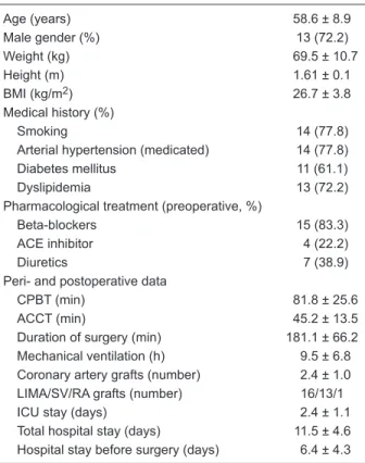
Table 1. Baseline, surgical and hospital data of the 18 patients studied.
Age (years) 58.6 ± 8.9
Male gender (%) 13 (72.2)
Weight (kg) 69.5 ± 10.7
Height (m) 1.61 ± 0.1
BMI (kg/m2) 26.7 ± 3.8
Medical history (%)
Smoking 14 (77.8)
Arterial hypertension (medicated) 14 (77.8)
Diabetes mellitus 11 (61.1)
Dyslipidemia 13 (72.2)
Pharmacological treatment (preoperative, %)
Beta-blockers 15 (83.3)
ACE inhibitor 4 (22.2)
Diuretics 7 (38.9)
Peri- and postoperative data
CPBT (min) 81.8 ± 25.6
ACCT (min) 45.2 ± 13.5
Duration of surgery (min) 181.1 ± 66.2
Mechanical ventilation (h) 9.5 ± 6.8
Coronary artery grafts (number) 2.4 ± 1.0
LIMA/SV/RA grafts (number) 16/13/1
ICU stay (days) 2.4 ± 1.1
Total hospital stay (days) 11.5 ± 4.6
Hospital stay before surgery (days) 6.4 ± 4.3
CPAP after coronary artery bypass grafting surgery 41
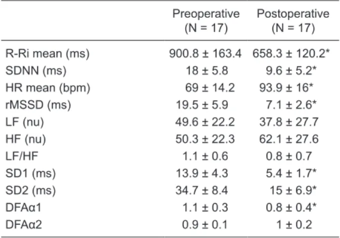
Heart rate variability and breathing pattern responses after CABG surgery
Table 2 shows the HRV of patients before and after
CABG surgery. There was a reduction of mean R-Ri and
SDNN, rMSSD, SD1, SD2, and DFAα1 indexes, with an
increase of mean HR in CABG PO compared to the preop-erative period. Table 3 presents the patients’ BP responses before and after CABG surgery. There was an increase of BF and PhRIB, PhREB indexes, as well as a reduction of Ti, Te and Tt in CABG PO, compared to the preoperative period. These analyses were carried out with 17 patients
in the final sample for data pairing since one patient’s data
during SB in the preoperative period presented poor quality of the stretch signal, being unsuitable for processing and analysis.
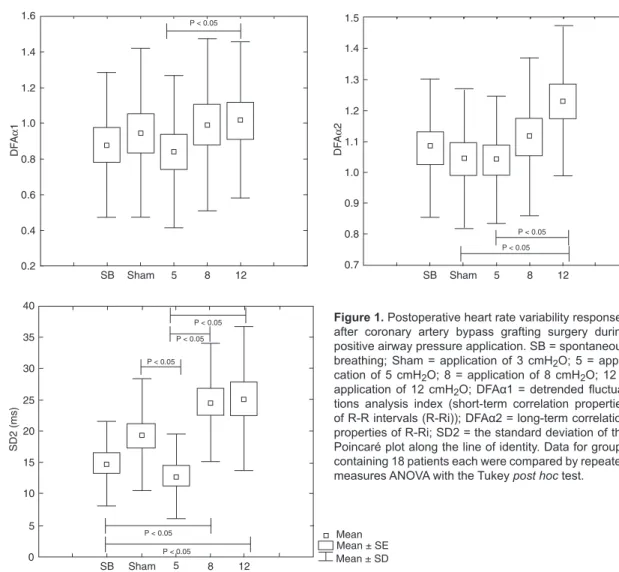
Heart rate variability responses during CPAP application
CPAP application on the 2nd PO day provoked
sig-nificant alterations of DFAα1, DFAα2 and SD2 (Figure 1). There was an increase of DFAα1 during application of 12
cmH2O compared to 5 cmH2O and an increase of DFAα2
index during 12 cmH2O compared to the sham condition
and 5 cmH2O. Regarding the SD2 index, we observed an
increase when the higher CPAP levels were applied (12 and 8 cmH2O), compared to SB and the lower CPAP level
(5 cmH2O). There was a decrease of SD2 during 5 cmH2O
compared to the sham condition.
Breathing pattern responses during CPAP application
Table 4 presents the ventilatory variables during SB and different levels of positive airway pressure applica-tion. There was BF reduction and an increase of Ti, Te and Tt during the sham condition and at all levels applied compared to SB. ViVol values were increased during 5, 8, and 12 cmH2O compared to sham ventilation and SB.
There was a decrease of %RCi during all levels of CPAP application and during the sham condition compared to SB, and a decrease of its values during 8 cmH2O compared
to the sham condition. However, 12 cmH2O provoked an
increase of %RCi compared to 8 cmH2O. The PhRIB index
was increased during 12 cmH2O compared to SB and 8
cmH2O. Peripheral oxygen saturation presented higher
val-ues during all levels applied (5, 8 and 12 cmH2O) compared
to the SB condition. Figure 2 shows correlations between ventilatory variables and HRV values of delta between 12 cmH2O and SB condition. There was a positive correlation
between DFAα1 index and Ti.
Discussion
The main finding of our study was that patients under -going CABG surgery presented important damage in HRV
and BP. To our knowledge, this is the first study to compare
the effects of different levels of CPAP on respiratory pattern (evaluated by respiratory inductive plethysmography) and autonomic nervous control of heart rate in patients submitted
Table 2. Heart rate variability variables in the preoperative and postoperative periods during spontaneous breathing.
Preoperative (N = 17)
Postoperative (N = 17)
R-Ri mean (ms) 900.8 ± 163.4 658.3 ± 120.2*
SDNN (ms) 18 ± 5.8 9.6 ± 5.2*
HR mean (bpm) 69 ± 14.2 93.9 ± 16*
rMSSD (ms) 19.5 ± 5.9 7.1 ± 2.6*
LF (nu) 49.6 ± 22.2 37.8 ± 27.7
HF (nu) 50.3 ± 22.3 62.1 ± 27.6
LF/HF 1.1 ± 0.6 0.8 ± 0.7
SD1 (ms) 13.9 ± 4.3 5.4 ± 1.7*
SD2 (ms) 34.7 ± 8.4 15 ± 6.9*
DFAα1 1.1 ± 0.3 0.8 ± 0.4*
DFAα2 0.9 ± 0.1 1 ± 0.2
Data are reported as means ± SD. R-Ri = R-R intervals; SDNN = standard deviation of all R-Ri; HR = heart rate; rMSSD = root mean square of the squares of the differences between succes-sive R-Ri; LF = low frequency, in normalized units (nu); HF = high frequency; LF/HF = low/high frequency ratio; SD1 = the standard deviation of the Poincaré plot perpendicular to the line of iden-tity; SD2 = the standard deviation of the Poincaré plot along the
line of identity; DFAα1= short-term correlation properties of R-Ri; DFAα2 = long-term correlation properties of R-Ri. *P < 0.05 com -pared to preoperative group (Student paired t-test).
Table 3. Respiratory inductive plethysmographic variables in the preoperative and postoperative periods during spontaneous breathing.
Preoperative (N = 17)
Postoperative (N = 17)
BF (breaths/min) 18.8 (15-22) 24.2 (19-30)*
ViVol (mL) 500 (350-900) 620 (500-800)
Ti (s) 1.2 (1.0-1.4) 0.9 (0.7-1.1)*
Te (s) 1.9 (1.6-2.6) 1.4 (1.2-1.9)*
Tt (s) 3.2 (2.7-4.0) 2.4 (2.0-3.0)*
Ti/Tt 0.4 (0.34-0.44) 0.4 (0.34-0.41)
%RCi 82 (72.3-89.9) 87.3 (69.7-97.0)
PhRIB (%) 5.0 (1.6-10.5) 11.1 (2.7-20.9)*
PhREB (%) 2.6 (0.9-9.5 ) 10.7 (2.7-28.2)*
Data are reported as median (first quartile-third quartile). BF =
Figure 1. Postoperative heart rate variability responses after coronary artery bypass grafting surgery during positive airway pressure application. SB = spontaneous breathing; Sham = application of 3 cmH2O; 5 = appli-cation of 5 cmH2O; 8 = application of 8 cmH2O; 12 = application of 12 cmH2O; DFAα1 = detrended fluctua -tions analysis index (short-term correlation properties
of R-R intervals (R-Ri)); DFAα2 = long-term correlation
properties of R-Ri; SD2 = the standard deviation of the Poincaré plot along the line of identity. Data for groups containing 18 patients each were compared by repeated measures ANOVA with the Tukey post hoc test.
Table 4. Respiratory inductive plethysmographic variables during the postoperative period.
SB Sham 5 cmH2O 8 cmH2O 12 cmH2O
BF (breaths/min) 24.3 (19-24) 22.9 (19-26)* 22.9 (20-25)** 22.9 (19-27)+ 23.4 (19-26)++
ViVol (mL) 518.5 (435-641) 558 (470-659) 569.5 (489-668)**‡ 573 (516-658)+€ 592 (529-680)++£
Ti (s) 0.94 (0.76-1.12) 1.00 (0.86-1.22)* 1.00 (0.88-1.16)** 0.98 (0.86-1.19)+ 1.00 (0.84-1.18)++
Te (s) 1.4 (1.2-1.8) 1.5 (1.2-1.9)* 1.5 (1.3-1.8)** 1.5 (1.3-1.8)+ 1.5 (1.3-1.8)++
Tt (s) 2.4 (2-3) 2.6 (2-3)* 2.6 (2-3)** 2.5 (2-3)+ 2.5 (2-3)++
Ti/Tt 0.38 (0.34-0.41) 0.39 (0.35-0.43) 0.39 (0.35-0.43) 0.39 (0.35-0.42) 0.39 (0.34-0.42)
%RCi 91.5 (70-96) 89.8 (68-95)* 89.6 (68-96)** 87.6 (69-96)+€ 88.2 (63-96)++∞
PhRIB (%) 10.3 (2-19) 12.1 (3-23) 13.9 (3-22) 11.6 (2-21) 12.5 (2-27)++∞
PhREB (%) 9.8 (3-27) 11.8 (2-22) 11.4 (4-23) 10.5 (3-19) 11.2 (3-26)
SpO2 (%) 88 (85-93) 88 (87-94) 90 (85-93)** 89 (83-94)+ 90 (86-93)++
CPAP after coronary artery bypass grafting surgery 43
to bypass grafting surgery. These results are important since short-term interventions are commonly used in respiratory therapy in these patients during hospitalization. We found that higher levels of positive pressure (8 and12 cmH2O)
applied by CPAP were able to positively modify the cardiac autonomic function and BP of these patients.
Important deleterious alterations of pulmonary function have been described after CABG (4). In agreement with our study, Kristjánsdóttir et al. (23) observed that CABG resulted in rib cage movements and BP worsening, as pre-viously observed with an instrument measuring respiratory movements. Atelectasis has been reported to occur after the onset of general anesthesia and the cardiopulmonary
bypass may markedly contribute to an inflammatory reac -tion in the lungs and postoperative atelectasis (24-27). This atelectasis, if persistent, may lead to postoperative pulmonary complications, includinghypoxemia, increased shunt fraction and work of breathing (28).
In this context, CPAP is important by producing airway dilation, improving arterial oxygenation, gas exchanges and indirectly the BP with a reduction of respiratory rate and an increase in tidal volume(8-10,29) in the first days after cardiac surgery. In the present study, we observed that CPAP decreased respiratory breathing and increased expiratory, inspiratory and total times, as well as peripheral oxygen saturation. Importantly as a novel result, our study showed that CPAP was able to reduce %RCi during all levels of positive pressure applied, contributing to a better respiratory synchronism, especially at the higher levels ap-plied. Celebi et al. (30) observed a lower atelectasis score in recruitment maneuvers with application of high levels of CPAP (40 cmH2O) and positive end-expiratory pressure
(PEEP) (20 cmH2O) during a short period of time with
subsequent decremental levels, when compared to those
achieved in patients receiving about 5 cmH2O PEEP. This
could be explained, according to the authors, by the levels applied since a PEEP level of 5 cmH2O is unable to open
the lung after surgery.
Regarding cardiac autonomic activity, it has been
re-ported that CABG surgery produces significant alterations in
cardiac autonomic function with reduction of HRV (31). It is known that the aging process is related to HRV (32) reduc-tion, and altered autonomic regulation after cardiac surgery can precipitate the appearance of cardiac arrhythmias and increase the risk of sudden death. Some investigators have observed HRV reduction after myocardial revascularization surgery, which can return to preoperative values within 2 months (3,33).
In our study, we also observed deleterious changes in HRV after CABG with a decrease of linear (in the time do-main) and nonlinear indexes, Poincaré plot, and DFA. These
modifications after CABG can be related to the acute effects of surgery, with injuries to the autonomic nerve fibers or to
the sinus node, and to the effects of anesthesia, surgical stress, pain, myocardial ischemia, and medications, as well as the effects of extracorporeal circulation, which is not a benign intervention and is associated with large numbers of adverse consequences (1,34).
Despite the important changes provoked by surgery, we observed that CPAP was able to positively modify HRV at the higher levels studied (8 and 12 cmH2O), as observed
by nonlinear indexes that may directly characterize the complexity, irregularity and predictability of the properties of biological systems and complement conventional measures of variability (35).
The effects of different levels of CPAP on the activity of the autonomic cardiovascular system have been poorly explored in the literature; nevertheless, it is known that higher levels can produce acute deleterious effects on hemodynamic properties (36). Important hemodynamic alterations have been reported to occur when high levels of CPAP (>15 cmH2O) are applied to healthy subjects (12).
In contrast, we used lower values when compared to the cited studies.
We observed in our study that moderate values of CPAP (8 and 12 cmH2O) acutely increased HRV, as evaluated by
nonlinear indexes in patients during the 2nd PO of CABG. An increase in SD2 index suggests a total HRV improvement (16). The decrease of DFA indexes may be associated with greater likelihood of cardiac events and mortality. Ksela et al.(37) observed a greater decline of nonlinear indexes (DFA) in patients who developed cardiac arrhythmias in the postoperative period compared with those who maintained stable sinus rhythm.
Our study focused on the acute effects of CPAP and pos-sibly there was only an acute cardiovascular stress, which could have decreased HRV due to a sympathetic response to a reduction of cardiac output. However, our results may be due to the levels of positive airway pressure applied
Figure 2. Correlation between detrended fluctuations analysis
since it has been shown that moderate CPAP levels (10 cmH2O) inhibit cardiac sympathetic nervous activity in heart
failure patients (38). Moreover, it has been demonstrated that immediately after cardiac surgery patients present an imbalance of vagal and sympathetic regulation, with vagal activity suppression (3,31). In this condition, CPAP applica-tion may have improved the activity of the parasympathetic
nervous system, reflecting an increase of HRV.
Regarding the correlations between the delta of the ventilatory variables and HRV, we observed that the greater
variation in inspiratory time, the greater the influence on
cardiac autonomic control, suggesting that BP can positively modulate HRV with higher levels of CPAP application.
In this way, although it is impossible for us to extend the present results to long-term CPAP application and to speculate about the effects on cardiac events, we did observe a real acute improvement of HRV and a reduc-tion of indexes associated with cardiac events in patients
undergoing CABG with short-term CPAP application. In this context, associated with the pulmonary results, we
could observe a greater and better influence of the higher levels of positive pressure applied. Thus, our findings may provide a mechanism for the beneficial actions of CPAP
in PO cardiac surgery as an adjunct therapy for an early cardiopulmonary rehabilitation of these patients during hospital convalescence.
Acknowledgments
The authors would like to thank FAPESP (#05/59427-7;
#07/53202-9; #09/54194-5) for financial support and the staff
of the Irmandade Santa Casa de Misericórdia Hospital of Araraquara for their enthusiastic participation in this project. More importantly, they are indebted to the patients for their effort and enthusiastic cooperation throughout the study.
References
1. Sellke FW. Vascular changes after cardiopulmonary bypass and ischemic cardiac arrest: roles of nitric oxide synthase and cyclooxygenase. Braz J Med Biol Res 1999; 32: 1345-1352.
2. Komatsu T, Kimura T, Nishiwaki K, Fujiwara Y, Sawada K,
Shimada Y. Recovery of heart rate variability profile in pa -tients after coronary artery surgery. Anesth Analg 1997; 85: 713-718.
3. Soares PP, Moreno AM, Cravo SL, Nobrega AC. Coronary artery bypass surgery and longitudinal evaluation of the autonomic cardiovascular function. Crit Care 2005; 9: R124-R131.
4. Borghi-Silva A, Mendes RG, Costa FdeS, Di Lorenzo V,
Oliveira CR, Luzzi S. The influences of positive end ex -piratory pressure (PEEP) associated with physiotherapy intervention in phase I cardiac rehabilitation. Clinics 2005; 60: 465-472.
5. Wynne R, Botti M. Postoperative pulmonary dysfunction in adults after cardiac surgery with cardiopulmonary bypass:
clinical significance and implications for practice. Am J Crit Care 2004; 13: 384-393.
6. Pantoni CBF, Mendes RG, Di Thommazo L, Catai AM, Sampaio LMM, Borghi-Silva A. Acute application of bilevel
positive airway pressure influences the cardiac autonomic
nervous system. Clinics 2009; 11: 1085-1092.
7. Pasquina P, Tramer MR, Walder B. Prophylactic respiratory physiotherapy after cardiac surgery: systematic review. BMJ
2003; 327: 1379-1384.
8. Matte P, Jacquet L, Van Dyck M, Goenen M. Effects of conventional physiotherapy, continuous positive airway pressure and non-invasive ventilatory support with bilevel positive airway pressure after coronary artery bypass graft-ing. Acta Anaesthesiol Scand 2000; 44: 75-81.
9. Carrera M, Marin JM, Anton A, Chiner E, Alonso ML, Masa JF, et al. A controlled trial of noninvasive ventilation for chronic obstructive pulmonary disease exacerbations. J Crit Care 2009; 24: 473.e7-e14.
10. Loeckinger A, Kleinsasser A, Lindner KH, Margreiter J, Keller C, Hoermann C. Continuous positive airway pressure at 10 cmH2O during cardiopulmonary bypass improves post-operative gas exchange. Anesth Analg 2000; 91: 522-527. 11. Frazier SK, Moser DK, Stone KS. Heart rate variability and
hemodynamic alterations in canines with normal cardiac function during exposure to pressure support, continuous positive airway pressure, and a combination of pressure support and continuous positive airway pressure. Biol Res Nurs 2001; 2: 167-174.
12. Valipour A, Schneider F, Kossler W, Saliba S, Burghuber OC.
Heart rate variability and spontaneous baroreflex sequences
in supine healthy volunteers subjected to nasal positive airway pressure. J Appl Physiol 2005; 99: 2137-2143. 13. Grossman P, Wilhelm FH, Brutsche M. Accuracy of
ventila-tory measurement employing ambulaventila-tory inductive plethys-mography during tasks of everyday life. Biol Psychol 2010; 20.
14. Task Force of the European Society of Cardiology and the North American Society of Pacing and Electrophysiology. Heart rate variability: standards of measurement, physi-ological interpretation and clinical use. Circulation 1996; 93: 1043-1065.
15. Kleiger RE, Stein PK, Bigger JT Jr. Heart rate variability: measurement and clinical utility. Ann Noninvasive Electro-cardiol 2005; 10: 88-101.
16. Guzik P, Piskorski J, Krauze T, Schneider R, Wesseling KH, Wykretowicz A, et al. Correlations between the Poincare plot and conventional heart rate variability parameters assessed during paced breathing. J Physiol Sci 2007; 57: 63-71.
CPAP after coronary artery bypass grafting surgery 45
19. Ksela J, Kalisnik JM, Avbelj V, Vidmar G, Suwalski P, Suwal-ski G, et al. Short- versus long-term ECG recordings for the assessment of non-linear heart rate variability parameters after beating heart myocardial revascularization. Comput Biol Med 2009; 39: 79-87.
20. Hsin HT, Yang CY, Yeih DF, Shieh JS, Li AH. The detrended
fluctuation analysis of acute-phase heart-rate variability in
acute coronary syndromes - a pilot study. Int J Cardiol 2010; 140: 252-255.
21. Stein PK, Reddy A. Non-linear heart rate variability and
risk stratification in cardiovascular disease. Indian Pacing Electrophysiol J 2005; 5: 210-220.
22. Rusconi F, Gagliardi L, Aston H, Silverman M. Respiratory inductive plethysmography in the evaluation of lower airway obstruction during methacholine challenge in infants. Pediatr Pulmonol 1995; 20: 396-402.
23. Kristjansdottir A, Ragnarsdottir M, Hannesson P, Beck HJ, Torfason B. Respiratory movements are altered three months and one year following cardiac surgery. Scand Car-diovasc J 2004; 38: 98-103.
24. Strandberg A, Tokics L, Brismar B, Lundquist H, Heden-stierna G. Constitutional factors promoting development of atelectasis during anaesthesia. Acta Anaesthesiol Scand
1987; 31: 21-24.
25. Kotani N, Hashimoto H, Sessler DI, Muraoka M, Wang JS, O’Connor MF, et al. Cardiopulmonary bypass produces
greater pulmonary than systemic proinflammatory cytokines. Anesth Analg 2000; 90: 1039-1045.
26. Hedenstierna G, Rothen HU. Atelectasis formation during anesthesia: causes and measures to prevent it. J Clin Monit Comput 2000; 16: 329-335.
27. Tenling A, Hachenberg T, Tyden H, Wegenius G, Hedensti-erna G. Atelectasis and gas exchange after cardiac surgery.
Anesthesiology 1998; 89: 371-378.
28. Magnusson L, Zemgulis V, Wicky S, Tyden H, Thelin S, Hedenstierna G. Atelectasis is a major cause of hypoxemia and shunt after cardiopulmonary bypass: an experimental study. Anesthesiology 1997; 87: 1153-1163.
29. Zarbock A, Mueller E, Netzer S, Gabriel A, Feindt P,
Kindgen-Milles D. Prophylactic nasal continuous positive airway pressure following cardiac surgery protects from postoperative pulmonary complications: a prospective, ran-domized, controlled trial in 500 patients. Chest 2009; 135: 1252-1259.
30. Celebi S, Koner O, Menda F, Korkut K, Suzer K, Cakar N. The pulmonary and hemodynamic effects of two different recruitment maneuvers after cardiac surgery. Anesth Analg
2007; 104: 384-390.
31. Bauernschmitt R, Malberg H, Wessel N, Kopp B, Schirm-beck EU, Lange R. Impairment of cardiovascular autonomic control in patients early after cardiac surgery. Eur J Cardio-thorac Surg 2004; 25: 320-326.
32. Melo RC, Santos MD, Silva E, Quiterio RJ, Moreno MA, Reis MS, et al. Effects of age and physical activity on the autonomic control of heart rate in healthy men. Braz J Med Biol Res 2005; 38: 1331-1338.
33. Kuo CD, Chen GY, Lai ST, Wang YY, Shih CC, Wang JH. Sequential changes in heart rate variability after coronary artery bypass grafting. Am J Cardiol 1999; 83: 776-779. 34. Paparella D, Yau TM, Young E. Cardiopulmonary bypass
induced inflammation: pathophysiology and treatment. An
update. Eur J Cardiothorac Surg 2002; 21: 232-244. 35. Papaioannou TG, Vlachopoulos C, Ioakeimidis N,
Alexopou-los N, Stefanadis C. Nonlinear dynamics of blood pressure variability after caffeine consumption. Clin Med Res 2006; 4: 114-118.
36. Nielsen J, Ostergaard M, Kjaergaard J, Tingleff J, Berthelsen PG, Nygard E, et al. Lung recruitment maneuver depresses central hemodynamics in patients following cardiac surgery.
Intensive Care Med 2005; 31: 1189-1194.
37. Ksela J, Suwalski P, Kalisnik JM, Avbelj V, Suwalski G, Ger-sak B. Assessment of nonlinear heart rate dynamics after beating-heart revascularization. Heart Surg Forum 2009; 12: E10-E16.
38. Kaye DM, Mansfield D, Aggarwal A, Naughton MT, Esler