Acute effects of Expiratory Positive Airway Pressure
(EPAP) on different levels in ventilation and electrical
activity of sternocleidomastoid and parasternal muscles
in Chronic Obstructive Pulmonary Disease (COPD)
patients: a randomized controlled trial
Dannuey M. Cardoso1,2,3, Guilherme A. F. Fregonezi4,5, Renan T. Jost2, Ricardo Gass3, Cristine L. Alberton6, Isabella M. Albuquerque7, Dulciane N. Paiva2,8, Sérgio S. M. Barreto9
ABSTRACT | Objective: To investigate the acute effects of EPAP on the activity of sternocleidomastoid (SCM), parasternal muscles and ventilatory parameters in COPD patients. Method: Twenty-four patients with COPD were studied using surface electromyography (sEMG) and a ventilometer. Patients were randomly assigned to EPAP 10 cmH2O-EPAP10 or 15 cmH2O-EPAP15 for 20 minutes. Results: The parasternal muscle sEMG activity increased during EPAP10 and EPAP15;
however, a greater and signiicant increase was observed with EPAP10 (mean between-group difference: 12.5% RMS, 95% CI: 9.5 to 15.4, p<0.001). In relation to the baseline, at 10 and 20 minutes and upon recovery, respectively parasternal
activity increased by 23.9%, 28.9% and 19.1% during EPAP10 and by 10.7% at 10 and 20 minutes and upon recovery, respectively, 11.4% and 6.9% during EPAP15 at 10 and 20 minutes and upon recovery, respectively. The sEMG activity
of SCM muscle showed an opposite pattern, increasing with EPAP15 and decreasing with EPAP10 (mean between-group
difference: 15.5% RMS, 95% CI: 12.6 to 18.4, p<0.001). SCM muscle activity during EPAP15, increased by 4.8% and
6.1% at 10 and 20 minutes and decreased by -4.0% upon recovery compared to decreases of –5.6%, –20.6% and –21.3%
during EPAP10 at 10, 20 minutes, and recovery. Ventilation at both EPAP intensities promoted signiicant reductions
in respiratory rate (RR) and dyspnea, more pronounced in EPAP15: RR (mean between-group difference: –3,8bpm, 95%CI: –7,5 to –0,2, p=0,015) and dyspnea (mean between-group difference: –1.01, 95%CI: –1.4 to –0.53, p=0.028).
Conclusion: In COPD patients, the use of EPAP10 was more effective in reducing accessory inspiratory activity and
increasing parasternal activity, which was accompanied by an improvement in ventilation and a reduction in dyspnea. Keywords: chronic obstructive pulmonary disease; electromyography; intercostal muscles; positive-pressure respiration;
respiratory muscles; physical therapy.
Clinical Trials Identiier: NCT01111487 (https://clinicaltrials.gov/ct2/show/NCT01111487).
BULLET POINTS
• In COPD patients, respiratory muscles showed different patterns depending on expiratory load;
• The parasternal muscle was more active during breathing against EPAP10;
• During EPAP15, COPD patients developed improper respiratory muscle recruitment patterns;
• EPAP 10 results in a better physiologic breathing pattern compared to EPAP15.
HOW TO CITE THIS ARTICLE
Cardoso DM, Fregonezi GAF, Jost RT, Gass R, Alberton CL, Albuquerque IM, et al. Acute effects of Expiratory Positive Airway
Pressure (EPAP) on different levels in ventilation and electrical activity of sternocleidomastoid and parasternal muscles in Chronic
Obstructive Pulmonary Disease (COPD) patients: a randomized controlled trial. Braz J Phys Ther. 2016 Nov-Dec; 20(6):525-534 . http://dx.doi.org/10.1590/bjpt-rbf.2014.0190
1Programa de Pós-graduação em Ciências Médicas, Universidade Federal do Rio Grande do Sul (UFRGS), Porto Alegre, RS, Brazil 2Departamento de Educação Física e Saúde, Universidade de Santa Cruz do Sul (UNISC), Santa Cruz do Sul, RS, Brazil 3Programa de Pós-graduação em Ciências Pneumológicas, UFRGS, Porto Alegre, RS, Brazil
4Laboratório de Desempenho PneumoCardioVascular & Músculos Respiratórios, Universidade Federal do Rio Grande do Norte (UFRN), Natal, RN, Brazil 5Laboratório PneumoCardioVascular, Hospital Universitário Onofre Lopes, Empresa Brasileira de Serviços (EBSERH), Natal, RN, Brazil
6Escola de Educação Física, Universidade Federal de Pelotas (UFPel), Pelotas, RS, Brazil
7Programa de Pós-graduação em Reabilitação Funcional, Departamento de Fisioterapia e Reabilitação, Universidade Federal de Santa Maria (UFSM),
Santa Maria, RS, Brazil
Introduction
Chronic obstructive pulmonary disease (COPD) is characterized by chronic obstruction of air low and reduced aerobic capacity of the peripheral muscles
secondary to alterations in ventilatory mechanics1.
A common feature in a COPD respiratory system is
the presence of dynamic hyperinlation that is frequent
at rest and increases with exercise2.
Another mechanical change in the respiratory
system is the lattening of the diaphragm resulting
from an increase in physiological dead space and
dynamic hyperinlation that contributes to respiratory mechanical ineficiency. This change results in greater energy expenditure required by the respiratory muscles,
and is also related to the presence of dynamic intrinsic positive end-expiratory pressure (i.e., dynamic intrinsic positive end-expiratory pressure [PEEPi])3,4.
The dynamic PEEPi is a positive end-expiratory alveolar pressure that is not extrinsically applied and
occurs at the beginning of inspiration in a volume above relaxation volume when inspiratory muscles
markedly reduce the pleural pressure. In this case, the
mechanical breathing that is necessary to generate the
ventilation demands more work. In COPD patients,
the metabolic cost of breathing is assigned to the
expiratory muscles that contract during exhalation5.
This phenomenon reduces the capacity of expiratory
muscles to increase ventilation because of an expiratory low limitation which promotes an imbalance in
respiratory muscle work5,6.
In the past, several devices have been used by
respiratory physical therapists to improve ventilation and to preserve physiological levels of lung volume. Expiratory pressure delivery systems employing valve devices (e.g. positive expiratory pressure [PEP]) or a face mask (e.g. expiratory positive airway pressure
[EPAP] devices) have been used in COPD patients,
particularly to assist in elimination of respiratory secretions7,8. Additionally, an expiratory pressure device increases the resistance during the expiratory phase and induces a reduction in minute ventilation (VE),
respiratory rate (RR) and physiological dead space. Furthermore, it improves the length/tension ratio of respiratory muscles, making them more eficient9.
The purpose of this study was to assess the acute effects of EPAP at two different intensities, 10 cmH2O (EPAP10) and 15 cmH2O (EPAP15), on the electrical activity levels of the sternocleidomastoid (SCM) and parasternal intercostal muscles and on ventilation in
patients with stable COPD. The hypotheses tested
was the two different loads of EPAP would improve
the coordination of the respiratory muscles studied (i.e.parasternal and SCM) and would enhance ventilation.
Method
Study design
A double blind randomized controlled trial was
conducted in the Pulmonology Service Unit at the
Porto Alegre University Hospital from November 2009 to September 2011. All patients were recruited from the COPD ambulatory unit of the same hospital.
At a clinical visit to a secondary clinical setting,
subjects went through an interview, an anthropometric
evaluation, a spirometry assessment, a respiratory muscle function test and an EPAP at different intensities along with surface electromyography (sEMG) testing of the sternocleidomastoid (SCM) and the third right intercostal muscle.
Patients
Eligibility criteria for the study included patients with COPD (GOLD stages II-III)1 who accepted the
study proposal and exhibited clinical stability of the disease with no signs of exacerbation during the 30 days
prior to enrollment. The exclusion criteria were the current use of supplemental oxygen, hemodynamic
instability, or a body mass index (BMI) >30 Kg/m2.
All subjects signed an informed written consent, which was approved by the Ethics Committee of Hospital
de Clínicas de Porto Alegre, Universidade Federal do
Rio Grande do Sul (UFRGS), Porto Alegre, RS, Brazil (protocol number 09-500). This trial was registered at www.ClinicalTrials.gov (Identiier: NCT01111487) in November 2009.
Randomization and blinding
A computer-generated list of random numbers was used, and a randomization sequence was created by the software Random Number Generator (Pro v2.00, Segobit, Issaquah, WA, USA). Each subject was
assigned to one of two groups (EPAP10 or EPAP15)
through a sequence that was stratiied accordance with the severity of disease, with a ratio of allocation of 1: 1 using random block sizes of 2 and 4. All participants received the intervention by two physical therapists blinded, which did not participate in the evaluation process. The assessments were also blinded and performed by only one physical therapist who was not involved in the randomization process. Randomization was performed by an external collaborator, without
the level of pressure selected was covered in the post end-expiratory pressure (PEEP) valve was covered
with duct tape by the physical therapist responsible
for the randomization. Thus the patient and the physiotherapist who applied the mask EPAP were not aware of the pressure level used.
Intervention
The anthropometric data, pulmonary function and respiratory muscle strength of the patients were
evaluated. Weight and height were assessed by a digital scale with a stadiometer (2096PP, Toledo, São Bernardo do Campo, SP, Brazil), and body mass index (BMI) was successively calculated on the irst day.
After screening for exclusion and inclusion criteria, the patients who gave informed consent were included in group that used the EPAP of 10 cmH2O (EPAP10) or 15 cmH2O (EPAP15). The following week, the patients returned for sEMG measurements that were recorded with the patient in a sitting position, where an initial measurement was taken during spontaneous
breathing (i.e. baseline or control situation), followed by a maximal voluntary isometric contraction (i.e.,
an inspiration maximum for signal normalization).
After the EMG signal was reestablished to near-basal
levels, 10 or 15 cmH2O EPAP was applied for 20 minutes via a face mask (Vital Signs, New Jersey, USA) containing a unidirectional valve with a PEEP-generating expiratory resistance mechanism (Vital Signs, New Jersey, USA). The sEMG signal was captured at 10 and 20 minutes of application and 10 minutes after the removal of the mask to determine whether there was a sustained effect on the muscles.
Measurements
In this study sEMG and ventilation (tidal volume)
were considered to be the primary outcomes. All the other’s variables were considered to be secondary
outcomes.
Surface EMG recordings
The sEMG was conducted using circular surface
electrodes with a radius of 15 mm, using a bipolar conigured Meditrace 100 pediatric electrode (Tyco Healthcare, QC, Canada). The signal was pre-ampliied
and connected to a differential surface sensor (model SDS500) with a clamp connection using a 100-fold
gain, ilter frequencies ranging from 0.1 to 500 Hz or 1000 Hz and a 2-pole Butterworth architecture. A 2-cm space was maintained between electrodes to
reduce crosstalk10. The signal was captured by a surface
electromyography device Miotool 400 (MIOTEC, RS, Brazil) composed of a 2-channel system with 14 bits of resolution, a sampling frequency of 2000 Hz per channel, a common mode rejection of 110 decibels (db), a low noise level below 2 Least Signiicant Bit (LSB), and a 100-fold gain ampliier.
To remove dead skin cells and enhance the EMG
signal, cotton swabs moistened with alcohol were used
to disinfect and roughen the skin11. The electrodes
containing a conductive gel were affixed with adhesive tape to the middle line of the muscle, with their detection surface perpendicular to the muscle
iber12. The location of the muscle of interest was based
on the palpation of the central portion for the right
SCM, 3 cm above its anterior head in the posterior triangle of the neck, during segmental neck lexion
against manual resistance. The participant was asked
to perform a brief isometric lexion contraction (3-5s) of the neck to conirm that the electrodes were in the
correct position13. Similarly, an electrode was placed in the third right intercostal space, near the sterna
border, for the right parasternal muscle14.
The curve corresponding to the EMG signal of
the inspiratory phase was obtained by observing the intra-mask pressure (MVD300, RS, Brazil). Speciically, the inspiratory phase was deined as the period beginning 1 second after the absence of
intra-mask expiratory pressure was detected and
ending 1 second before the presence of any pressure
level was detected within the mask15. The root mean
square (RMS) value of the EMG signal consisting of 2 minutes in the middle of a 4-minute window was
used for analysis.
The captured signal by the Miograph software
(MIOTEC, RS, Brazil) was then exported for
analysis (SAD 32) by a fourth-order Butterworth high-pass ilter with a cutoff frequency of 20 Hz, a third-order Butterworth low-pass ilter with a cutoff frequency of 1000 Hz, and a 50 to 60 Hz band-pass ilter15,16. The EMG activity values during the maximal
inspiratory pressure (MIP) measurement were used to normalize the SCM and the parasternal muscle
activity, and the values of the EMG were described as
the maximum activation percentage of the respective
muscle (%RMS).
Pulmonary function
using a 3-L syringe, according to ambient temperature
conditions, as well as standardizations for measures,
were in accordance with the Brazilian Society of
Tisiology and Pneumology17. A minimum of 3 and
a maximum of 8 tests were conducted with a 1-min interval between each test to get 3 reproduceable curves. A spirometer (Jaeger-v4.31, Wuerzburg,
Germany) was used to measure forced expiratory volume in 1 second (FEV1) and forced vital capacity
(FVC). Three reproducible tests were performed, and the best curve was considered for the study. Results were expressed both as absolute and as
percent-of-predicted values18.
Respiratory muscle strength
The respiratory muscle strength was measured
using a MVD 300 digital manometer (MDI, RS,
Brazil). The tests were conducted with patients in
the sitting position and the patients made an effort
to blow out against the occluded valve19. Before
each test, the patients were thoroughly instructed
regarding the procedures, and the results obtained were assessed in their absolute values. The igure
considered for data analysis was the highest value
among the three tests if it did not differ more than 10%
from the second highest value in descending order.
Maximum expiratory pressure (MIP) was obtained with the patient breathing in from residual volume (RV) to total lung capacity (TLC), and maximal expiratory pressure (MEP) was recorded from TLC to RV. MIP and MEP were expressed in both absolute
and percent-of-predicted values using reference values
obtained for the Brazilian population20.
Tidal volume and vital signs
The tidal volume (VT) assessment was performed
using an analog ventilometer (RM121 Respirometer, Ohmeda, Tokyo, Japan), while respiratory rate (RR) was determined by counting the breaths per minute. The VT and RR data were visually checking on the
device and captured at 10 and 20 minutes of the test for
1 minute. According to the equipment’s manufacturers, the device is capable of good precision in current ventilation, with 3-4% variation (continuous low) depending on the volume LPM (liters per minute): 3% for volumes greater than 5 LPM and 4% by volume 4 LPM. Additionally, it has a low resistance
of approximately 2 cmH2O21-25.
The peripheral oxygen saturation (SpO2) and heart
rate (OxiMax, Puritan Bennett™, MA, USA) were measured, and patients were also asked about the
sensation of dyspnea, measured using the modiied Borg scale26. These variables were evaluated under
spontaneous breathing conditions, i.e., before applying
EPAP and immediately after its removal.
Statistical analysis
The statistics analysis was performed using SPSS software, version 20.0. The data were screened for
normality using the Shapiro-Wilk test. The physiological variables, VT and the Borg scale were compared
using ANCOVA. The effect of applying the EPAP
on the SCM and parasternal muscles over time was
compared between the groups using a two-way ANOVA, considering the intensities, time, and
interaction factors with the Bonferroni post-hoc test. The anthropometric, physiological and demographic characteristics and lung function (pre-intervention)
were compared between groups using Student’s
t-test, except for gender, which was compared using
the chi-square test (p<0.05). The software G*Power
(version 3.1.9.2) was used to calculate the sample
size and power of the study. A t-test for independent
variables and a signiicance level of 5% were applied. The data from a pilot study of 4 subjects, with means
and standard deviations, were used in each group. Standard deviation of sEMG signal from the parasternal muscle after 20 minutes of EPAP application was
used. A sample of 6 subjects in each group showed a power of 99% to detect a signiicant difference in the
sEMG signal of parasternal muscle after 20 minutes
of an EPAP application of 10 between the groups.
Results
Thirty-ive patients were enrolled in the study.
Seven patients were not randomized to a treatment
group because they did not meet the inclusion criteria
during the screening procedures and seven others were lost during the follow-up to application of EPAP (Flowchart, Figure 1). The power achieved with a post-hoc test was calculated with the sEMG
data from the parasternal muscle after 20 minutes
of EPAP, and an effect size of d=6.35 and a power
(1-ß err prob)= 1.0 were found. Patient demographics
are described in Table 1.
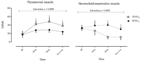
Effect of EPAP on sEMG of parasternal and sternocleidomastoid muscles
An increase of sEMG activity of the parasternal
muscle during the use of EPAP was observed in both
between EPAP10 and EPAP15 was observed because
of the inluence of intensities and time interaction
(p=0.008). The sEMG activity of the parasternal muscle
exhibited a further increase during EPAP10. Post-hoc
Bonferroni analysis showed a signiicant difference
between 10 minutes, 20 minutes, and recovery between
EPAP10 and EPAP15 (p<0.001). In relation to baseline,
normalized values during EPAP10, parasternal muscle
activity increased by 23.9% at 10 minutes, 28.9% at 20 minutes, and 19.1% upon recovery compared to increases of 10.7% at 10 minutes, 11.4% at 20 minutes and 6.9% upon recovery during EPAP15.
Figure 1. Flowchart of study. COPD: Chronic Obstructive Pulmonary Disease; BMI: Body mass index; EPAP: Expiratory Positive
Airway Pressure.
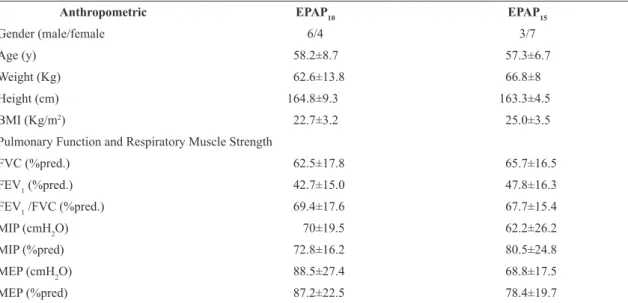
Table 1. Baseline characteristics by 2 groups of COPD patients, one group receiving EPAP10 & the other EPAP15.
Anthropometric EPAP10 EPAP15
Gender (male/female 6/4 3/7
Age (y) 58.2±8.7 57.3±6.7
Weight (Kg) 62.6±13.8 66.8±8
Height (cm) 164.8±9.3 163.3±4.5
BMI (Kg/m2) 22.7±3.2 25.0±3.5
Pulmonary Function and Respiratory Muscle Strength
FVC (%pred.) 62.5±17.8 65.7±16.5
FEV1 (%pred.) 42.7±15.0 47.8±16.3
FEV1 /FVC (%pred.) 69.4±17.6 67.7±15.4
MIP (cmH2O) 70±19.5 62.2±26.2
MIP (%pred) 72.8±16.2 80.5±24.8
MEP (cmH2O) 88.5±27.4 68.8±17.5
MEP (%pred) 87.2±22.5 78.4±19.7
BMI: Body mass index; FVC: Forced vital capacity; FEV1: Forced expiratory volume in the irst second; FVC/FEV1: Ratio between forced
Regarding the sEMG activity of the SCM muscle, the
pattern that was observed was relatively different from that observed in the parasternal muscle. The electrical
activity increased during EPAP15 and decreased during EPAP10 (Figure 2). A signiicant difference
between EPAP10 versus EPAP15 was observed because
of inluence of EPAP levels and time interaction
(p<0.001). Bonferroni post-hoc analysis showed a
signiicant difference between 10 minutes, 20 minutes and recovery between EPAP10 and EPAP15 (p<0.001).
In relation to baseline values, during EPAP15, the
SCM muscle activity increased by 4.8% at 10 minutes and 6.1% at 20 minutes and decreased by –4.0%
upon recovery compared to decreases of –5.6% at
10 minutes, –20.6% at 20 minutes and –21.3% upon
recovery during EPAP10.
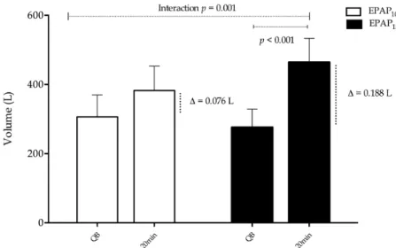
Effect of EPAP on ventilation parameters The application of EPAP induced a signiicant
increase in VT because of interaction between EPAP and time (p=0.001, Figure 3). The variation between quiet
breathing and 20 minutes of EPAP10 and EPAP15 were
∆=0.076 L and ∆=0.188 L, respectively. Bonferroni
post-hoc analysis showed a signiicant difference between quiet breathing, and 20 minutes of EPAP15
use (p=0.001).
The VE showed no signiicant changed in either group (EPAP10=5.5±0.4 L to 5.8±0.5 L, versus EPAP15=5.2±0.5 L to 5.5±0.6 L, p=0.081). In contrast,
there was a signiicant change in SpO2 and RR in
EPAP10 group but just in the RR during EPAP15.
The decrease in RR was accompanied by a reduction in dyspnea in both groups (Table 2).
Discussion
The hypothesis that the two different loads of EPAP would proportionally improve the coordination of the respiratory muscles studied and would enhance
ventilation was tested. The main indings of this study
were partially in agreement with the hypothesis.
With an increase in expiratory load, it was expected
that the inspiratory muscles would act in coordination, increasing their activity to cope with the expiratory
resistance and to improve ventilation. Nevertheless,
the results indicated different patterns of sEMG
activity in the parasternal and SCM muscles during the use of EPAP10 and EPAP15. While the effect of EPAP10 and EPAP15 exhibited a similar pattern of increased sEMG activity in the parasternal muscles which was more pronounced in EPAP10, the effect of EPAP10 and EPAP15 was different in the SCM muscle. A slight increase in the sEMG activity of the SCM
muscle was observed during EPAP10 while a decrease
in the sEMG activity was observed during EPAP15.
Ventilation at both intensities of EPAP promoted a signiicant reduction of RR accompanied by an
increased tidal volume (VT). It is important to state that the increases in ventilatory parameters during EPAP15 were greater than those seen during EPAP10. However, the different (opposite) pattern of the SCM muscle recruitment during EPAP15 use compared to that seen during EPAP10 use, appears to be related
to the activity employed by the respiratory system
to cope with the increase in expiratory load and to promote ventilation. It was not clear why these different responses to EPAP occurred. There were
Figure 2. The sEMG activity of the parasternal and sternocleidomastoid (SCM) muscles in COPD Patients during the use of EPAP10
no differences in disease severity among the groups
at baseline. Although both absolute and % predicted maximum expiratory volume (MEP) differed between
the groups, the differences in SCM activity could not
be attributed to the slight decrease in expiratory muscle strength based on maximal static effort. However, it might be possible that parasternal muscle was unable to improve its activity, as observed during EPAP10.
As previously described, the EMG activity of
the parasternal muscle during application of EPAP15 might result from an overload of the inspiratory muscles27. This effect was previously described by Simkovitz et al.28, who demonstrated that applying positive end expiratory pressure (PEEP) levels greater than 10 cmH2O during mechanical ventilation could
compromise inspiratory muscle function and increase
the operation volumes above levels imposed by the expiratory low limitation. This would be particularly
true if the operation volumes were great enough that the inspiratory muscles function at shorter lengths, a
condition that would be less favorable to ventilatory
mechanics. However, the EPAP10 group showed
signiicant activity in this muscle during therapy and after mask removal. This inding was also observed by O’Donoghue et al.29, who examined the effect
of extrinsic PEEP (ePEEP) via the application of 1 to 10 cmH2O CPAP, on the inspiratory muscle effort
of nine patients with severe and stable COPD. These
authors found that applying ePEEP in their patients increased the sEMG activity in the parasternal muscle,
Figure 3. Changes in tidal volume (VT) in 2 groups of COPD patients during the use of EPAP10 and EPAP15.
Table 2. Behavior of physiological variables in 2 groups of COPD patients in the application of EPAP10 and EPAP15.
Variables
EPAP10 EPAP15
Between group differences (95% CI) p-value* Quite
Breathing 20 minutes p-value
Quite
Breathing 20 minutes p-value
RR (bpm) 18.5±4.1 15.1±2.1 0.013 19.3±3.1 12.0±3.1 <0.001 –3.8 (–7.53 to –0.21) 0.015
SpO2 (%) 94.6±2.9 96.0±2.8 0.017 94.7±3.1 96.6±3.4 0.011 –0.5 (–3.23 to 2.22) 0.181
Heart Rate (bpm) 73.1±9.2 80.1±6.4 0.097 76.4±14.1 81.4±12.5 0.043 1.9 (–6.7 to 10.5) 0.649
SBP (mmHg) 118.3±13.1 125.5±20.7 0.413 125.3±18.3 123±15.7 0.541 9.4 (–7.71 to 26.6) 0.261
DBP (mmHg) 77.0±12.1 77.7±12.5 0.759 76.1±6.9 77.1±7.0 0.798 –0.4 (–10 to 9.23) 0.937
Borg’s scale 3.0±0.5 1.5±0.3 0.003 3.1±0.4 0.6±0.2 <0.001 –1.01 (–1.4 to –0.53) 0.028
RR: Respiratory rate; SpO2: Saturation peripheral oxygen; SBP: Systolic blood pressure; DBP: Diastolic blood pressure. Data presented as the
in addition to reducing the indices of muscle effort,
most likely at the expense of a substantial increase
in lung volume and expiratory muscle recruitment. Our study demonstrated that EPAP10 reduced SCM
muscle activity. This inding was assumed to be the
result of the capacity of ePEEP to reduce iPEEP, inspiratory threshold load and respiratory work, even in
the absence of a signiicant increase in lung volume30,31.
The waterfall theory32 hypothesized that ePEEP could
reduce iPEEP without aggravating hyperinlation only if the latter was caused by expiratory low limitation,
which may have occurred in the present study. The EPAP15 group exhibited greater SCM muscle
activity during EPAP therapy. This observation conirmed the results presented by our group in an
earlier study33, which assessed the effect of EPAP15 on the electrical activity of the SCM and scalene muscles in patients with COPD. As in the present study, there was a tendency for greater SCM muscle activity during the application of EPAP, which may
have occurred in an attempt to maintain an adequate
inspiratory pressure34. However, in this study, a
signiicant reduction of SCM electrical activity after the
withdrawal of EPAP15 was not found, a fact that may
have been a result of the insuficient time application
of the therapy. Thus, it is also important to consider that the SCM muscle participated in non-respiratory functions, including the maintenance of posture and head movement. De Troyer et al.35 found an EMG
silent period in the SCM during spontaneous breathing
in patients with severe COPD in the supine position, suggesting that respiratory function was the primary function of this muscle.
As expected, VT increased signiicantly with the application of EPAP10 and EPAP15. This increase in VT was also observed in a previous study34, during the use of 5 and 15 cmH2O EPAP applied to healthy
subjects. The authors suggested that the higher VT
was associated with the level of expiratory pressure that was imposed34. This may occur under conditions of higher ventilatory demand, resulting from the
greater need for oxygen production or higher carbon
dioxide concentration. Conversely8, when the effect of 5 cmH2O EPAP was assessed in eight men with
moderate to severe COPD during exercise; no signiicant
alteration in VT was found, although they suggested that the increase in VT could have been caused by a
prolonged expiratory time. These indings are similar to results of our study, where the reduction of RR in both groups was responsible for the increase in VT
because the VM did not change signiicantly.
The indings of the present study could have practical
implications and could open a new perspective for EPAP prescription in COPD. There is currently no
consensus regarding the adequate pressure levels
used as to when the application of elevated PEEP might aggravate ventilatory dysfunction. However, it is important to mention some limitations of this study. The results of VT and RR must be interpreted with caution. It is important to consider that while
mechanical respirometers are portable, relatively low
cost, precise and accurate devices with worldwide use, they are designed for assessments over short time
periods. End expiratory lung volumes (EELVs) were not assessed which is one marker of hyperinlation
that could impair ventilation in COPD patients; nonetheless, worsening symptoms, such as dyspnea,
were not observed in our study. Additionally, due
to the heterogeneity among patients with COPD,
new studies with greater subject numbers should be
performed. Considering the complexity of respiration, determining the effects of different intensities of expiratory loads on patient comfort and respiratory mechanics through more complex analyses of the
chest wall and the symptoms experienced would be
appropriate.
In conclusion, in COPD patients, the EPAP10 for at least 10 minutes reduces the accessory inspiratory activity of the SCM muscle and increases parasternal
muscle activity, which was accompanied by an
improvement in ventilation and a reduction in the sensation of dyspnea. The EPAP15 improves ventilation more than EPAP10, however, the parasternal muscle activity during EPAP15 was lower than that observed during EPAP10. Patients with COPD adopt different strategies to maintain their ventilation, and the reasons
for these differences require further investigation. Based on our results, it is possible to state that EPAP10
use, as demonstrated in our study, appears to be more
advantageous to patients with COPD.
References
1. Global Initiative for Chronic Obstructive Lung Disease –
GOLD. Pocket guide to COPD diagnosis, management, and prevention. 2014 [cited 2015 Mar 1]. Available from: http://
www.goldcopd.org/uploads/users/files/GOLD_Report2014_ Feb07.pdf.
2. Gary TF. Why does the lung hyperinflate? Proc Am Thoracic Soc. 2006;3(2):176-9. PMid:16565428.
3. Aliverti A, Stevenson N, Dellaca RL, Lo MA, Pedotti A, Calverley PM. Regional chest wall volumes during
2004;59(3):210-6. http://dx.doi.org/10.1136/thorax.2003.011494.
PMid:14985554.
4. Jones A, Dean E, Chow C. Comparison of the oxygen cost
of breathing exercises and spontaneous breathing in patients with stable chronic obstructive pulmonary disease. Phys Ther. 2003;83(5):424-31. PMid:12718708.
5. Ninane V, Rypens F, Yernault JC, De Troyer A. Abdominal
muscle use during breathing in patients with chronic airflow obstruction.Am Rev Respir Dis. 1992;146(1):16-21. http://
dx.doi.org/10.1164/ajrccm/146.1.16. PMid:1385684.
6. Grimby G, Goldman M, Mead J. Respiratory muscle action
inferred from rib cage and abdominal V-P partitioning. J Appl Physiol. 1976;41(5 pt.1):739-51. PMid:993162.
7. Lien T, Wang J, Chang M, Kuo CD. Comparison of BiPAP nasal ventilation and ventilation via iron lung in severe
stable COPD. Chest. 1993;104(2):460-6. http://dx.doi.
org/10.1378/chest.104.2.460. PMid:8339634.
8. van der Schans C, de Jong W, de Vries G, KaanWA, Postma DS, Koëter GH, et al. Effects of positive expiratory pressure
breathing during exercise in patients with COPD. Chest.
1994;105(3):782-9. http://dx.doi.org/10.1378/chest.105.3.782.
PMid:8131541.
9. Su C, Chiang L, Chiang T, Yu CT, Kuo HP, Lin HC. Domiciliary positive expiratory pressure improves pulmonary function
and exercise capacity in patients with chronic obstructive
pulmonary disease. J Formos Med Assoc. 2007;106(3):204 -11. http://dx.doi.org/10.1016/S0929-6646(09)60241-2.
PMid:17389164.
10. Winter DA, Fuglevand AJ, Archer SE. Crosstalk in surface
electromyography: theoretical and practical estimates. J Electromyogr Kinesiol. 1994;4(1):15-26. http://dx.doi.
org/10.1016/1050-6411(94)90023-X. PMid:20870543. 11. Leduc D, De Troyer A. The effect of lung inflation on the
inspiratory action of the canine parasternal intercostals. J Appl Physiol. 2006;100(3):858-63. http://dx.doi.org/10.1152/
japplphysiol.00739.2005. PMid:16293705.
12. NormanRW. Standardizing biomechanical testing in sport.
Res Q Exerc Sport. 1987;58:286-7.
13. Falla D, Dall’Alba P, Rainoldi A, Merletti R, Jull G. Location of innervation zones of sternocleidomastoid and scalene
muscles--a basis for clinical and research electromyography
applications. Clin Neurophysiol. 2002;113(1):57-63. http://
dx.doi.org/10.1016/S1388-2457(01)00708-8. PMid:11801425.
14. Gross RD, Atwood CW Jr, RossSB, Olszewski JW, Eichhorn
KA. The coordination of breathing and swallowing in
chronic obstructive pulmonary disease.Am J Respir Crit
Care Med. 2009;179(7):559-65. http://dx.doi.org/10.1164/
rccm.200807-1139OC. PMid:19151193.
15. Hug F, Raux M, Prella M, Morelot-Panzini C, Straus C, Similowski T. Optimized analysis of surface electromyograms of the
scalenes during quiet breathing in humans.Respir Physiol Neurobiol. 2006;150(1):75-81. http://dx.doi.org/10.1016/j.
resp.2005.04.008. PMid:16448935.
16. Hermens HJ, Freriks B, Disselhorst-Klug C, Rau G. Development of recommendations for SEMG sensors and sensor placement procedures. J Electromyogr Kinesiol. 2000;10(5):361-74.
http://dx.doi.org/10.1016/S1050-6411(00)00027-4. PMid:11018445.
17. Brazilian Society of Pneumology and Tisiology. Guidelines for pulmonary function tests. Braz J Pneumol. 2002;28(3):237. [in Portuguese]
18. Pereira CAC, Sato T, Rodrigues SC. New reference values for forced spirometry in white adults. Braz J Pneumol.
2007;33(9):397-406.
http://dx.doi.org/10.1590/S1806-37132007000400008. [in Portuguese] PMid:17982531.
19. Souza RB. Maximum static respiratory pressures: e Guidelines for pulmonary function tests. Braz J Pneumol. 2002;28(Supl 3):S155-65. [in Portuguese]
20. Neder JA, Andreoni S, Lerario MC, NeryLE. Reference values for lung function tests. II. Maximal respiratory pressures and voluntary ventilation. Braz J Med Biol Res.
1999;32(6):719-727.
http://dx.doi.org/10.1590/S0100-879X1999000600007. PMid:10412550.
21. Hall KD, Reeser FH Jr. Calibration of wright spirometer. Anesthesiology. 1962;23(1):126-9. http://dx.doi.
org/10.1097/00000542-196201000-00020. PMid:13903878. 22. Daykin AP, Nunn GF, WrightBM. The measurement
of vital capacity and minute volume with the wright respirometer. Br J Dis Chest. 1978;72(4):333-5. http://dx.doi.
org/10.1016/0007-0971(78)90063-3. PMid:728358.
23. Ilsley AH, Hart JD, WithersRT, Roberts JG. Evaluation
of five small turbine-type respirometers used in adult
anesthesia. J Clin Monit. 1993;9(3):196-201. http://dx.doi.
org/10.1007/BF01617028. PMid:8345373.
24. Nemer SN, Barbas CS, Caldeira JB, Cárias TC, Santos
RG, Almeida LC, et al. A new integrative weaning index of discontinuation from mechanical ventilation. Crit Care. 2009;13(5):R152. http://dx.doi.org/10.1186/cc8051.
PMid:19772625.
25. Boniatti VM, Boniatti MM, Andrade CF, Zigiotto CC, Kaminski P, Gomes SP, et al. The modified integrative weaning
index as a predictor of extubation failure.Respir Care.
2014;59(7):1042-7. http://dx.doi.org/10.4187/respcare.02652.
PMid:24282317.
26. WilsonRC, Jones PW. A comparison of the visual analogue
scale and modified Borg scale for the measurement of
dyspnoea during exercise. Clin Sci. 1989;76(3):277-82.
http://dx.doi.org/10.1042/cs0760277. PMid:2924519.
27. Duiverman ML, van Eykern LA, Vennik PW, Koëter GH, Maarsingh EJ, Wijkstra PJ. Reproducibility and
responsiveness of a noninvasive EMG technique of the
respiratory muscles in COPD patients and in healthy
subjects. J Appl Physiol. 2004;96(5):1723-9. http://dx.doi.
org/10.1152/japplphysiol.00914.2003. PMid:14660508.
28. Simkovitz P, BrownK, Goldberg P, Milic-Emili J, Gottfried SI. Interaction between intrinsic and externally applied PEEP during mechanical ventilation. Am Rev Respir Dis.
1987;135:202.
29. O’Donoghue FJ, Catcheside PG, Jordan AS, Bersten AD, McEvoy RD. Effect of CPAP on intrinsic PEEP, inspiratory
effort, and lung volume in severe stable COPD. Thorax. 2002;57(6):533-9. http://dx.doi.org/10.1136/thorax.57.6.533.
PMid:12037230.
30. Legrand A, Schneider E, Gevenois P, De Troyer A. Respiratory effects of the scalene and sternomastoid muscles in humans. J Appl Physiol. 2003;94(4):1467-72. http://dx.doi.org/10.1152/
31. Appendini L, Patessio A, Zanaboni S, Carone M, Gukov
B, Donner CF, et al. Physiologic effects of positive end-expiratory pressure and mask pressure support
during exacerbations of chronic obstructive pulmonary
disease. Am J Respir Crit Care Med. 1994;149(5):1069
-76. http://dx.doi.org/10.1164/ajrccm.149.5.8173743.
PMid:8173743.
32. Tobin MJ, LodatoRF. PEEP, auto-PEEP, and waterfalls. Chest.
1989;96(3):449-51. http://dx.doi.org/10.1378/chest.96.3.449.
PMid:2670461.
33. Cardoso DM, Paiva DN, Albuquerque IM, Jost RT, Paixão AV. Effects of expiratory positive airway pressure on the electromyographic activity of accessory inspiratory muscles in COPD patients. Braz J Pulm. 2011;37(1):46
-53. http://dx.doi.org/10.1590/S1806-37132011000100008.
PMid:21390431.
34. van der Schans C, de Jong W, de Vries G, Postma DS, Koëter GH, van der Mark TW. Effect of positive expiratory pressure
on breathing pattern in healthy subjects.Eur Respir J.
1993;6(1):60-6. PMid:8425596.
35. De Troyer A, Peche R, Yernault JC, Estenne M. Neck muscle
activity in patients with severe chronic obstructive pulmonary
disease. Am J Respir Crit Care Med. 1994;150(1):41-7. http://
dx.doi.org/10.1164/ajrccm.150.1.8025770. PMid:8025770.
Correspondence
Dannuey Machado Cardoso Rua Thomaz Flores, 1064/101 B2