Effect of treadmill gait on bone
markers and bone mineral density
of quadriplegic subjects
1Departamento de Ortopedia e Traumatologia, 2Departamento de Patologia Clínica,
Faculdade de Ciências Médicas, 3Divisão de Patologia Clínica, Hospital Universitário,
Universidade Estadual de Campinas, Campinas, SP, Brasil
4Departamento de Engenharia Elétrica, Universidade de São Paulo, São Carlos,
SP, Brasil D.C.L. Carvalho1,
C.R. Garlipp2, P.V. Bottini3,
S.H. Afaz3, M.A. Moda3
and A. Cliquet Jr.1,4
Abstract
Quadriplegic subjects present extensive muscle mass paralysis which is responsible for the dramatic decrease in bone mass, increasing the risk of bone fractures. There has been much effort to find an efficient treatment to prevent or reverse this significant bone loss. We used 21 male subjects, mean age 31.95 ± 8.01 years, with chronic quadriple-gia, between C4 and C8, to evaluate the effect of treadmill gait training using neuromuscular electrical stimulation, with 30-50% weight re-lief, on bone mass, comparing individual dual-energy X-ray absorpti-ometry responses and biochemical markers of bone metabolism. Subjects were divided into gait (N = 11) and control (N = 10) groups. The gait group underwent gait training for 6 months, twice a week, for 20 min, while the control group did not perform gait. Bone mineral density (BMD) of lumbar spine, femoral neck, trochanteric area, and total femur, and biochemical markers (osteocalcin, bone alkaline phosphatase, pyridinoline, and deoxypyridinoline) were measured at the beginning of the study and 6 months later. In the gait group, 81.8% of the subjects presented a significant increase in bone formation and 66.7% also presented a significant decrease of bone resorption mark-ers, whereas 30% of the controls did not present any change in markers and 20% presented an increase in bone formation. Marker results did not always agree with BMD data. Indeed, many individuals with increased bone formation presented a decrease in BMD. Most indi-viduals in the gait group presented an increase in bone formation markers and a decrease in bone resorption markers, suggesting that gait training, even with 30-50% body weight support, was efficient in improving the bone mass of chronic quadriplegics.
Correspondence
D.C.L. Carvalho
Departamento de Ortopedia e Traumatologia
FCM, UNICAMP Rua Alexander Fleming, 181 13083-970 Campinas, SP Brasil
Fax: +55-19-3788-7505 E-mail:dclcarvalho@yahoo.com.br
Research supported by FAPESP (Nos. 2005/53530-0, 2003/05856-9 and 1996/12198-2).
Received March 3, 2006 Accepted August 21, 2006
Key words
•Quadriplegic gait
•Electrical stimulation •Osteoporosis
•Rehabilitation
Introduction
Many investigations (1-3) have been car-ried out in order to find methods for gait recovery after spinal cord injury. However, subjects who suffer a spinal cord injury
contraction and mechanical load results in decreased BMD below the injury level (11). Previous studies (12-17) have evaluated the effect of neuromuscular electrical stimu-lation (NMES) on bone density. Mohr et al. (12) and Bloomfield et al. (13), observed an improvement in BMD after NMES-cycling intervention. However, Leeds et al. (14) and Eser et al. (15) did not observe any differ-ences after treatment. Different responses have been attributed to the different training protocols used (18).
Dual-energy X-ray absorptiometry (DEXA) has been extensively used to evalu-ate BMD and to monitor the effects of treat-ment on bone sites (19). Despite the high precision, low irradiation dose and fast scan-ning time, studies have suggested that even with a low precision error (20,21), depend-ing on the expected rate of bone gain, a 1- to 2-year interval between exams would be required for an accurate identification of changes in bone mass due to the fact that densitometry is slow in revealing bone alter-ations (22-25). However,it is possible to use DEXA at short testing intervals to assess bone changes following spinal cord injury since the rate of bone loss is typically rapid and large relative to the precision error. Many studies have analyzed the effect of short periods of treatment on bone density in spi-nal cord-injured subjects using DEXA (13, 14,26).
Biochemical markers of bone turnover have been used to evaluate bone metabolism (27-29) since they respond to treatment more rapidly than bone density (30-32). Signifi-cant differences can be noted within 3-6 months of treatment (33). Bone markers measure the rate of bone turnover when the events of bone resorption and formation are coupled. However, when the events are un-coupled they reflect the increase in either bone formation or bone resorption (34). Hence, the uncoupled increase of osteocal-cin, for instance, represents an increase in bone formation rate (30).
NMES allows the activation of paralyzed muscles in order to perform physical activi-ties such as pedaling on a bicycle ergometer and gait (35,36). For quadriplegic subjects, gait training can be achieved using body weight support (BWS), which provides trunk stability and allows the control of the weight that can be supported by the lower limbs of each patient, thus reducing the risk of frac-ture in osteoporotic bones (3,37).
In view of the lack of studies on this topic, the purpose of the present investiga-tion was to evaluate the effect of treadmill gait training with 30-50% of weight relief associated with NMES on the bone mass of quadriplegic subjects and to determine whether individual responses are similar when BMD measured by DEXA is com-pared with biochemical markers of bone metabolism.
Material and Methods
fractures, joint degeneration changes or clini-cal joint instability.
The individuals were divided into two groups: GG and CG. GG individuals (N = 11) were submitted to treadmill gait training provided by NMES for 6 months, twice a week, 20 min per session, whereas CG indi-viduals (N = 10) were not submitted to gait training. GG subjects had their quadriceps and tibialis anterior muscles stimulated for at least 5 months before beginning gait train-ing (twice a week) in order to be able to walk for 20 min, as well as to support at least 50% of their body weight through knee extension provided by NMES (pregait training). A 4-channel electrical stimulator delivered a 25-Hz signal with monophasic rectangular pulses of 300-µs duration and a maximum intensity of 200 V (1 kΩ load).
BWS was provided by a harness hanging from an overhead support, and the support vest allowed free movement of the lower limbs. The body weight support was 30 to 50%. The four-channel electrical stimulator was used to provide the stance gait phase through quadriceps muscle activation and the swing phase by the withdrawal reflex (stimuli to the common peroneal nerve). Manual assistance during treadmill walking was provided to all patients in order to allow a safe and closer to normal pattern of gait.
Surface electrodes (5 x 9 cm2) were placed
on the quadriceps and over the common peroneal nerve (round electrodes 3.2 cm in diameter). The stimulation unit was trig-gered by hand switches controlled by the staff.
BMD (g/cm2) was determined by DEXA
using a DPX-ALPHA Lunar Apparatus (DeWitt, MI, USA). The BMD of the lumbar spine (L2-L4), proximal femur (femoral neck and trochanteric area) and total femur was analyzed. The precision error was 1.0% for the lumbar spine, 1.37% for the femoral neck, 1.7% for the trochanteric area, and 1.5% for the femur. The least significant changes (LSC) were 2.77% for the lumbar
spine, 3.80% for the femoral neck, 4.59% for the trochanteric area, and 4.15% for the total femur. The coefficient of variation was determined from three repeated measure-ments of the 5 spinal cord-injured subjects included in the study on 3 separate days (95% confidence interval) (12,13,15). All exams were performed by the same techni-cian at the beginning of the study and 6 months later. Biochemical markers of bone turnover were also evaluated. Serum osteo-calcin (OC) and bone alkaline phosphatase (B-ALP), both markers of bone formation, and free pyridinoline (PYD) and free deoxy-pyridinoline (DPD), both markers of bone resorption, were analyzed. PYD and DPD were corrected for creatinine and are re-ported as nmol/mmol. Bone markers were measured by an immunoassay method (Me-tra Biosystems, Mountain View, CA, USA). Serum and urine samples were collected between 8:00 and 10:00 am after an over-night fast and stored at -80ºC until analysis. The measurements were made at the begin-ning of the study and 6 months later.
Data were analyzed separately for each individual, because each subject presented different bone responses, since BMD is in-fluenced by many factors as nutrition, bone condition before lesion, physical activity, and body weight, which is an important factor for bone mass variability (15-32%) (38).
Bone markers and BMD results were considered to be significant when differ-ences between the initial and final values were higher than the LSC (34). Bone marker values were analyzed based on the study of Hannon et al. (33), who observed that the LSC, within a single individual, was 21% from baseline values for OC, 28% for B-ALP, 36% for PYD, and 26% for DPD (95% confidence interval).
Results
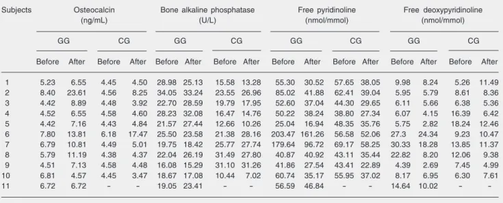
each GG and CG subjects. The results showed a significant increase in bone formation mark-ers after gait training in 81.8% (9 als) of the subjects, with 66.7% (8 individu-als) presenting a significant decrease in bone resorption markers. However, in many cases, despite the increased bone formation rate and decreased bone resorption markers, re-sorption marker values were higher than reference values. Reference values were 3.7-10.0 ng/mL for OC, 12-41 U/L for B-ALP, 12-37 nmol/mmol for PYD/Cr, and 2-7 nmol/ mmol for DPD/Cr. Moreover, one subject presented a decrease in bone turnover and one presented a decrease in bone resorption rate.
In the CG, no alterations were observed in 30% of the individuals (3 individuals), while 20% (2 individuals) presented a sig-nificant increase in bone formation markers. CG individuals who presented an increase of formation markers increased their indepen-dence during daily activities (they managed to obtain driver licenses and became less dependent on accompanying persons) dur-ing the 6-month period. A decrease in bone resorption markers was noted in 30% of subjects (3 individuals). One subject showed
increased bone resorption markers and an-other showed decreased bone formation markers.
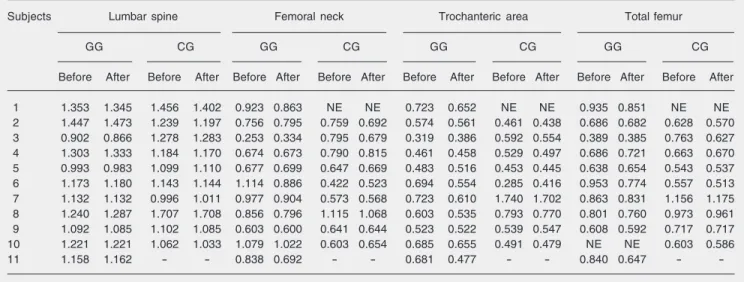
Table 2 presents the results of BMD (g/ cm2) at the beginning and after 6 months of
GG and CG. The results obtained with bio-chemical markers were not always repro-duced in BMD. Of the individuals who pre-sented an increase in bone formation mark-ers (N = 9), 3 presented a BMD gain at most of the sites analyzed (individuals 3, 4, and 5), 4 presented loss of BMD at most of the sites analyzed (individuals 1, 6, 7, and 8), 1 (individual 9) presented maintenance of BMD except for total femur, which lost bone mass, and 1 (individual 2) presented mainte-nance of BMD except for the femoral neck, which gained bone mass. The total femur of individual 10 was not evaluated.
In the CG, at the beginning of the study and 6 months later, one subject could not have his femur evaluated because of a flexor contracture of his hip. Of the two individuals who presented a significant increase in bone formation markers, one presented an increase and the other a decrease in BMD. Even among those who did not present any altera-tion of bone markers (when comparing
ini-Table 1. Biochemical markers of bone formation and resorption for each subject of gait group (GG, N = 11) and control group (CG, N = 10) before and after 6 months.
Subjects Osteocalcin Bone alkaline phosphatase Free pyridinoline Free deoxypyridinoline (ng/mL) (U/L) (nmol/mmol) (nmol/mmol)
GG CG GG CG GG CG GG CG
Before After Before After Before After Before After Before After Before After Before After Before After
-tial and final values), some presented a gain of BMD and some lost or maintained their BMD.
Discussion
The present results show that treadmill gait training was efficient in increasing the rate of bone formation, even with 30-50% of BWS, since most individuals presented a significant increase of OC (a bone formation marker) and a decrease of PYD and/or DPD (bone resorption markers). Since the results for the bone markers presented a dissocia-tion of bone resorpdissocia-tion and formadissocia-tion events, they represented an increase of bone forma-tion rate (30,34). The increase in bone for-mation rate is associated with gait training and not with pregait training since NMES used to provide knee extension without any resistance has been shown not to improve bone density in many studies (13,39).
All individuals had sustained their inju-ries at least 25 months before the study and, based on the literature, bone loss occurs dramatically during the first 3-4 months post-injury (22-27% depletion) and bone mass achieves a new steady state by 16 months
after the injury, with approximately 37% bone depletion. Garland et al. (40) did not observe any significant differences in bone mass between 16 months and 10 years post-injury.
Some individuals included in our study (6 months between DEXA measurements) presented a significant decrease of bone mass, i.e., a reduction of 20.46% in the femoral neck and of 29.95% in the trochanteric area. These dramatic decreases of bone mass may reflect the imprecision of DEXA due to the short time between measurements or due to the problems that occur during densitometry exams in spinal cord-injured persons (spas-ticity and lack of reproducibility of lower limb position).
Bloomfield et al. (13) also observed that some quadriplegic individuals submitted to NMES-cycle ergometer training presented a bone loss at one or more bone sites analyzed during 9 months of physical activity. They hypothesized that insufficient calcium in-take could have been responsible for the increased bone loss since the maintenance of mineral homeostasis (extracellular calcium ion concentration) is essential for body func-tion.
Table 2. Bone mineral density (BMD) values (g/cm2) obtained before and after 6 months for each subject of gait group (GG, N = 11) and control
group (CG, N = 10) for lumbar spine (L2-L4), femoral neck, trochanteric area, and total femur.
Subjects Lumbar spine Femoral neck Trochanteric area Total femur
GG CG GG CG GG CG GG CG
Before After Before After Before After Before After Before After Before After Before After Before After
1 1.353 1.345 1.456 1.402 0.923 0.863 NE NE 0.723 0.652 NE NE 0.935 0.851 NE NE 2 1.447 1.473 1.239 1.197 0.756 0.795 0.759 0.692 0.574 0.561 0.461 0.438 0.686 0.682 0.628 0.570 3 0.902 0.866 1.278 1.283 0.253 0.334 0.795 0.679 0.319 0.386 0.592 0.554 0.389 0.385 0.763 0.627 4 1.303 1.333 1.184 1.170 0.674 0.673 0.790 0.815 0.461 0.458 0.529 0.497 0.686 0.721 0.663 0.670 5 0.993 0.983 1.099 1.110 0.677 0.699 0.647 0.669 0.483 0.516 0.453 0.445 0.638 0.654 0.543 0.537 6 1.173 1.180 1.143 1.144 1.114 0.886 0.422 0.523 0.694 0.554 0.285 0.416 0.953 0.774 0.557 0.513 7 1.132 1.132 0.996 1.011 0.977 0.904 0.573 0.568 0.723 0.610 1.740 1.702 0.863 0.831 1.156 1.175 8 1.240 1.287 1.707 1.708 0.856 0.796 1.115 1.068 0.603 0.535 0.793 0.770 0.801 0.760 0.973 0.961 9 1.092 1.085 1.102 1.085 0.603 0.600 0.641 0.644 0.523 0.522 0.539 0.547 0.608 0.592 0.717 0.717 10 1.221 1.221 1.062 1.033 1.079 1.022 0.603 0.654 0.685 0.655 0.491 0.479 NE NE 0.603 0.586 11 1.158 1.162 - - 0.838 0.692 - - 0.681 0.477 - - 0.840 0.647 -
References
1. Wickelgren I. Teaching the spinal cord to walk. Science 1998; 279: 319-321.
2. Field-Fote EC, Tepavac D. Improved intralimb coordination in people with incomplete spinal cord injury following training with body weight support and electrical stimulation. Phys Ther 2002; 82: 707-715. 3. Field-Fote EC. Combined use of body weight support, functional
electric stimulation, and treadmill training to improve walking ability in individuals with chronic incomplete spinal cord injury. Arch Phys
Med Rehabil 2001; 82: 818-824.
4. Figoni SF. Exercise responses and quadriplegia. Med Sci Sports
Exerc 1993; 25: 433-441.
5. Dallmeijer AJ, Hopman MT, van As HH, van der Woude LH. Physi-cal capacity and physiPhysi-cal strain in persons with tetraplegia; the role of sport activity. Spinal Cord 1996; 34: 729-735.
6. Kiratli BJ, Smith AE, Nauenberg T, Kallfelz CF, Perkash I. Bone mineral and geometric changes through the femur with immobiliza-tion due to spinal cord injury. J Rehabil Res Dev 2000; 37: 225-233. 7. Frey-Rindova P, de Bruin ED, Stussi E, Dambacher MA, Dietz V. Bone mineral density in upper and lower extremities during 12 months after spinal cord injury measured by peripheral quantitative computed tomography. Spinal Cord 2000; 38: 26-32.
8. Carvalho DCL, Carvalho MM, Cliquet A Jr. Disuse osteoporosis: Its relationship to spine cord injured patient rehabilitation. Acta Ortop Bras 2001; 9: 34-43.
9. Zehnder Y, Luthi M, Michel D, Knecht H, Perrelet R, Neto I, et al. Long-term changes in bone metabolism, bone mineral density, quan-titative ultrasound parameters, and fracture incidence after spinal cord injury: a cross-sectional observational study in 100 paraplegic men. Osteoporos Int 2004; 15: 180-189.
10. Wilmet E, Ismail AA, Heilporn A, Welraeds D, Bergmann P. Longitu-dinal study of the bone mineral content and of soft tissue composi-tion after spinal cord seccomposi-tion. Paraplegia 1995; 33: 674-677. 11. Carvalho DC, Rosim GC, Gama LO, Tavares MR, Tribioli RA, Santos
IR, et al. Non-pharmacological treatments in the stimulation of os-teogenesis. Rev Saude Publica 2002; 36: 647-654.
12. Mohr T, Podenphant J, Biering-Sorensen F, Galbo H, Thamsborg G, Kjaer M. Increased bone mineral density after prolonged electrically induced cycle training of paralyzed limbs in spinal cord injured man.
Calcif Tissue Int 1997; 61: 22-25.
13. Bloomfield SA, Mysiw WJ, Jackson RD. Bone mass and endocrine adaptations to training in spinal cord injured individuals. Bone 1996; 19: 61-68.
14. Leeds EM, Klose KJ, Ganz W, Serafini A, Green BA. Bone mineral density after bicycle ergometry training. Arch Phys Med Rehabil
1990; 71: 207-209.
15. Eser P, de Bruin ED, Telley I, Lechner HE, Knecht H, Stussi E. Effect of electrical stimulation-induced cycling on bone mineral
den-sity in spinal cord-injured patients. Eur J Clin Invest 2003; 33: 412-419.
16. de Bruin ED, Frey-Rindova P, Herzog RE, Dietz V, Dambacher MA, Stussi E. Changes of tibia bone properties after spinal cord injury: effects of early intervention. Arch Phys Med Rehabil 1999; 80: 214-220.
17. Belanger M, Stein RB, Wheeler GD, Gordon T, Leduc B. Electrical stimulation: can it increase muscle strength and reverse osteopenia in spinal cord injured individuals? Arch Phys Med Rehabil 2000; 81: 1090-1098.
18. Jiang SD, Dai LY, Jiang LS. Osteoporosis after spinal cord injury.
Osteoporos Int 2006; 17: 180-192.
19. Seeley DG, Browner WS, Nevitt MC, Genant HK, Scott JC, Cum-mings SR. Which fractures are associated with low appendicular bone mass in elderly women? The Study of Osteoporotic Fractures Research Group. Ann Intern Med 1991; 115: 837-842.
20. Biering-Sorensen F, Bohr H, Schaadt O. Bone mineral content of the lumbar spine and lower extremities years after spinal cord lesion.
Paraplegia 1988; 26: 293-301.
21. Demirel G, Yilmaz H, Paker N, Onel S. Osteoporosis after spinal cord injury. Spinal Cord 1998; 36: 822-825.
22. Maimoun L, Couret I, Micallef JP, Peruchon E, Mariano-Goulart D, Rossi M, et al. Use of bone biochemical markers with dual-energy X-ray absorptiometry for early determination of bone loss in persons with spinal cord injury. Metabolism 2002; 51: 958-963.
23. Johnston CC, Melton LJ III. Bone densitometry. In: Riggs BL, Melton LJ III (Editors), Osteoporosis: etiology, diagnosis, and management. 2nd edn. New York: Lippincott-Raven; 1995. p 275-297.
24. Wasnich RD. Epidemiology of osteoporosis. In: Favus MJ (Editor), Primer
on the metabolic bone diseases and disorders of mineral metabolism. 4th
edn. Philadelphia: Lippincott-Raven; 1999. p 257-259.
25. Garnero P, Delmas PD. Biochemical markers of bone turnover. Applications for osteoporosis. Endocrinol Metab Clin North Am 1998; 27: 303-323.
26. Kunkel CF, Scremin AM, Eisenberg B, Garcia JF, Roberts S, Martinez S. Effect of “standing” on spasticity, contracture, and os-teoporosis in paralyzed males. Arch Phys Med Rehabil 1993; 74: 73-78.
27. Adachi JD. The correlation of bone mineral density and biochemical markers to fracture risk. Calcif Tissue Int 1996; 59: 16-19. 28. Watts NB. Clinical utility of biochemical markers of bone remodeling.
Clin Chem 1999; 45: 1359-1368.
29. Inoue M, Tanaka H, Moriwake T, Oka M, Sekiguchi C, Seino Y. Altered biochemical markers of bone turnover in humans during 120 days of bed rest. Bone 2000; 26: 281-286.
30. Garnero P, Delmas PD. New developments in biochemical markers for osteoporosis. Calcif Tissue Int 1996; 59 (Suppl 1): S2-S9.
As shown in Table 1, it is clear that there was a reduction in bone marker resorption in the GG subjects, meaning that the calcium intake was not the reason for the bone loss observed by DEXA.
The present results show that treadmill
31. Harris ST, Gertz BJ, Genant HK, Eyre DR, Survill TT, Ventura JN, et al. The effect of short term treatment with alendronate on vertebral density and biochemical markers of bone remodeling in early post-menopausal women. J Clin Endocrinol Metab 1993; 76: 1399-1406. 32. Mundy GR. Bone remodeling and its disorders. 2nd edn. London:
Martin Dunitz; 1999.
33. Hannon R, Blumsohn A, Naylor K, Eastell R. Response of biochemi-cal markers of bone turnover to hormone replacement therapy: impact of biological variability. J Bone Miner Res 1998; 13: 1124-1133.
34. Seibel MJ. Biochemical markers of bone metabolism in the assess-ment of osteoporosis: useful or not? J Endocrinol Invest 2003; 26: 464-471.
35. Petrofsky JS, Phillips CA, Heaton HH, Glaser RM. Bicycle ergom-eter for paralyzed muscle. J Clin Eng 1984; 9: 13-19.
36. Sepulveda F, Granat MH, Cliquet A Jr. Gait restoration in a spinal cord injured subject via neuromuscular electrical stimulation
con-trolled by an artificial neural network. Int J Artif Organs 1998; 21: 49-62.
37. Carvalho DC, de Cassia ZM, Sereni JM, Cliquet A. Metabolic and cardiorespiratory responses of tetraplegic subjects during treadmill walking using neuromuscular electrical stimulation and partial body weight support. Spinal Cord 2005; 43: 400-405.
38. Gerdhem P, Ringsberg KA, Akesson K, Obrant KJ. Influence of muscle strength, physical activity and weight on bone mass in a population-based sample of 1004 elderly women. Osteoporos Int
2003; 14: 768-772.
39. Pacy PJ, Hesp R, Halliday DA, Katz D, Cameron G, Reeve J. Muscle and bone in paraplegic patients, and the effect of functional electrical stimulation. Clin Sci 1988; 75: 481-487.