Printed version ISSN 0001-3765 / Online version ISSN 1678-2690 http://dx.doi.org/10.1590/0001-3765201720160479
www.scielo.br/aabc | www.fb.com/aabcjournal
Evaluation of the anti-osteoporotic effect of
Ginkgo biloba
L. in Wistar rats
with glucocorticoid-induced-osteoporosis by bone densitometry using
dual-energy x-ray absorptiometry (DEXA) and mechanical testing
LEDA M.F. LUCINDA¹, BEATRIZ J.V. AARESTRUP¹, MAYCON M. REBOREDO¹, THAIS D.A. PAINS³, RAPHAEL Z. CHAVES³, JOÃO E.P. REIS¹, MÁRIO J.Q. LOUZADA² and MARTHA O. GUERRA¹
¹Centro de Biologia da Reprodução, Universidade Federal de Juiz de Fora/UFJF, São Pedro, Caixa Postal 328, 36001-970 Juiz de Fora, MG, Brazil
²Faculdade de Medicina Veterinária de Araçatuba, Universidade do Estado de São Paulo/ UNESP, Rua Clovis Pestana, 793, 16050-680 Araçatuba, SP, Brazil
³Faculdade de Medicina, Universidade Federal de Juiz de Fora/UFJF, Avenida Eugênio do Nascimento, s/n, Caixa Postal 328, 36001-970 Juiz de Fora, MG, Brazil
Manuscript received on July 29, 2016; accepted for publication on January 1, 2017
ABSTRACT
Evaluate the effect of the extract of Ginkgo biloba in the bone alkaline phosphatase, bone mineral density, in the mechanical properties of the tibia in rats with glucocorticoid-induced-osteoporosis. After osteoporosis induction, the rats were divided into five groups: Osteoporosis; EGb1 (28 mg/Kg); EGb2 (56 mg/Kg); alendronate (0.2 mg/animal) and control. The animals were treated during 20 and 30 days. The control group was compared with the osteoporosis’s (Student’s t-test), while the other were analyzed by ANOVA test followed by Tukey/Dunnett’T3 (p<0.05). In the osteoporosis group the bone alkaline phosphatase, bone mineral density, the bone stiffness, the maximum load and the resilience were reduced. The bone alkaline phosphatase values increased in the EGb1 and EGb2 groups (30 days). In addition, in the EGb2 and alendronate groups (20 and 30 days) the bone mineral density increased. The extract of Ginkgo biloba
restored bone alkaline phosphatase and bone mineral density using dual-energy x-ray absorptiometry.
Key words: bone density, DEXA scan, Ginkgo biloba, osteoporosis.
Correspondence to: Leda Marília Fonseca Lucinda E-mail: [email protected]
INTRODUCTION
The low bone mass is associated with genetic, nutritional and lifestyle factors, as well as estrogen levels and the use of some drugs (Johnell 1996). Among these drugs, glucocorticoids have potent anti-inflamatory effects and were used for decades in the treatment of chronic diseases, as well as in
posttransplantation immunotherapy (McLaughlin
et al. 2002). The current use of oral corticosteroids is associated with serious side effects, including osteoporosis and consequently an increase in
fractures (Henneicke et al. 2011). Despite the
clinical recognition that glucocorticoids can
cause bone loss, many patients receiving
long-term glucocorticoid therapy are not evaluated for
prophylaxis or treatment (Cruse et al. 2006, Feldstein et al. 2005).
Glucocorticoids act directly on bone cells and one of their principal actions is to reduce osteoblasts function and number by apoptosis (Chang et al. 2009, Hock et al. 2001). The Bax expression by ostoblasts increase in the glucocorticoid-induced osteoporosis (GIO) as showed by Lucinda et al. (2013). It`s well known that apoptosis is regulated by an intrinsic process involving activation of genes that can promote cell death (Bras et al. 2005). Likewise, the Bcl-2 gene family encodes a large number of proteins, including Bax, a pro-apoptotic protein member of the Bcl-2 gene family that participates in programmed cell death (Verborgt et al. 2002).
The biochemical markers are also important in the management of osteoporosis, one of them being the bone alkaline phosphatase (BAP) which is a specific enzyme released from osteoblasts into the blood during the process of bone formation and believed to be indicative of the bone mineralization process (Brown et al. 2009).
Biphosphonates are widely used in the prevention or in the treatment of osteoporosis (Russell 2006), however, some side effects like gastrointestinal intolerance (Szejnfeld 2000) and osteonecrosis of the jaw have been reported (Hoff et al. 2008, Pozzi et al. 2007); thus, the necessity to search for new approaches to the current therapies. Phytoestrogens showed significant results in the treatment of osteoporosis (Bawa 2010). Similarly, Trivedi et al. (2009) suggest that the phytoestrogens components of EGb, kaempferol and quercetin, are involved in the beneficial outcomes on bone.
Ginkgo biloba L. (family Ginkgoaceae) is a plant currently used by the population in different therapeutical approaches. The main compounds of this plant comprise 6% of terpenoids (ginkgolides and bilobilides), less than 5 ppm of ginkgolic acid, and 24% of phytoestrogens (Kaempferol, ishorhamnetin and quercetin) (Oh and Chung 2004, Van Beek 2002). Notwithstanding the fact
that there is lack of pre-clinical studies as well as absence of clinical ones using Ginkgo biloba in the treatment of osteoporosis, the few existing pre-clinical studies have showed promising results in the management of osteoporosis (Lucinda et al. 2010a, b, Oh et al. 2008, Trivedi et al. 2009).
Among the major effects of Ginkgo biloba
extract (EGb) that could be important in the treatment of osteoporosis, the antiapoptotic properties of this extract have been reported by some authors (Smith et al. 2002, Smith and Luo 2004, Lucinda et al. 2013). Specifically, Brayboy et al. (2001) reported that EGb was effective in protecting the osteoblasts from death when they were exposed to the action of free radicals in vitro.
Consequently, Ginkgo biloba was effective in stimulating osteoblast differentiation and increase in the bone formation (Brayboy et al. 2001, Oh et al. 2008). In our previous studies the EGb improved periodontal bone support and the percentual of alveolar bone in the mandible and also in the mandible it improved the expression of Bcl-2 and decreased the expression of Bax by osteoblasts. Additionally, the EGb improved the trabecular bone in femur of rats with glucocorticoid-induced osteoporosis (Lucinda et al. 2010a, b, 2013). Similarly, Trivedi et al. (2009) reported that EGb improved bone mineral density (BMD) in ovariectomized rats.
Based on our previous results, the present study was designed to evaluate the effect of EGb in the biochemical marker BAP, in the BMD using dual-energy x-ray absorptiometry (DEXA) and in the mechanical properties of the tibia of Wistar rats with GIO.
MATERIALS AND METHODS
follows the international principles in ethics for animal experimentation.
PLANT MATERIAL
The aqueous EGb was supplied by JR Pharma (Lot no. 20091112). The EGb used was composed of 28.2% ginkgoflavonglicosides: 8.3% of terpenolactones; 15% of quercetin glycosides; 10.9% of kaempferol glycosides; 2.3% of ishorhamnetin glycosides and less than 5 ppm of ginkgolic acids.
SODIUM ALENDRONATE
The solution of sodium alendronate was supplied by JR Pharma (India- lot no AS/004/08/2008-050309A). Alendronate of sodium in the present study was used to evaluate the efficacy of EGb. This drug is currently indicated for the treatment of glucocorticoid-induced osteoporosis and acts directly in the osteoclasts activity improving their apoptosis.
ANIMALS
Female Wistar rats (50 days old and weighing approximately 100-150 g) were obtained from the vivarium of the Federal University of Juiz de Fora, where they were born and bred. Groups of three animals were housed in clear plastic cages with stainless steel wire lids and pinewood shavings as bedding and kept in an animal room with controlled environmental conditions (12-h light/12-h dark cycle, temperature 22 ºC) on closed ventilated shelves. The animals were fed on rat chow pellets (an average of 25 g daily) and received water ad libitum.
OSTEOPOROSIS INDUCTION
The osteoporosis induction was done in the 50 day-old rats through the intramuscular administration of dexamethasone disodium phosphate (Decadron ® 4 mg/ml) at the dose level of 7 mg/Kg of body
weight, once a week, during five weeks in all
groups, except in the control group (Lucinda et al. 2010a, b, 2013).
BIOASSAY
In this experiment, after the end of osteoporosis
induction, 60 animals, 85 day-old, were selected
at random and divided evenly into five groups (n=6): Osteoporosis group (dexamethasone only); EGb1 group (Extract of Ginkgo biloba 28 mg/Kg);
EGb2 group (Extract of Ginkgo biloba 56 mg/Kg);
Alendronate group (sodium alendronate 0.2 mg/
animal/day) and Control group. The Control group
was submitted to neither osteoporosis induction nor
any treatment. The Alendronate, EGb1 and EGb2
groups were treated intragastrically, once a day,
during 20 days (n=30) and 30 days (n=30), after the
osteoporosis induction. The choice of EGb doses
was based on previous studies (Lucinda et al. 2010a,
b, 2013). The 105 day-old animals were euthanized
on the 21st (n=30) and the 115 day -old animals on
the 31st (n=30) days. First they were anaesthetized intraperitoneally with Xylazine and Ketamine in doses of 180 mg/Kg and 10 mg/kg respectively, the blood samples (5 ml) collection was performed by
cardiac puncture. Following blood collection the
animals were euthanized by total exsanguination. The blood samples were centrifuged at 3000 rpm
for 10 min and the serum was stored at -80 ºC
during three
months. Serum bone specific alkaline phosphatase (BAP) was measured using AccessOstase assay (Beckman access, Beckman Coulter
Inc. Fullerton, CA, USA) (Gao et al. 2011). The right tibias were removed and stored at -20 ºC for
30 days until the analysis, afterward they were
thawed at room temperature for four hours. Finally,
the tibias were placed centrally in their anatomicalBONE MINERAL DENSITY (BMD) ANALYSIS
The BMD of the tibia was measured using DPX-ALPHA LUNARTM1 by bone densitometry using dual-energy x-ray absorptiometry (DEXA). The analyses were performed using the software for small animals and bones were evaluated individually (Estanislau et al. 2010).
BIOMECHANICAL PARAMETERS
The isolated right tibias were assessed for the three-point-bending test using the universal machine EMIC, model DL 3000. Universal machine was fitted with a load cell of 2000 N capacity at a loading rate of 5 mm/min. The graphic register enabled assays according to parameters and unities previously defined.
In the bending test of the right tibia diaphysis,
the distance between two points of the support was
standardized at two thirds of the specimen and
load application was equidistant from each end.
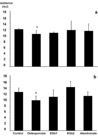
Tests were performed considering the following
structural-mechanical properties: resilience (mJ) which was defined as the energy that the specimen admits in the elastic phase; maximum load (N) and stiffness (103 N/m) which was defined as
the relationship between load and deformation
indicating the rigidity of the structure (Estanislau
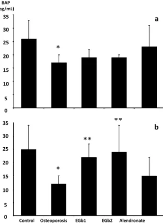
et al. 2010). Figure 1 - The levels of bone alkaline phosphatase (BAP).
a - 20 days of treatment. b - 30 days of treatment. *p<0.05 when the control group was compared to the osteoporosis group (Student`s t-test). **p<0.05 when all groups, except the control, were compared to the osteoporosis group (Dunnett T3 test).
STATISTICAL ANALYSIS
The data were expressed by (mean± standard
deviation) and were analyzed for statistical significance using one-way analysis of variance (ANOVA), followed by Tukey/Dunnett’T3
post-hoc test except for the control group. The control
group was compared with the osteoporosis group
using the Student’s t-test. p<0.05 was considered significant.
RESULTS
The values of BAP in the present study were significantly reduced in the osteoporosis group and in the EGb1, EGb2 groups (thirty days) the levels of BAP increased significantly (figure 1).
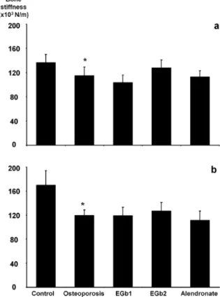
Figure 2 shows that the osteoporosis group had a significant lower BMD than the control group. The EGb2 and alendronate groups at the end of 20 and 30 days significantly recovered the BMD. The results of three-point bending test are reported in figures 3, 4 and 5. The osteoporosis group revealed a significant reduction of the bone stiffness, maximum load and resilience values when compared to the control group.
DISCUSSION
To our knowledge, a few pre-clinical studies have been conducted to evaluate the effects of EGb in osteoporosis; the majority of them, however, employed as a model for osteoporosis induction, ovariectomized rats. We are the first group that evaluated the effects of EGb in the GIO. The aim of
Figure 3 - Mechanical properties of the three-point bending test- Maximum load. a - 20 days of treatment. b - 30 days of treatment. *p<0.05 when the control group was compared to the osteoporosis group (Student`s t-test).
Figure 4 - Mechanical properties of the three-point bending
test-bone stiffness. a - 20 days of treatment. b - 30 days of
this study was to assess the effect of the treatment with EGb in recovering the osteoporosis induction by glucocorticoids trough mechanical tests and BMD using DEXA.
Our previous results were basically based on bone biopsy following by histomorphometric and immunohistochemestry analysis, in the present study the BMD was evaluated by DEXA that is the gold standard method for the clinical evaluation of osteoporosis.
We observed that the use of glucocorticoids reduced the BAP, BMD, the maximum load, resilience and stiffness. The treatment with EGb was effective in recovering the levels of BAP, BMD, suggesting that the extract increased bone mass in those animals, probably reducing the apoptosis of osteoblasts as showed in our previous studies (Lucinda et al. 2013).
Glucocorticoid is typically associated with decreased BMD, number of osteoblasts, bone formation rate (Chang et al. 2009, Manolagas and Weinstein 1999, Weinstein et al. 1998) trabecular bone (Jee and Yao 2001, Lucinda et al. 2010b) and biomechanical parameters (Wang et al. 2005).
Biochemical markers, such as BAP, reflect the bone destruction in conditions that affect bone metabolism (Melton et al. 1997). The values of BAP in the present study were significantly reduced in the osteoporosis group (figure 1) showing an alteration in the process of bone formation and bone mineralization.
A relevant finding was that the EGb increased significantly the levels of BAP (figure 1), and such increase is a response to an anabolic therapy (Miyauchi et al. 2008) that is directly linked with osteoblastic activity (Duque and Rivas 2007). In accordance with the present study results, studies
in vitro showed that Ginkgo biloba (100µg/ml) increased alkaline phosphatase levels in 147.2% when compared to the culture of control cells. Similarly, the EGb components, kaempferol and quercetin, were also shown to stimulate alkaline phosphatase activity in cultures of human osteoblastic cells (Oh et al. 2008, Prouillet et al. 2004).
In the current, study we evaluated the whole right tibias with DEXA, which is currently the method of first choice for measuring BMD (Ralston 2005). The higher dose of EGb was effective in recovering the BMD of the tibia as well as the alendronate, a drug that is widely used in the treatment of osteoporosis. Trivedi et al. (2009) reported an increase of the BMD in ovariectomized rats treated with EGb. The authors based their results in the improvement of the osteoblasts function and number, which was confirmed by the increase in the osteogenic genes and osteocalcin. We also believe that the EGb has an important role in increasing osteoblast function and number, once in our previous results the extract reduced the Figure 5 - Mechanical properties of the three-point bending
pro apoptotic protein suggesting a decrease in the osteoblasts death (Lucinda et al. 2013).
B o n e f r a g i l i t y c a n b e d e f i n e d b y biomechanical parameters and it is influenced by several components including bone turnover, microarchiteture, mineralization, microdamage, collagen crosslink and mineral crystal structure (Friedman 2006, Turner 2002). Glucocorticoids have been associated with rapid and significant bone loss as well as increased risk in bone fractures. In the present study the GIO showed not only a significant reduction in BMD but also reduction in the biomechanical parameters of bone like the bone stiffness, maximum load and resilience.
Despite the fact that EGb and alendronate increased the BMD, no significant improvement in the biomechanical parameters of the tibia occurred. Sliwiński et al. (2004) showed an increase in the mineral content of femoral diaphysis in the treatment with alendronate; nevertheless, it had a less significant effect on mechanical properties of the femoral diaphysis. In addition, they reported that the femoral neck, which is an area with the predominance of trabecular bone, had a significant increase of load when it was compared to the ovariectomized rats. These results may be indicative of a bigger influence of alendronate and EGb on the trabecular bone despite the cortical bone present in the diaphysis of long bones, and also lack of correlation between bone density and risk of fractures, as postulated by some researchers (Ferretti et al. 1993).
Although the measurement of BMD is one of the most important tools in the diagnosis of osteoporosis it has become increasingly clear that it cannot reflect all components of bone strength. Several osteoporosis therapies, such as biphosphonates, have shown improvement in BMD; however, those changes provide only information about the quantity of bone loss or its gain and cannot fully account for changes in mechanical properties which include the architectural changes
occurring in bone (Cummings et al. 2002, Watts et al. 2004). For example, the increase in BMD in patients treated with risedronate was found to be independent of fracture incidence (Watts et al. 2004).
Likewise, our data showed that EGb and alendronate did not affect the biomechanical properties, apart from the BMD improvement. To our knowledge, the present study is the first one to evaluate the effect of EGb in the biomechanical parameters of tibial diaphysis and the results suggest that the EGb increase the biomechanical parameters but not in a significant way. We may speculate that the period of treatment and the dose of EGb could not be enough to recover the cortical bone of the tibia, despite the promising results with alveolar bone (Lucinda et al. 2010a, b). However, Trivedi et al. (2009) reported an increase in tibial cortical bone of ovariectomized rats with EGb in a higher dose (100mg/kg/day) and with a longer period of treatment (five weeks of treatment).
In summary, the treatment with GIO reduced BMD and the biomechanical parameters of bone like the bone stiffness, maximum load and resilience. We showed that EGb restored BMD evaluated by DEXA, however no improvement in the biomechanical parameters of the tibia occurred (Lucinda et al. 2013).
ACKNOWLEDGMENTS
This work was financed by Fundação de Amparo à Pesquisa do Estado de Minas Gerais (FAPEMIG)
(APQ-00154-11 and 173/08). We would like to thank Cassiana M. Boya for the English review of the manuscript.
REFERENCES
BRAS M, QUEENAN B AND SUSIN S. 2005. Programmed cell death via mitochondria: different modes of dying. Biochemistry 70: 231-239.
BRAYBOY JR, CHEN XW, LEE YS AND ANDERSON JJB. 2001. The protective effects of Ginkgo biloba extract (EGb 761) against free radical damage osteoblast-like bone cells (MC3T3-E1) and the proliferative effects of EGb 761 on these cells. Nutr Res 21: 275-285.
BROWN JP ET AL. 2009. Bone turnover markers in the management of postmenopausal osteoporosis. Clin Biochem 42: 929-942.
CHANG JK, LI CJ, LIAO HJ, WANG CK, WANG GJ AND HO
ML. 2009. Anti-inflammatory drugs suppress proliferation and induce apoptosis through altering expressions of cell cycle regulators and pro-apoptotic factors in cultured human osteoblasts. Toxicology 258: 148-156.
CRUSE LM, VALERIANO J, VASEY FB AND CARTER JD. 2006. Prevalence of evaluation and treatment of glucocorticoid-induced osteoporosis in men. J Clin Rheumatol 12: 221-225.
CUMMINGS SR, KARPF DB, HARRIS F, GENANT HK, ENSRUD K, LACROIX AZ AND BLACK DM. 2002.
Improvement in spine bone density and reduction in risk of vertebral fractures during treatment with antiresorptive drugs. Am J Med 112: 281-289.
DUQUE G AND RIVAS D. 2007. Alendronate has an anabolic effect on bone through the differentiation of mesenchymal stem cells. J Bone Miner Res 22: 1603-1611.
ESTANISLAU CA, RAHAL SC, MULLER SS, LOUZADA
MJQ AND ARAÚJO FAP. 2010. Evaluation of femur of orchiectomized guinea pigs by bone densitometry using dual-energy x-ray absorptiometry (DXA) and mechanical
testing. Vet and Zootec 17: 104-112.
FELDSTEIN AC, ELMER PJ, NICHOLS GA AND HERSON M. 2005. Practice patterns in patients at risk for glucocorticoid-induced osteoporosis. Osteoporos Int 16: 2168-2174.
FERRETTI JL, CAPOZZA RF, MONDELO N AND ZANCHETTA JR. 1993. Interrelationships between
densitometric, geometric, and mechanical properties of rat femora: Inferences concerning mechanical regulation of bone modeling. J Bone Miner Res 8: 1389-1396.
FRIEDMAN AW. 2006. Important determinants of bone strength: beyound bone mineral density. J Clin Rheumatol 12: 70-77.
GAO SG, LI KH, XU M, JIANG W, SHEN H, LUO W, XU
WS, TIAN J AND LEI GH. 2011. Bone turnover in passive smoking female rat: relationship to change in bone mineral density. BMC Musculoskelet Disord 12: 131.
HENNEICKE H ET AL. 2011. Corticosterone selectively
targets endocortical surfaces by an osteoblast-dependent mechanism. Bone 49: 733-742.
HOCK JM, KRISHNAN V, ONYIA JE, BIDWELL JP, MILAS
J AND STANISLAUS D. 2001. Osteoblast Apoptosis and Bone Turnover. J Bone Miner Res 16: 975-984.
HOFF AO ET AL. 2008. Frequency and risk factors associated with osteonecrosis of the jaw in cancer patients treated with intravenous bisphosphonates. J Bone Miner Res 23: 826-836.
JEE WSS AND YAO W. 2001. Overview: animal models of osteopenia and osteoporosis. J Musculoskel Neuron Interact 1: 193-207.
JOHNELL O. 1996. Advences in osteoporosis: better identification of risk factors can reduce morbity and mortality. J Intern Med 239: 229-304.
LUCINDA LM, DE OLIVEIRA TT, SALVADOR PA, PETERS VM, REIS JE AND GUERRA M DE O. 2010a. Radiographic evidences of mandibular osteoporosis improvement in Wistar rats treated with Ginkgo biloba. Phytother Res 24: 264-267.
LUCINDA LM, VIEIRA BJ, OLIVEIRA TT, SÁ RC, PETERS VM, REIS JE AND GUERRA M DE O. 2010b. Evidences of osteoporosis improvement in Wistar rats treated with
Ginkgo biloba extract: a histomorphometric study of mandible and femur. Phytother Res 81: 982-987.
LUCINDA LM, VIEIRA BJ, PETERS VM, REIS JEP, OLIVEIRA RSMF AND GUERRA M DE O. 2013. The effect of the Ginkgo biloba extract in the expression of Bax, Bcl-2 and bone mineral content of Wistar rats with glucocorticoid-induced osteoporosis. Phytother Res 27: 515-520.
MANOLAGAS SC AND WEINSTEIN RS. 1999. New developments in the pathogenesis and treatment of steroid-induced osteoporosis. J Bone Miner Res 14: 1061-1066.
MCLAUGHLIN F, MACKINTOSH J, HAYES BP,
MCLAREN A, UINGS IJ, SALMON P, HUMPHREYS J, MELDRUM E AND FARROW SN. 2002. Glucocorticoid-induced osteopenia in the mouse as assessedby histomorphometry, microcomputed tomography, and biochemical markers. Bone 30: 924-930.
MELTON LJ 3RD, KHOSLA S, ATKINSON EJ, O’FALLON
WM AND RIGGS BL. 1997. Relationship of bone turnover to bone density and fractures. J Bone Miner Res 12: 1083-1091.
MIYAUCHI A, MATSUMOTO T, SHIGETA H, TSUJIMOTO
M, THIEBAUD D AND NAKAMURA T. 2008. Effect
of teriparatide on bone mineral density and biochemical markers in Japanese women with postmenopausal osteoporosis: a 6-month dose-response study. J Bone Miner Metab 26: 624-634.
OH SM AND CHUNG KH. 2004. Estrogenic activities of
Ginkgo biloba extracts. Life Sci 74: 1325-1335.
OH SM, KIM HR AND CHUNG KH. 2008. Effects of Ginkgo
POZZI S ET AL. 2007. Bisphosphonate associated
osteonecrosis of the jaw: a review of 35 cases and an evaluation of its frequence in multiple myeloma patients. Leuk Lynphoma 48: 1852-1854.
PROUILLET C, MAZIÈRE J, MAZIÈRE C, WATTEL A, BRAZIER M AND KAMEL S. 2004. Stimulatory effect of
naturally occurring flavonols quercetin and kaempferol on alkaline phosphatase activity in MG-63 human osteoblasts
through ERK and estrogen receptor pathway. Biochem
Pharmacol 67: 1307-1313.
RALSTON SH. 2005. Bone densitometry and bone biopsy. Best Pract Res Clin Rheumatol 19: 487-501.
RUSSELL RG. 2006. Bisphosphonates: from bench to bedside. Ann NY Acad Sci 1068: 367-401.
SLIWIŃSKI L, JANIEC W, PYTLIK M, FOLWARCZNA J, KACZMARCZYK-SEDLAK I, PYTLIK W, CEGIEŁA U AND NOWIŃSKA B. 2004. Effect of administration
of alendronate sodium and retinol on the mechanical properties of the femur in ovariectomized rats. Pol J Pharmacol 56: 817-824.
SMITH JV, BURDICK AJ, GOLIK P, KHAN I, WALLACE
D AND LUO Y. 2002. Antiapoptotic properties of Ginkgo biloba extract EGb761 in differentiated PC12 cells. Cell Mol Biol 48: 699-707.
SMITH JV AND LUO Y. 2004. Studies on molecular mechanisms of Ginkgo biloba extract. Appl Microbiol Biotechnol 64: 465-472.
SZEJNFELD VL. 2000. Clinical manifestations. Osteoporosis:
diagnostic and treatment. 1st
ed., São Paulo: Sarvier, 406 p.
TRIVEDI R, KUMAR A, GUPTA V, KUMAR S, NAGAR GK, ROMERO JR, DWIVEDI AK AND
CHATTOPADHYAY N. 2009. Effects of Egb 761 on bone mineral density, bone microstructure, and osteoblast function: Possible roles of quercetin and kaempferol. Mol Cell Endocrinol 302: 86-91.
TURNER CH. 2002. Biomechanics of bone: determinants of skeletal fragility and bone quality. Osteoporos Int 13: 97-104.
VAN BEEK TA. 2002. Chemical analysis of Ginkgo biloba
leaves and extracts. J Chromatogr A 967: 21-55.
VERBORGT O, TATTON NA, MAJESKA RJ AND
SCHAFFLER MB. 2002. Spatial distribution of Bax and Bcl-2 in osteocytes after bone fatigue: Complementary roles in bone remodeling regulation. J Bone Miner Res 17: 907-914.
WANG FS, LIN CL, CHEN YJ, WANG CJ, YANG KD,
HUANG YT, SUN YC AND HUANG HC. 2005. Secreted Frizzled-Related Protein 1 Modulates Glucocorticoid Attenuation of Osteogenic Activities and Bone Mass. Endocrinology 146: 2415-2423.
WATTS NB, COOPER C, LINDSAY R, EASTELL R, MANHART MD, BARTON IP, VAN STAA TP AND ADACHI JD. 2004. Relationship between changes in bone mineral density and vertebral fracture risk associated with risedronate: greater increases in bone mineral density do not relate to greater decreases in fracture risk. Clin Densitom 7: 255-261.
WEINSTEIN RS, JILKA RL, PARFITT AM AND