Interleukin-6 promoter polymorphisms
(-174 G/C) in Malaysian patients with
systemic lupus erythematosus
K.H. Chua
1,
B.P. Kee
1, S.Y. Tan
2and L.H. Lian
11
Department of Molecular Medicine,
2Department of Medicine, Faculty of Medicine, University of Malaya,
Kuala Lumpur, Malaysia
Correspondence to: K.H. Chua, Department of Molecular Medicine, Faculty of Medicine, University of
Malaya, 50603 Kuala Lumpur, Malaysia
Fax: +60-3-7967-6600. E-mail: khchua@um.edu.my
Systemic lupus erythematosus (SLE) is a chronic autoimmune disease that involves the inflammation of various organs upon deposition of immune complexes and is characterized by uncontrolled B cell hyperactivity. Despite intensive research on the etiology of the disease, the exact cause of the onset of SLE is unknown. The pathogenesis of the disease has been proposed to be associated with the imbalance of T helper type 1 (Th1) and Th2 cytokine activities. Elevated serum levels of interleukin-6 (IL-interleukin-6), a Th2 cytokine with various functions in the regulation of human biological systems, are observed in SLE patients. In the present study, 100 Malaysian SLE patients and 100 controls were evaluated in order to determine the association of polymorphisms existing in the promoter region of the IL-6 gene with the onset of SLE. The homozygous G genotype was found to be significant in SLE patients (χ2 = 33.754; P = 0.00000000625), whereas the heterozygous G/C genotype was significant in
the controls (χ2 = 25.087; P = 0.000000548). We suggest that the C allele might have a masking effect on the G allele when both
alleles are present in heterozygous individuals. However, we did not observe any significant association of the homozygous C allele with the onset of SLE or with protection from the disease (χ2 = 1.684; P = 0.194366).
Key words: Interleukin-6; Systemic lupus erythematosus; Promoter; Polymorphisms
Research supported by the BM271, FP047/2005C, and 36-02-03-6006 grants.
Received March 16, 2008. Accepted December 11, 2008
Introduction
Systemic lupus erythematosus (SLE) is a well-known autoimmune disease that often involves multiple organ inflammation. Generally, the organs affected by SLE are the heart, the skin, the brain, joints, lungs, and blood vessels (1). These organs could be damaged upon depo-sition of immune complexes by the body’s own immune system. During the early stage of the disease, SLE pa-tients often present a variety of symptoms that are similar to those of other diseases, with difficulty in diagnosis by physicians, leading to a consequent delay in the onset of treatment. Thus, there is a great deal of interest in the identification of biomarkers that are responsible for the
onset and progression of the disease. The common symp-toms of SLE are extreme fatigue, painful or swollen joints, unexplained fever, skin rashes, and malaise (2). Photo-sensitivity is also present in some patients. The butterfly rash is one of the characteristic features of SLE. This rash usually occurs over the bridge of the patient’s nose and resembles the shape of a butterfly. The fatal outcome of SLE patients is often due to infections and renal disorders. Despite much effort by scientists, there is still no cure for SLE. However, treatments are available to control the symptoms of the disease. For example, immunosuppres-sants can be used to prevent flares (3).
age (4). This could possibly be due to differences between genders in terms of hormone levels and life styles. Ethnic-ity might also play a role in the predisposition to disease development (5). The exact etiology of the onset of SLE has yet to be confirmed. It is suggested that both environ-mental and genetic factors play a role in triggering the onset of the disease. Environmental factors such as ultra-violet light, external stress, and infectious agents interact with complex genes and ultimately lead to the uncontrolled production of autoantibodies (3). Genetic variation is also believed to have an effect on the susceptibility to SLE. Despite intensive investigations carried out by research-ers worldwide, there is still no single gene shown to be responsible for the onset of the disease.
It has been proposed that the functional disequilibrium of T helper type 1 (Th1) and Th2 cells is possibly associated with immune responses in different kinds of immune disor-ders (6). Th1 cytokines [interferon gamma (IFN-γ) and inter-leukin-2 (IL-2)] activate macrophages and promote the pro-duction of antibodies that are responsible for complement fixing and opsonization. Th2 cytokines (4, 5, 6, IL-10, and IL-13), on the other hand, induce antibody produc-tion and mast cell activaproduc-tion (7). It has been reported that Th1 levels are decreased, whereas Th2 levels are elevated, in SLE patients (8). Thus, alteration of Th1 and Th2 lympho-cyte functions can result in the up-regulation of autoantibody production by B cells that can lead to the pathogenesis of SLE. Additionally, it has been suggested that the ratio of Th1 and Th2 could serve as a strong determinant predicting the histopathology of lupus nephritis (9).
IL-6 is a multifunctional Th2 cytokine involved in acute inflammation by the production of acute phase proteins and by the modulation of specific immune responses. IL-6 is responsible for the transformation of IL-4-preactivated B cells to immunoglobulin-secreting plasma cells (10). In addition, IL-6 is also involved in the regulation of several biological processes such as bone metabolism and the production of platelets (11). Linker-Israeli and colleagues (12) have reported that elevated plasma levels of IL-6 messenger-RNA and proteins could be detected in SLE patients.
The IL-6 gene has been mapped to the short arm of chromosome 7 at location 21 to 15 (13). The entire IL-6 gene is organized into five exons and four introns. The translation of IL-6 RNA and post-translational processing result in the formation of a 21- to 28-kDa protein with 128 amino acids (14). In the present study, we investigated whether there is an association between SLE susceptibil-ity and single nucleotide polymorphisms located in the promoter region of the IL-6 gene (-174 G/C) in our Malay-sian SLE patients, although many negative correlation
results have been observed in various studies in the world (10,15,16).
Material and Methods
Our study included 100 SLE patients (6 males and 94 females) and 100 matched healthy control individuals, who were recruited from the University Malaya Medical Center, located in Kuala Lumpur (Ethics Approval No. 380.1). In general, the patients ranged in age from 16 to 50 years and their clinical manifestations were renal disorder with proteinuria (>0.5 g/day), malar rash, arthritis, and photosensitivity with production of anti-dsDNA at >200 IU/ mL. Blood samples were collected into tubes containing EDTA. A conventional phenol-chloroform genomic DNA extraction method was employed as described previously (17). Primer sequences (forward primer G: 5'-GCACTTTT CCCCCTAGTTGTGTCTTACG-3'; forward primer C: 5'-GACGACCTAAGCTTTACTTTTCCCCCTAGTTGTGT CTTGAC-3'; reverse primer: 5'-ATAAATCTTTGTTGG AGGGTGAGG-3') and PCR cycling parameters (95°C, 10 min [1 cycle]; 94°C, 30 s, 66°C, 45 s, 72°C, 45 s [40 cycles]; 72°C, 7 min [1 cycle]) were used based on the study of Schotte et al. (10). Each PCR assay contained 10 ng genomic DNA in a final volume of 20 μL containing 10 pmol of each primer (1st Base Pte. Ltd., Singapore), 1 U Taq DNA polymerase (Fermentas, USA), 0.1 mM dNTP mix (Promega, USA), 2.0 μL Taq buffer with KCl, and 1.0 mM MgCl2. PCR was carried out in a thermal cycler
(Mastercycler gradient, Eppendorf, Germany). The ampli-fied products were then visualized by 18% polyacrylamide gel electrophoresis (PAGE). Genotype distribution and allelic frequencies of the polymorphisms were generated in all samples. In addition, χ2 and odds ratio (OR) tests with
95% confidence intervals (95%CI) were performed to test for any significant association.
Results
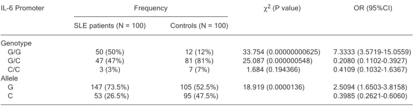
Figure 1 shows the band patterns observed for the PCR products in the study of the IL-6 promoter polymor-phism (-174 G/C) on 18% (w/v) polyacrylamide gel. Three genotypic combinations were observed in the study, i.e., homozygous G, heterozygous G/C, and homozygous C. A 121-bp fragment denotes the G allele, whereas the C allele is indicated by a 136-bp band on the polyacrylamide gel.
Pattern 1
Pattern 2
Pattern 3
Pattern 4
Lane B Lane
A
300 bp
200 bp
121 bp 100 bp
300 bp 250 bp 200 bp
150 bp
136 bp 100 bp
50 bp
Figure 1. Figure 1.Figure 1. Figure 1.
Figure 1. PCR products of IL-6 promoter polymorphism (-174 G/C) were shown by ethidium bromide-stained 18% polyacrylamide gels run at 140 V for 2 h. Lane A, 100-bp DNA marker (Fermentas Life Sciences, USA). Pattern 1: Homozygous G (121 bp). Pattern 2 : Heterozygous G/C (121 and 136 bp). Pattern 3: Homozygous C (136 bp). Pattern 4: DNA blank. Lane B, 50-bp DNA marker (Fermentas Life Sciences, USA).
group. The homozygous C genotype, however, did not present any significant association.
Both the homozygous G and heterozygous G/C geno-types are present at almost equal frequencies in SLE patients, i.e., 50 and 47%, respectively. In contrast, the heterozygous G/C genotype was observed most frequently in the control group (81%) compared to the homozygous G genotype (12%). However, the homozygous C genotype was the least commonly observed in both the SLE patients (3%) and the control group (7%).
The allelic frequencies are noted to be significant in our study. The OR value for allele C was 0.3985, a 95%CI from 0.2621 to 0.6060. In SLE patients, the G allele was up to three times more frequent than the C allele. However, both alleles were observed equally in the healthy control group.
Discussion
IL-6 is a multifunctional cytokine produced in response to inflammatory stimuli to regulate the human immune response against infection (18). IL-6 levels are increased in patients with SLE, especially in those with active dis-ease (18). It was believed that the over-production of IL-6 in SLE patients led to the pathogenesis of the disease (18). The IL-6 gene expression products may play a role in the onset of SLE. Thus, the polymorphic gene can be an important biomarker for predisposition to SLE.
In the present study, the homozygous G genotype was observed significantly in SLE patients, whereas the het-erozygous G/C genotype was significant in the controls. The gene expression products from the G allele may affect
Table 1. Table 1.Table 1. Table 1.
Table 1. Genotypic distribution and allelic frequencies of the IL-6 promoter polymorphism (-174 G/C) in patients with systemic lupus erythematosus (SLE) and controls.
IL-6 Promoter Frequency χ2 (P value) OR (95%CI)
SLE patients (N = 100) Controls (N = 100)
Genotype
G/G 50 (50%) 12 (12%) 33.754 (0.00000000625) 7.3333 (3.5719–15.0559) G/C 47 (47%) 81 (81%) 25.087 (0.000000548) 0.2080 (0.1102–0.3927) C/C 3 (3%) 7 (7%) 1.684 (0.194366) 0.4109 (0.1032–1.6367) Allele
G 147 (73.5%) 105 (52.5%) 18.919 (0.0000136) 2.5094 (1.6503–3.8158)
the susceptibility of an individual to SLE. Individuals with the homozygous G genotype were observed to be predis-posed to SLE development. Individuals with the heterozy-gous G/C genotype could be protected from developing SLE. Thus, it is suggested that the C allele has a masking effect over the G allele in the heterozygous G/C genotype, which may be due to a complex interaction of both alleles when present co-dominantly. Under such conditions, it is postulated that the C allele induces a protective response that prevents the individuals from developing SLE. How-ever, we did not observe any significant association of the homozygous C genotype with the onset of SLE or with protection from developing SLE. This observation further suggests the protective role initiated by the interaction of both G and C alleles in heterozygous individuals. Overall, the suppressor effect of the C allele can only be suggested for female SLE patients since the number of male SLE patients screened in this study was small.
In a study of Caucasian German SLE patients carried out by Schotte et al. (10) in 2001, the IL-6 promoter polymorphism (-174 G/C) did not reveal any significant association with susceptibility to SLE. However, the G allele was found to be responsible for the presence of discoid skin lesions and for the production of anti-histone antibodies in SLE patients (10). Based on their study, we suggest that the G allele may be involved in the pathogen-esis of SLE and in the development of certain clinical manifestations in SLE patients.
Another study by Fishman et al. (19) showed that IL-6 promoter polymorphism is significantly associated with chronic juvenile arthritis, an autoimmune disease that af-fects the joints. They observed that the C allele was signifi-cantly less common in systemic juvenile chronic arthritis in the Anglo-Saxon Caucasian population studied (19). It was then reported that the C allele is responsible for the suppression of IL-6 transcriptional activity. Thus, the ho-mozygous C genotype is suggested to reduce the levels of IL-6 in plasma and to play a role against the development of systemic juvenile chronic arthritis. The homozygous C genotype has also been linked to other clinical conditions, for example Gaucher disease (20). Gaucher disease is an autosomal recessive lysosomal storage disorder caused by mutations in the gene coding for β-glucocerebrosidase, which involves a characteristic increase of plasma IL-6 levels in patients (20). It was reported that patients with Gaucher disease carrying the homozygous C genotype can have low plasma levels of IL-6 (20).
In the present study, however, the homozygous C genotype did not show a significantly protective role against SLE. This observation again suggests the role of the complex interaction of multiple genes and the involvement of other factors in the development of SLE. Overall, our results do not agree with several other studies carried out on other world populations (10,15,16) and these contradic-tory results could be due to the genetic heterogeneity of SLE in different ethnicities.
References
1. Katsiari CG, Tsokos GC. Systemic lupus erythematosus: From disease pathogenesis to therapy. Drug Discov Today Dis Mech 2006; 3: 185-192.
2. Manson JJ, Rahman A. Systemic lupus erythematosus. Orphanet J Rare Dis 2006; 1: 6.
3. Franshin G, Peeva E, Diamond B. Pathogenesis of SLE: implications for rational therapy. Drug Discov Today Dis Mech 2004; 1: 303-308.
4. Danchenko N, Satia JA, Anthony MS. Epidemiology of sys-temic lupus erythematosus: a comparison of worldwide dis-ease burden. Lupus 2006; 15: 308-318.
5. Wang F, Wang CL, Tan CT, Manivasagar M. Systemic lupus erythematosus in Malaysia: a study of 539 patients and comparison of prevalence and disease expression in different racial and gender groups. Lupus 1997; 6: 248-253. 6. Funauchi M, Yu H, Sugiyama M, Ikoma S, Ohno M, Kinoshita K, et al. Increased interleukin-4 production by NK T cells in systemic lupus erythematosus. Clin Immunol 1999; 92: 197-202.
7. Csiszar A, Nagy G, Gergely P, Pozsonyi T, Pocsik E. In-creased interferon-gamma (IFN-gamma), IL-10 and
de-creased IL-4 mRNA expression in peripheral blood mono-nuclear cells (PBMC) from patients with systemic lupus erythematosus (SLE). Clin Exp Immunol 2000; 122: 464-470.
8. Chun HY, Chung JW, Kim HA, Yun JM, Jeon JY, Ye YM, et al. Cytokine IL-6 and IL-10 as biomarkers in systemic lupus erythematosus. J Clin Immunol 2007; 27: 461-466. 9. Nakashima H, Akahoshi M, Masutani K. Th1/Th2 balance of
SLE patients with lupus nephritis. Rinsho Byori 2006; 54: 706-713.
10. Schotte H, Schluter B, Rust S, Assmann G, Domschke W, Gaubitz M. Interleukin-6 promoter polymorphism (-174 G/C) in Caucasian German patients with systemic lupus erythe-matosus. Rheumatology 2001; 40: 393-400.
11. Papassotiropoulos A, Hock C, Nitsch RM. Genetics of inter-leukin 6: implications for Alzheimer’s disease. Neurobiol Aging 2001; 22: 863-871.
13. Chen WF, Fischer M, Frank G, Zlotnik A. Distinct patterns of lymphokine requirement for the proliferation of various sub-populations of activated thymocytes in a single cell assay. J Immunol 1989; 143: 1598-1605.
14. May LT, Santhanam U, Tatter SB, Bhardwaj N, Ghrayeb J, Sehgal PB. Phosphorylation of secreted forms of human beta 2-interferon/hepatocyte stimulating factor/interleukin-6. Biochem Biophys Res Commun 1988; 152: 1144-1150. 15. Guarnizo-Zuccardi P, Lopez Y, Giraldo M, Garcia N,
Rodri-guez L, Ramirez L, et al. Cytokine gene polymorphisms in Colombian patients with systemic lupus erythematosus. Tis-sue Antigens 2007; 70: 376-382.
16. Huang CM, Huo AP, Tsai CH, Chen CL, Tsai FJ. Lack of association of interleukin-6 and interleukin-8 gene polymor-phisms in Chinese patients with systemic lupus erythemato-sus. J Clin Lab Anal 2006; 20: 255-259.
17. Sambrook J. Molecular cloning: a laboratory manual. New York: Cold Spring Harbor Laboratory Press. Vol. 2. 2001. 18. Ripley BJ, Goncalves B, Isenberg DA, Latchman DS,
Rahman A. Raised levels of interleukin 6 in systemic lupus erythematosus correlate with anaemia. Ann Rheum Dis 2005; 64: 849-853.
19. Fishman D, Faulds G, Jeffery R, Mohamed-Ali V, Yudkin JS, Humphries S, et al. The effect of novel polymorphisms in the interleukin-6 (IL-6) gene on IL-6 transcription and plasma IL-6 levels, and an association with systemic-onset juvenile chronic arthritis. J Clin Invest 1998; 102: 1369-1376.