Patch-clamp detection of
macromolecular translocation
along nuclear pores
Departamento de Fisiologia, Faculdade de Medicina de Ribeirão Preto, Universidade de São Paulo, Ribeirão Preto, SP, Brasil
J.O. Bustamante and W.A. Varanda
Abstract
The present paper reviews the application of patch-clamp principles to the detection and measurement of macromolecular translocation along the nuclear pores. We demonstrate that the tight-seal ‘gigaseal’ be-tween the pipette tip and the nuclear membrane is possible in the presence of fully operational nuclear pores. We show that the ability to form a gigaseal in nucleus-attached configurations does not mean that only the activity of channels from the outer membrane of the nuclear envelope can be detected. Instead, we show that, in the presence of fully operational nuclear pores, it is likely that the large-conductance ion channel activity recorded derives from the nuclear pores. We conclude the technical section with the suggestion that the best way to demonstrate that the nuclear pores are responsible for ion channel activity is by showing with fluorescence microscopy the nuclear translocation of ions and small molecules and the exclusion of the same from the cisterna enclosed by the two membranes of the enve-lope. Since transcription factors and mRNAs, two major groups of nuclear macromolecules, use nuclear pores to enter and exit the nucleus and play essential roles in the control of gene activity and expression, this review should be useful to cell and molecular biolo-gists interested in understanding how patch-clamp can be used to quantitate the translocation of such macromolecules into and out of the nucleus.
Correspondence J.O. Bustamante Departamento de Fisiologia FMRP, USP
Av. Bandeirantes, 3900 14049-900 Ribeirão Preto, SP Brasil
Fax: 55 (016) 633-0017 E-mail: [email protected] and [email protected]
Presented at the XII Annual Meeting of the Federação de Sociedades de Biologia Experimental, Caxambu, MG, Brasil, August 27-30, 1997.
J.O. Bustamante and W.A. Varanda were recipients of grants from MRC, Canada and FAPESP.
Received August 8, 1997 Accepted November 11, 1997
Key words
•Nuclear pores •Nuclear envelope •Cell nucleus
•Nucleocytoplasmic transport •Macromolecular transport •Gene activity
•Gene expression •Transcriptional control •Ion channels
•Patch-clamp •Measurement
Introduction
In the present review we attempt to ex-plain how the patch-clamp technique can be used to detect and measure macromolecular translocation (MMT) by using both qualita-tive and quantitaqualita-tive analysis of gated ion flow along the nuclear pore complex (NPC). In doing so, we hope that the concepts used can be extrapolated to other systems such as those of channels from other organellar mem-branes. The mechanisms of NPC-mediated
patch-clamp recording of the large-conduc-tance ion channel activity at the nuclear envelope (NE) represents gating of the NPC channel (NPCC, the central NPC hole) and how this measurement can be used to inves-tigate NPC-MMT. We hope that this balanc-ing-act, tight-rope approach, although trivial at times for one or another group of scientists (i.e., electrophysiologists or cell biologists), contributes to the development of new meth-odologies for the study of the mechanisms of gene control by imported transcription fac-tors and of gene expression by the export of messenger RNAs (mRNAs). Both of these processes are relevant to the maintenance of normal cell function and to the development of various cellular pathologies and pro-grammed cell death. Space limitations re-strict the number of publications cited in the present review. For this reason, we refer the reader to further reading on these special-ized areas.
Nuclear pore ion channel behavior
Ion channels and ion channel activity
Here we review some basic concepts on ion channels. The more demanding reader may consult other sources (e.g., 1). The simplest channel system is one that switches in a binary scheme between two states: open and closed, with values of, say, 1 and 0 (picoamperes, picosiemens, etc.). In the steady state, this simple two-state system exhibits open and closed
probabili-ties, po and pc, such that:
po + pc = 1 (Eq. 1)
Although a popular term, ion channel activity has both qualitative and quantitative connotations. Channel activity can be taken to be the set of all those parameters which determine the observable or measured quan-tity which, in patch-clamp, is the time-de-pendent electrical current, i(t), carried out by the ions moving along the channel. At each point in time, the current will be determined by the value of the conductance, γ:
i(V,t) = γ(V,t) V(t) (Eq. 2) where the parentheses next to the ‘depend-ent’ variables indicate that the variable is a function of the ‘independent’ variables (e.g.,
V(t) indicates that V is a function of t). In a first, gross approximation, if we think of the channel as a hollow cylinder, then the channel conductance is related to the cylinder geometry and electrolyte resis-tivity, ρ,
γ = A/(lρ)or γ = πr2/(lρ) (Eq. 3) where A is the cross-sectional area of the cylinder and r and l are the radius and length of the cylinder. Equation 3 summarizes the relationship between conductance, area, length and resistivity of the channel hole. The wider and shorter the channel, the larger the conductance. But, what equation 3 does not describe is the gating mechanism, which could be represented as a multiplying term whose values are either 0 or 1. We could use
Abbreviations and acronyms:
AP-1:activator protein-1, a transcription factor complex
consisting of c-Fos and c-Jun
AP-1/c-Jun:the c-Jun component of AP-1
ATP and GTP:adenosine and guanosine triphosphate
cAMP:cyclic adenosine monophosphate
CFTR:cystic fibrosis transmembrane conductance regulator
B-PE and FITC:B-phycoerythrin and fluorescein
isothiocyanate; two fluorescent probes
INM and ONM:inner and outer nuclear membranes of the
nuclear envelope
IP3:inositol 1,4,5-trisphosphate
mAb414:monoclonal antibody 414; raised against the
nuclear pore
MMT:macromolecular translocation
NE:nuclear envelope
NF-κB:the transcription factor named nuclear factor kappa B
NLS:nuclear localization signal
NPC and NPCC:nuclear pore complex and its central
channel or hole
NPC-MMT:NPC-mediated MMT
PKA:cAMP-dependent protein kinase
Ran/TC4:a small GTPase involved in NPC-MMT
RNA and mRNA:ribonucleic acid and messenger RNA
SP1:a transcription factor
a time-dependent Dirac’s delta function, δ =
δ(t), widely used to represent 0 everywhere except at a single point (which we envision as that representing the proper gating condi-tions):
γ = δ(t) [A/(lρ)] or γ = δ(t) [πr2/(lρ)] (Eq. 4) In a random, stochastic system such as an ion channel switching between the open and closed states, the macroscopic behavior of the system is represented by time average quantities which give a measure of how the macroscopic system (the collection of single, microscopic units) works. For example, the time average channel conductance, <γ> would be given by:
<γ> = <δ(t)> [A/(lρ)] or
<γ> = <δ(t)> [πr2/(lρ)] (Eq. 5) which can also be written as:
<γ> = po [A/(lρ)] or <γ> = po [πr2/(lρ)]
(Eq. 6)
Thus, the average ion current can be written as:
<i(V)> = <γ> V (Eq. 7) where V = V(t) is the applied voltage, which in patch-clamp is most of the time conve-niently set to be constant. If po, and V, as well
as channel geometry and resistivity of envi-ronment (i.e., ρ), do not change with time or voltage, then:
<i(t)> = poγV (Eq. 8)
Together, these microscopic and macro-scopic parameters give a measure of channel
activity, function and behavior. The more frequently the channel is open (i.e., the higher the po), and/or the greater the conductance
and voltage, γ and V, the greater the value of the average current, <i>. Here lies the power of patch-clamp. While patch-clamp allows precise determination of the parameters that determine <i> changes, macroscopic meas-urements (such as whole-nucleus voltage-clamp which detects whole nucleus ion cur-rent or fluorescence microscopy which de-tects integrated changes in probe concentra-tion) do not permit such determination be-cause a larger value of <i> can be obtained with either greater po, γ, or V, individually.
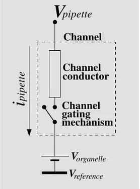
Figure 1 illustrates an equivalent circuit for an organellar ion channel with negligible reversal potential (Vrev, the value of voltage
at which the ion current reverses direction). The channel components are delimited by the rectangular box formed by the discontin-ued line. The channel is represented by a resistor or conductor (i.e., the vertical, solid line rectangular box represents the channel cylinder) plus a gating mechanism (a switch). The organelle electrical potential, Vorganelle,
is represented by the pair of thin-and-thick horizontal lines near the bottom. The voltage in the system is measured with reference to the bath potential, Vreference or Vbath
(repre-sented as the thick horizontal line at the bottom). The pipette potential, Vpipette, is
ap-plied at one side of the channel. The meas-ured quantity, the pipette current, ipipette, is
given by:
ipipette = γ (Vpipette - Vorganelle) (Eq. 9)
Variables:
i and γ:current and conductance of a single ion channel;
microscopic variables
I and Γ:current and conductance of a channel population;
macroscopic variables
N:number of functional units in the ion channel population
po and pc:open and close probabilities of an ion channel
Po and Pc:open and close probabilities of the ion channel
population
τ:time constant of decay
R, V and t:electrical resistance, voltage and time
Vrev:reversal potential, voltage at which the ion current
changes direction
A, r, l and ρ:cross-sectional area, radius, length and
resistivity of a cylinder-like channel
ncarrier and ninert:number of electrical charge carriers and inert particles inside the NPCC
vNPCC:NPCC volume
vcarrierand vinert:volume occupied by the electrical charge
carriers and inert particles inside the NPCC
ρcharge:electrical charge density per unit time
Xcarrierand Xinert:generic designation of the electrical
Essential features of the NPC
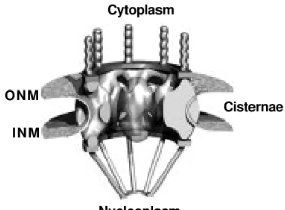
The NPC is the only direct pathway con-necting nucleus and cytoplasm (e.g., 2-6). Under electron microscopy, the NPC is one of the most conspicuous features of the NE (e.g., 7,8). The NPC stands out as the most prominent structure in topological imaging of the NE with scanning electron micros-copy (e.g., 9) and atomic force microsmicros-copy (e.g., 10). This supramolecular structure con-sists of many proteins which together have a mass estimated at about 124 MDa (megadal-tons, e.g., 8). Figure 2 shows a 3-D recon-struction model generated with electron mi-croscopy data (from Ref. 8). The figure dis-plays the putative plug in the center in a fuzzy, transparent fashion to indicate that the concept of the plug is not clear (8). Cytoplasmic filaments (vertical rod-like fila-ments) as well as nucleoplasmic ones (con-vergent at the bottom) are shown. The NPC spans the two membranes of the NE: the outer and the inner nuclear membranes (ONM and INM, respectively). This feature is
es-sential to the electrophysiological discus-sion that will follow. The space delimited by ONM and INM is a cisterna used by many processes to store proteins, molecules, etc. It is there that Ca2+ is thought to mediate NPC-MMT (e.g., 11,12). Note that the outer mem-brane of the NE is contiguous with the rough endoplasmic reticulum. Therefore, the ONM and rough endoplasmic reticulum, although distinct structures, share many properties. This contiguity may cause arguments when it comes to identifying the source of ion channel activity. Therefore, it is not suffi-cient to simply place the patch-clamp pipette at the outside of the nucleus but one is re-quired to prove that the activity is not de-rived from the rough endoplasmic reticu-lum. In a recent paper, it was shown that the large-conductance ion channel activity re-corded in cardiac myocyte nucleus-attached patches does not derive from the endoplas-mic reticulum (13). That this was the case was demonstrated by the inefficacy of vari-ous maneuvers known to affect protein-con-ducting channels of the endoplasmic reticu-lum (14).
The patent diameter of the NPC
Many electron and fluorescence micros-copy studies indicate that small molecules, ions and particles placed on one side of the NE reach the other side (e.g., 7,15,16). These observations have been interpreted to mean that the NPCs have a ‘patent’ diameter (a sort of effective cross-section for the electro-lyte-filled hole) of about 10 nm. The value of the patent diameter can vary from a few to several tens of nanometers according to the cell type and cycle (e.g., 7,16). This large patent diameter leads to the inescapable con-clusion that monoatomic ions flow freely along the NPCs and, consequently, that any observed heterogeneous concentration dis-tribution of small ions and molecules must result from ‘cytoplasmic exclusion’ (17) caused by the compartmentalized
distribu-Figure 1 - Equivalent electrical circuit for an organelle ion chan-nel. The model is drawn for an organelle with a simple mem-brane (vis à vis a dual memmem-brane like the nuclear envelope). The channel itself is delimited by the discontinued line. The channel is represented by a resistor or conductor (i.e., the rectangular box represents the channel cyl-inder) plus a gating mechanism (a switch). Allowance has been made for organelle electrical
po-tential, Vorganelle (represented by
the pair of thin-and-thick horizon-tal lines near the bottom). The voltage in the system is meas-ured with reference to the bath
potential, Vreference (represented
as the thick horizontal line at the bottom). The pipette potential, Vpipette, is applied to one side of the channel and the resulting
current, ipipette, measured. The
reversal potential, Vrev, for the
ion current has been assumed negligible (see 31).
i
pipette
V
organelleV
referenceChannel
gating
mechanism
Channel
conductor
Channel
tion of water- and molecular-binding sites (e.g., 7,15,18) from another (or more) non-NE mechanism(s), or from technical arti-facts. The ‘cytoplasmic exclusion’ principle states that because the state of water is dif-ferent in the difdif-ferent cellular compartments, the solubility, and thus the concentration, of a solute is compartmentalized (in our case, between the nucleus and cytoplasm). This exclusion principle resembles the ‘associa-tion-induction hypothesis’ introduced at the same time to explain the resting potential of cells without the need for a plasmalemmal Na-K pump (e.g., 19,20). A discussion of nuclear resting potentials is outside the scope of this review.
Agreement between patch-clamp and non-electrophysiological data
The patent diameter view of unrestricted flow of ions along the NPCC is in apparent contradiction with the electrophysiological data indicating that the NPC behaves as an ion channel (e.g., 13,21-24). However, this discrepancy is eliminated when both qualita-tive and quantitaqualita-tive analyses are performed (15). Qualitatively, at the present time only the patch-clamp is capable of detecting live single NPC conformational changes through the measurement of NPCC ion-conductive properties. Not even the most sophisticated electron or light microscope techniques are capable of giving a measurement of the live function of a single NPC and, although atomic force microscopy and other scanning probe microscopies hold this promise, reports have yet to appear with the time-resolution re-quired. All we can say with any kind of microscopy is that the particles have moved from one side to the other within an approxi-mate time interval of minutes, whereas with the patch-clamp we can have time resolution down to the millisecond range or even less. Thus, we can say that 10-kDa dextran mol-ecules have passed from the outside of the nucleus to the inside within, say, 15 min, but,
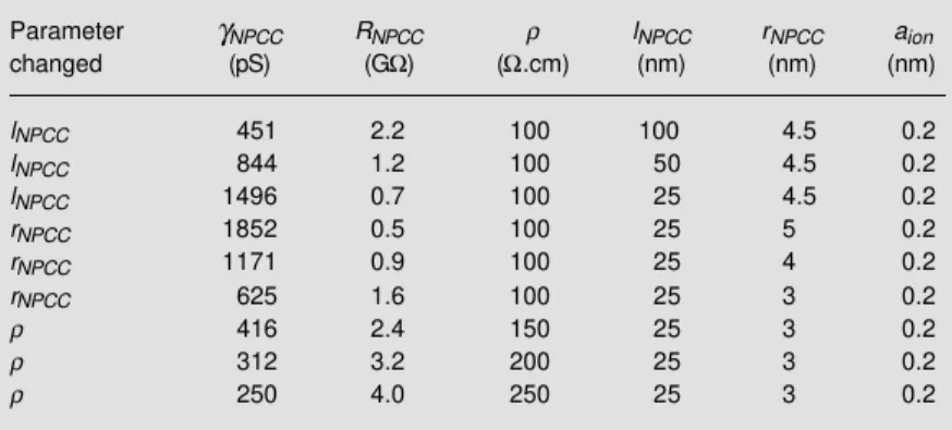
in physical terms, this does not mean that the NPCC is permanently open as commonly concluded. What patch-clamp measurements show is that the NPC gates behave like any other gate controlling the traffic of large quantities. Quantitatively, one may neglect particle-particle and particle-NPCC wall in-teractions, and simply use the geometrical parameters reported for the NPCC to esti-mate the single channel conductance of the NPCC, γNPCC. Table 1 gives the values of
γNPCC assuming that the hydrated radius, aion,
of the current carrying ions (e.g., K+) is 0.2 nm (15), and that the NPC has a hole of 3-10-nm radius, rNPCC, 25-100-nm length, lNPCC,
(to accommodate published data; see 8,15) and filled with an electric conductor (the electrolyte) of specific resistivity, ρ, of 100-250 Ω.cm (around that of cytoplasm and nucleoplasm, plus or minus small inert con-taminants, not medium-sized or larger mac-romolecules). The values are calculated when the resistance of each NPCC, RNPCC, is
ap-proximated by the following equation (15):
RNPCC = {ρ[lNPCC + (1.64 rNPCC)]}/{π(rNPCC)2
[1-(aion/rNPCC)]6} (Eq. 10)
Table 1 shows examples of RNPCC values
calculated with equation 10 and covers pub-lished values (25-29). In consideration of the NPC variation with cell type and cell cycle phase as well as the measurement errors (instrument, sample preparation, etc.), we can say that there is good agreement be-tween electrophysiological and
non-electro-Figure 2 - 3D-Reconstruction model of the nuclear pore com-plex (NPC) generated from elec-tron microscopy data. Cyto- and nucleoplasmic filaments are shown pointing to their respec-tive compartments. The outer and inner nuclear membranes (ONM and INM, respectively) of the nuclear envelope (NE) cre-ate a space which serves as cis-ternae for the storage of impor-tant factors, one of which is
Ca2+. Note that the putative
central plug is shaded to indi-cate the uncertain nature of the concept. (From Ref. 5, with per-mission).
Cytoplasm
ONM INM
Nucleoplasm
physiological data. For example, avian eryth-rocytes display a channel activity with a cationic channel conductance of 800 pS (23), coconut endosperm cells with one of about 1,000 pS (30), and murine cardiac myocytes show conductance values between 106 and 532 pS (22-36oC), with higher values corre-sponding to temperatures closer to that of this warm-blooded species (31). This tem-perature dependence of γNPCC demonstrates
the complexity of the biochemical processes involved in NPCC gating (e.g., ATPases and GTPases). Reports of nuclear ion channels which could be attributed to NPCCs have been published (e.g., 23,24,30,32). How-ever, these did not include demonstrations that the NPCCs were either functional or permanently closed, blocked or plugged. Other ion channels which are putatively not-NPCCs have also appeared. These conclude that the recorded channel activity can be attributed to Ca2+-activated K+ channels (33; see also 34), IP3-sensitive Ca2+-channels (35-37; see also 34) and even now the Cl- -channel known as the cystic fibrosis trans-membrane conductance regulator or CFTR (38). Despite the assertion by the authors that these channels were not NPCCs, no proof was given that the NPCs were closed or plugged. We will discuss the importance of providing such proof in the following paragraphs (see section Identification of NPCs as a source of ion channel activity).
Patch-clamp principles
The patch-clamp demonstration that the NPCC opens probabilistically (13) is con-sistent with non-electrophysiological obser-vations. Briefly, the NPCCs switch between open and closed states with open and close probabilities (po and pc, respectively). These
probabilities are regulated by cytosolic and nucleosolic factors such as GTPases, and localization signal receptors (e.g., 13,25,26). Therefore, despite the large diameter of the NPCC, the time average of its opening is less than the geometrical diameter of the NPC hole (what we are calling here the NPCC) unless, of course, po is 1. As a corollary,
conditions that favor the closed state will reduce the average nucleocytoplasmic ion flow. The more the NPCC dwells in either the closed or the plugged state (where ion flow/current is zero), the lower the po and the
smaller the macroscopic ion current. A re-duced macroscopic NE ion current favors the build-up of nucleocytoplasmic gradients of small ions, molecules and particles. Such generation of nucleocytoplasmic gradients (e.g., during Ca2+ transients, see 39) lend themselves to be misinterpreted as a reduced diameter of the NPCC but, instead, they are a reflection of the lower than unity open probability (i.e., po<1).
Early patch-clamp investigations sug-gested that ion channel activity recorded from the NE was attributable to NPCs (e.g., 23,24,30-32,40). In pure saline solutions containing high [K+], these NPCC candi-dates have a linear current-voltage relation-ship and, therefore, they have constant single channel conductance, γNPCC (e.g.,
23,24,30-32,40). To date, only the large-conductance cardiac channel activity has been directly demonstrated to derive from NPCs (13; see below). Like some of the NE channels in other nuclear preparations, these cardiac channels show a preference for cations (40). Cardiac myocyte NPCCs display two modes of operation labeled ‘stationary’ and
‘inacti-Table 1 - NPCC conductance, γNPCC, calculated from equation 10 with the
ion-conductive hole parameters: length (lNPCC), radius (rNPCC) and resistivity (ρ).
Parameter γNPCC RNPCC ρ lNPCC rNPCC aion
changed (pS) (GΩ) (Ω.cm) (nm) (nm) (nm)
lNPCC 451 2.2 100 100 4.5 0.2
lNPCC 844 1.2 100 50 4.5 0.2
lNPCC 1496 0.7 100 25 4.5 0.2
rNPCC 1852 0.5 100 25 5 0.2
rNPCC 1171 0.9 100 25 4 0.2
rNPCC 625 1.6 100 25 3 0.2
ρ 416 2.4 150 25 3 0.2
ρ 312 3.2 200 25 3 0.2
vating’ (31). This behavior has been ob-served also in hepatocytes (41). In the ‘sta-tionary’ or steady-state mode, the NPCCs open and close with a binomial behavior similar to an electron spin ensemble. That is, not only do all the NPCCs appear to be identical but they also switch between the open and closed states with open probabili-ties which are well fitted by a binomial mo-del:
Po(n) = N! pon (1-po)N-n/[n! (N-n)] (Eq. 11)
where Po(n) is the probability of finding n
channels simultaneously open, po is, as
be-fore, the single channel open probability, and N is the total number of ion-conducting channels in the patch (31). Note that equa-tion 2 does not contain reference to time. This is because the system is in steady state and its stochastic characteristics are the same at any given time. Also note that, as indi-cated at the beginning of this review, for a perfect binomial system, po + pc = 1. In the
‘inactivating’ mode of operation, NPCCs respond to voltage stimulation (equivalent to electrochemical gradient stimulation; see Ref. 42) with an exponential decay in Po(n). That
is, Po(n) is time-dependent.
Po(n,t) = Po(n,0) e-t/τ (Eq. 12)
with t being time - the independent variable,
τ the decay time constant, and Po(n,0) the
initial value of Po. Note that Po does not
necessarily follow the binomial behavior described in equation 10. The term inactivat-ing was assigned to this mode of operation because there are instances where the chan-nels can no longer open upon successive application of voltage pulses but require a time for recovery (31). This time, although not always the same, is equivalent to the refractory period of nerves associated with sodium channel inactivation (see 42). The lack of constancy of the refractory period may be indicative of the much greater com-plexity of the NPCC mechanism of opera-tion. For example, GTPase may play an
im-portant role in the inactivating machinery (see 40). Similarly to classical inactivation, the time constants for this putative inactiva-tion are reduced with voltage (see 42). At voltages between -10 mV and +10 mV, car-diac NPCCs showed no measurable inacti-vation: they were open all the time (31). This suggests that, at least in quiescent, non-stim-ulated cardiac myocytes (where the reversal potential is negligible), the NPCCs offer little opposition to ion flow. However, when the voltage gradient across the NE increased (i.e., increase in electrochemical gradient across the NE), the probability of finding open channels decreased. This dependence of NPCC gating on voltage (electrochemical gradient) is consistent with the fluorescence microscopy observation that the NE becomes a Ca2+-barrier when the cytosolic [Ca2+] in-creases over 300 nM (39). This can also be due to Ca2+-induced Ca2+ release from the NE cisternae which, in turn, leads to NPCC plugging by macromolecules (see below). Since live cardiac myocytes in situ are never at rest, it is likely that NPCCs play a signifi-cant role in determining the signals that cross to one or the other side of the NE.
Basic concepts in the identification of NPCs as a source of ion channel activity
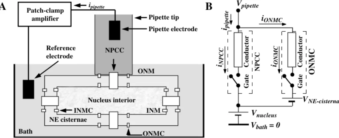
The idea that NE ion channel activity derives from NPCCs is very appealing from the electrophysiological point of view and, indeed, efforts have been made towards dem-onstrating that this activity derives from NPCCs (e.g., Innocenti and Mazzanti (46) used osmotic shock). However, patch-clamp investigations complemented with laser-scan-ning confocal fluorescence microscopy, transmission electron microscopy, field-emis-sion scanning electron microscopy, and atomic force microscopy showed a direct correlation between the large-conductance ion channel activity recorded from cardiac myocytes and myocyte NPCs (13). Both in the classical microelectrode studies of Loewenstein and coworkers (reviewed in 47) and in the first patch-clamp investiga-tions of NE ion channel activity (23,24), it was proposed that the NPC was the structure underlying the recorded activity. However, this proposition was hindered by patch-clamp investigations indicating that ion channels different from NPCCs also exist in either of the two membranes delimiting the NE. Such is the case for standard and CFTR chloride channels (38,48), for Ca2+-sensitive K+ chan-nels (33), for Ca2+- and Zn2+-channels (49, see 34), IP3-sensitive Ca2+-channels (34-37), etc. (50). That non-NPCCs do exist at the NE is further suggested by the demonstration that ion channel-forming proteins have been isolated from NE preparations (e.g., 34,51). Indeed, even a chloride ion channel protein which localizes primarily in the nucleus has been cloned and characterized (e.g., 52). Despite the non-NPCCs identified or yet to be identified at the NE, the NPCCs continue to stand out as the sole structures providing the direct pathway for the exchange of mate-rial between the nuclear and cytoplasmic compartments. For this reason, when the patch-clamp technique is used in the nucleus-attached mode (i.e., the pipette tip forming a
gigaseal with the outer membrane of the NE), and when it is demonstrated that small and medium-sized (e.g., <10 kDa) molecules enter the nucleus, then it must be unavoid-ably concluded that the NPCCs are effective electrical shunts for any non-NPCCs be-tween the recording patch-clamp pipette and bath electrodes (note that all non-NPPCs are located on either membrane of the NE whereas the NPCCs join the two membranes). Therefore, from an electrophysiological point of view, NPCCs should be the natural candi-dates for the source of large conductance channel activity. Figure 3 aims at explaining these concepts with the equivalent circuit. For the sake of clarity, we will only discuss the nucleus-attached patch-clamp mode of recording. Figure 3 summarizes the three groups of channels found in the NE prepara-tion. They are the NPCCs and the outer and the inner nuclear membrane channels (ONMC, and INMC, respectively). In nucleus-attached patches, the ONM and the INM can be thought of as two membranes in series. Since the pipette tip only touches the ONM, only the pipette voltage (Vpipette) is
applied to the NPCCs and the ONMCs. The voltage of the NE cisternae (VNE-cisternae) does
not change appreciably and, consequently, there is no voltage drop across the INM and the INMCs. The NPCCs and ONMCs form a system of parallel conductors through which the measured pipette current (ipipette) is
dis-tributed into essentially two branches: the NPCC current (iNPCC) and the ONMC
cur-rent (iONMC). That is: ipipette = iNPCC + iONMC.
Each current component is determined by the value of the conductance of the corre-sponding channel and the voltage applied to the channel. In cardiac myocytes, Vnucleus is
negligible in buffered 150 mM KCl (31). That is, the nuclear interior is short-circuited to the bath. Thus, Vnucleus equals the
refer-ence or bath voltage (Vreference). For this
rea-son, the voltage sensed by the NPCCs and the ONMCs equals Vpipette.
high (e.g., 7,16), the patch-clamp pipette always contains a few or more NPCCs. Fur-thermore, because inside the patch there are many NPCCs with γNPCC of several hundreds
of pS, it is clear that NPCCs are the major contributors to the recorded ion channel activ-ity. Therefore, the only available explana-tion against the NPCCs being responsible for the observed ion channel activity is that the NPCCs are permanently closed (or plugged) due to the conditions used in patch-clamp studies (e.g., saline not supplemented with MMT substrates). This counter-argu-ment remains to be proven. Furthermore, fluorescence microscopy data clearly dem-onstrate that under saline conditions that prevent MMT, the small monoatomic ions and molecules diffuse freely between the nucleus and cytosol (e.g., 53). That is, even in the most unfavorable conditions for MMT, the NPCs still allow ion transport. There-fore, a corollary of this discussion is that any report claiming non-NPCC activity should be accompanied by a direct demonstration
that the NPCCs are either closed, plugged or both.
Peripheral channels of the NPC
Only one computer-assisted 3-D recon-struction work, based on electron micros-copy data, shows that the eight-fold geom-etry of the NPC contains eight parallel path-ways for the transport of ions which are independent of the large central channel of the NPC (54). If these pathways behaved like ion channels, then one should see tens to hundreds of channels in a single nucleus-attached patch because the NPC density per unit area ranges from a few NPCs to tens of NPCs per square micrometer or several chan-nels with identical substates. However, this observation has never been reported and the 3-D reconstruction work has not been dupli-cated. Nevertheless, it should be noted that efforts to find a large-conductance ion chan-nel which shows eight substates have been recently published (41). Therefore, the only
Figure 3 - Equivalent circuit for the ion channels at the nuclear envelope (NE). A, Schematics of the
nucleus-attached patch-clamp pipette system. B, Equivalent circuit. Three groups of channels are represented: NPC
channels (NPCC), outer nuclear membrane channels (ONMC), and inner nuclear membrane channels (INMC). Under nucleus-attached patch-clamp configuration, the NPCCs and ONMCs form a system of parallel conductors
through which the measured pipette current (ipipette) is distributed: the NPCC current (iNPCC) and the ONMC
current (iONMC). There is no contribution of INMC current (iINMC). That is: ipipette = iNPCC + iONMC. Each current
component is determined by the value of the conductance of the corresponding channel and the voltage applied to the channel. The applied voltage for NPCCs equals the difference between the pipette and nucleus voltages (Vpipette - Vnucleus). The applied voltage for ONMCs and INMCs is (Vpipette - VNE-cisternae) and (VNE-cisternae -Vnucleus), respectively, where VNE-cisternae is the voltage at the NE cisternae. That large inert particles (e.g., colloidal gold and spherical dendrimers) go into the nuclear interior but not into the NE cisternae which indicates that the
ONMC conductance is much smaller than γNPCC. Vrev is assumed negligible.
A
Patch-clampB
amplifier
ipipette
Pipette tip Pipette electrode
Reference
electrode NPCC ONM
Nucleus interior
INMC INM
NE cisternae
ONMC Bath
Vpipette
ipipette
iONMC
iNPCC iONMC
Gate Conductor
NPCC
ONMC
VNE-cisternae
Vnucleus
Vbath = 0
Gate Conductor
Vpipette
ipipette
iNPCC NPCC
Vnucleus
Vbath = 0
iperipheral
Conductor
iperipheral
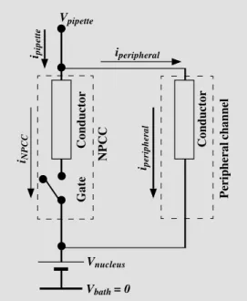
remaining possibility is that these pathways are not ion channels in the electrophysi-ological sense but, instead, permanently open peripheral holes as reported in the electron microscopy studies (54). However, if this were the case then the baseline current in patch-clamp recordings should be much greater than the zero value reported for all patch-clamp work. A simple computation demonstrates this fact:
ipipette = iNPCC + iperipheral = γNPCC Vpipette +
8γperipheral Vpipette (Eq. 13)
where the measured pipette current equals the sum of the current through the NPCC and the current through the peripheral channels (indicated by the subscripts). γNPCC and γ pe-ripheral are the conductance values for the
NPCC and one peripheral channel, respec-tively. Figure 4 shows the equivalent circuit for this concept. The measured pipette cur-rent, ipipette, is the sum of the currents flowing
through the central and peripheral channels. That is, ipipette = iNPCC + Niperipheral, where N is
the number of peripheral channels (8 in most cases, see Ref. 8). The putative peripheral channels should cause a baseline current to be recorded. However, patch-clamp
investi-gations have reported no such current, sug-gesting that the peripheral channels do not have access to the cytosol.
Technical issues
A question commonly asked is whether it is possible to attain a gigaseal in prepara-tions that have functional NPCs. In other words, do open NPCCs prevent gigaseal for-mation? The answer is no, they do not. An essential condition that must be technically attained prior to patch-clamp recording is the formation of the tight seal between the pipette tip and the membrane surface (in our case, the outer NE membrane). This seal has (or must have for a successful recording to be made) an electrical resistance, Rseal, greater
than 1 GΩ and, for this reason, it is called the gigaseal. The gigaseal is assessed indirectly by reading the current flowing through the pipette, ipipette, elicited with small current
pulses (resulting from the application of small voltage pulses to the pipette electrode). If the membrane contains many open NPCCs, it is likely that the investigator may think that a gigaseal has not been reached. However, this interpretation is erroneous. Say that under the pipette tip there are 10 permanently open NPCCs and that their single channel conduc-tance, γNPCC, is 500 pS. This would cause the
pipette tip to see a patch conductance not lower than 5,000 pS or 5 nS. This is equiva-lent to 0.2 GΩ. The investigator could mis-takenly take this to suggest that a gigaseal has not been formed. However, the fact is that the seal resistance between the rim of the pipette tip and the outer NE membrane,
Rseal, cannot be measured directly with the
patch-clamp technique. All that can be said is that the measured resistance, Rmeasured, is
not greater than that of either the NE patch directly under the pipette tip, Rpatch, or the
seal resistance, Rseal. The relationship among
these three quantities is:
1/Rmeasured = (1/Rpatch) + (1/Rseal) (Eq. 14) Figure 4 - Equivalent circuit for
the proposed NPC system formed by the central and pe-ripheral channels. The measured
pipette current, ipipette, is split
into the current for the central
and peripheral channels (iNPCC
and iperipheral, respectively). That is, ipipette = iNPCC + Niperipheral,
where N is the number of
pe-ripheral channels (8 most of the times, see Ref. 8). The peripher-al channels should cause a baseline current to be recorded in nucleus-attached experi-ments. However, this is yet to be reported, suggesting that the peripheral channels have no ac-cess to the cytosol (i.e., the side where the pipette is attached).
Vrev is assumed to be negligible.
Note that the contribution of other resis-tances (e.g., pipette, bath) are negligible and, thus, ignored in this discussion. There-fore, one can have a gigaseal and yet have a patch of NE containing many NPCs. The value of the gigaseal is revealed during mo-ments of simultaneous NPCC closure ob-served during prolonged periods of record-ing (up to 72 h), when the NPCs seem to exhaust the substrates for their gating mech-anism (Bustamante JO, unpublished obser-vations). Thus, the initial current recorded from a nucleus-attached patch may suggest that a gigaseal has not been reached and, consequently, the experimenter may discard the preparation. However, after many min-utes of observation one may capture a si-multaneous closure of the channels (Bustamante JO, unpublished results). Clearly, if the gigaseal had not been at-tained, this could be considered a poor pi-pette-membrane seal. To further confirm that the tight seal is retained, one may plug the NPCCs by adding diluted amounts of nuclear proteins (those which have nuclear-targeting sequences) without transport sub-strates (i.e., all those substances that par-ticipate in the successful NPC-MMT: ATP, GTP, cytosolic and nucleosolic receptors, etc.). In this manner, it is possible to reduce the number of unplugged NPCCs and, therefore, to clearly differentiate and count the NPCCs in the NE patch (Bustamante JO, unpublished results). It is also possible to obtain a gigaseal between the pipette tip and the NE but not see ion channel activity (Bustamante JO, unpublished observa-tions). This, however, cannot be taken as indicative that the NPCs are not present or that they are not functioning, for if given a sufficient amount of time (15-60 min, de-pending on the availability of NPC-MMT substrates), NPCC activity will appear as a consequence of macromolecular unplug-ging. As discussed below, during transloca-tion, macromolecules plug the NPCC for a period of time that will depend on the
ex-perimental conditions. Since most patch-clamp studies are carried out in pure saline solutions, macromolecular transport has the least favorable conditions. Therefore, those NPCCs plugged during nucleus isolation will remain that way for a very long time or forever, depending on the specific method-ologies used. The longer the isolated nuclei are exposed to saline, the less transport sub-strates are left in the preparation and the greater the number of plugged channels (Bustamante JO, unpublished observa-tions). This observation can be used to the investigator’s advantage because it provides a means of controlling the number of un-plugged, ion-conducting NPCCs. This is ad-vantageous because for the analysis of single channel activity it is best to have as low a number of gating channels as pos-sible (in the best case, only one). Therefore, with some fine tuning, a method can be developed that provides such a favorable situation for the statistical analysis of single channel activity (e.g., 13,31,40).
Nucleocytoplasmic gradients
We indicated above that electrophysi-ological and non-electrophysielectrophysi-ological data are not adversaries (see also Table 1) but, does NPCC activity explain nucleocytoplas-mic gradients of small ions and other mol-ecules? On the basis of NPCC activity alone (i.e., po, γNPCC and N), the amount of
monoatomic ion flow exchanged per cardiac myocyte patch was estimated at leading to a concentration change of 1 mM/s (see Ap-pendix in Ref. 31). Briefly, the maximal rate of change in ion concentration caused by the NPCC current in an NE patch is calculated by dividing the ionic current by the nuclear volume (e.g., about 500 µm3 for a small spherical nucleus of 5 µm radius, rnucleus).
would be several-fold greater. For example, for a pipette tip of 0.5 µm radius, rpipette, the
minimal NE patch area (corresponding to a flat plane and not to an Ω-shaped surface because no suction is applied) would be 0.8 µm2 orπ (r
pipette)2. The whole NE surface for
the 5-µm radius nucleus in our example has an area of 314 µm2 or 4π (r
nucleus)2.
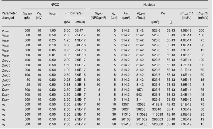
There-fore, the estimated concentration change in 1 s for the whole NE is 393 mM. This estimate demonstrates that NPCC behavior does not oppose the concept of diffusion, although the latter is usually assumed to indicate that the NPCs do not switch between open and closed states. Table 2 shows calculations for various changes in the parameters of the nucleus and NPCC.
NPC-mediated macromolecular translocation
Although it has been demonstrated by fluorescence microscopy that nucleocyto-plasmic ion flow is maintained even in iso-lated nuclei under pure saline conditions (e.g., 53), one arguable flaw in most patch-clamp studies is the lack of simultaneous tests for the basic transport properties of NPCs. In those studies favoring NPCs as the source for channel activity, there is no visual demonstration of translocation. Whereas in those assuming that the activity derives from structures different from NPCs, there is ab-sence of visual demonstration that the NPCs are closed. To resolve this problem, one can
Table 2 - Computed changes in nuclear ion concentration ∆Cion in 1 s, ∆Cion/∆t, caused by a 10 mV electrochemical gradient applied to the whole
nuclear envelope (i.e., a voltage drop between the cytoplasmic and the nucleoplasmic layers in the immediate vicinity of the NE). Definitions:
γNPCC is the ion conductance of a single NPC channel or NPCC; VNE is the voltage applied to the NE; popen is the probability of opening of a single
NPCC; <Flow Rate> is the mean rate of flow measured in terms of charges; DNPC is the NPC surface density; rN is the radius of the nucleus; ANE
is the total NE area; NNPC is the total number of NPCs in the nucleus; VN is the volume of the nucleus; ∆mion/∆t is the rate of change in the mass
of the total number of ions. Numbers are represented in the computer version of scientific notation, with x10n being indicated as En (e.g., x10-17
is given as E-17).
NPCC Nucleus
Parameter γNPCC VNE popen <Flow rate> DNPC rN ANE NNPC VN ∆mion/∆t ∆Cion/∆t
changed (pS) (mV) (NPC/µm2) (µm) (µm2) (Total) (mol/s) (mM/s)
(pA) (mol/s) (µm3) (l)
popen 500 10 1.00 5.00 5E-17 10 5 314.2 3142 523.6 5E-13 1.5E-13 300
popen 500 10 0.50 2.50 2.5E-17 10 5 314.2 3142 523.6 5E-13 7.9E-14 150
popen 500 10 0.25 1.25 1.3E-17 10 5 314.2 3142 523.6 5E-13 4.0E-14 75
popen 500 10 0.10 0.50 5.0E-18 10 5 314.2 3142 523.6 5E-13 1.6E-14 30
popen 500 10 0.05 0.25 2.5E-18 10 5 314.2 3142 523.6 5E-13 7.9E-15 15
popen 500 10 0.01 0.05 5.0E-19 10 5 314.2 3142 523.6 5E-13 1.8E-15 3
γNPCC 400 10 0.50 2.00 2.0E-17 10 5 314.2 3142 523.6 5E-13 6.3E-14 120
γNPCC 300 10 0.50 1.50 1.5E-17 10 5 314.2 3142 523.6 5E-13 4.7E-14 90
γNPCC 200 10 0.50 1.00 1.0E-17 10 5 314.2 3142 523.6 5E-13 3.1E-14 60
γNPCC 100 10 0.50 0.50 5.0E-18 10 5 314.2 3142 523.6 5E-13 1.6E-14 30
γNPCC 50 10 0.50 0.25 2.5E-18 10 5 314.2 3142 523.6 5E-13 7.9E-15 15
γNPCC 25 10 0.50 0.13 1.3E-18 10 5 314.2 3142 523.6 5E-13 3.9E-15 8
DNPC 500 10 0.50 2.50 2.5E-17 5 5 314.2 1571 523.6 5E-13 3.9E-14 75
DNPC 500 10 0.50 2.50 2.5E-17 3 5 314.2 942 523.6 5E-13 2.4E-14 45
DNPC 500 10 0.50 2.50 2.5E-17 1 5 314.2 314 523.6 5E-13 7.9E-15 15
rN 500 10 0.50 2.50 2.5E-17 10 10 1257 12566 4188.8 4E-12 3.1E-13 75
rN 500 10 0.50 2.50 2.5E-17 10 20 5027 50266 33510 3E-11 1.3E-12 38
rN 500 10 0.50 2.50 2.5E-17 10 30 11310 113098 113098 1E-10 2.8E-12 25
rN 500 10 0.50 2.50 2.5E-17 10 40 20106 201062 268083 3E-10 5.0E-12 19
test the NPC ion transport properties with fluorescent probes (e.g., 53). Either 4-kDa FITC-dextran (Molecular Probes, Eugene, OR) or 5.4-nm FITC-labeled StarburstTM dendrimers (Polysciences, Warrington, PA) can be used (Bustamante JO, unpublished results). Starburst dendrimers appear a bet-ter choice as they have an almost perfect spherical shape (see Ref. 55). Laser-scan-ning confocal fluorescent microscopy meas-urements performed during electrophysi-ological investigations show that both 4-kDa FITC-dextran and 5.4-nm FITC-dendrimers translocate under simple, high-K saline con-ditions (13; Bustamante JO, Geibel JP, Liepins A, Hanover JA and McDonnell TJ, and also Bustamante JO, unpublished re-sults). This observation is consistent with the demonstration that ≤10-kDa FITC-dex-tran molecules FITC-dex-translocate across the NE under simple saline conditions that normally prevent MMT (53). These results demon-strate that the nuclei utilized in those electro-physiological studies conformed to the preva-lent concept that NPCs allow translocation of not only monoatomic ions but also of small-to-medium-sized molecules and par-ticles (53). More importantly, the nuclei uti-lized in one electrophysiological laboratory have been shown to retain their macromo-lecular transport capacity (13; Bustamante JO, Geibel JP, Liepins A, Hanover JA and McDonnell TJ, unpublished results). This capacity was demonstrated by constructing a nuclear protein probe with the fluorescent B-phycoerythrin (B-PE, 240 kDa) conjugated to the nuclear localization signal (NLS) of the SV40 large T antigen (13). Readers inter-ested in the details of the mechanisms re-quired for NPC-MMT are encouraged to consult excellent reviews which have ap-peared elsewhere (e.g., 2-6,45). For this rea-son, we focus here on the highlights of MMT. Briefly, various mechanisms have been iden-tified for several classes of molecules. For proteins, the molecules carrying the NLS must first bind to a cytosolic receptor, then
bind to a docking site at the NPC, and then translocate in an ATP-dependent process. The process depends on other factors, in-cluding Ran/TC4 and phosphorylation of some of the substrates involved. Note that the conjugation of B-PE to the NLS was required to make the complex recognizable by the NPC-mediated mechanism for MMT (13). Without conjugation to an NLS, B-PE will not enter the nucleus. Thus, the cardiac myocyte investigations demonstrated that ion channel activity was recorded in a prepara-tion which retained the NPC capacity for ion and macromolecule transport. An advantage of conjugating a large fluorescent molecule to an NLS rather than conjugating a nuclear protein to a fluorescent probe such as FITC is that the large fluorescent probe will not enter the nucleus if not conjugated to the NLS whereas if the FITC conjugation of the nuclear protein is not strong enough, then the probe may enter the nucleus and give a false reading. Recent observations on changes of fluorescence response of fluorochromes caused by nuclear compartmentalization are important for absolute value determinations but not for relative, qualitative or semi-quan-titative observations (56).
The identification of NPCs as a source of ion channel activity
anti-myosin, and the mAb414 isotype con-trol (13). The blocking effect of mAb414 on channel activity was independently con-firmed (57). The effect of mAb414 on NPCC activity parallels that seen in the mitochon-drial multiple conductance channel upon the application of an antibody to a protein im-port component of the mitochondrial inner membrane (58). That mAb414 did not plug the NPCC, but instead blocked it, was shown by the unidirectional blockade (i.e., block-ade seen with only voltage of one sign) seen in some preparations (13). The reader should note that this unidirectional blockade is more than a simple voltage-dependent blockade because 100% block occurred with only one voltage polarity. Taken together, the results support the conclusion that the NPC, like other organellar and junctional ion channels, has an intrinsic ion channel behavior with high conductance at high [K+]. That a su-pramolecular structure may have an intrinsic ion channel activity (switching between the open and closed states) and at the same time may account for a large ion permeability is exemplified by the many reports on organellar ion channels. For example, at these high [K+] values, both the ryanodine receptor Ca2+ channel (reviewed in Ref. 59) and the mito-chondrial multiple conductance channel (e.g., 60) have a single channel conductance of the order of 103 pS. In analogy to other ion channels, NPCC activity depends on the volt-age across the NE (and thus, the electro-chemical potential). Note, however, that while the open probability decays faster (smaller time constants) at larger electro-chemical potentials, the single channel con-ductance remains constant.
Electrophysiological arguments favoring NPCs as ion channels
In addition to these supportive data, one can use electrical circuit theory alone to de-duce that in nucleus-attached patches of car-diac myocytes (with negligible resting
poten-tial, Vnucleus) the NPCCs are the source for the
recorded electrical activity. First, the hole of each NPC (i.e., the NPCC) allows a significant amount of ion flow and is solely responsible for the nucleocytoplasmic exchange of elec-trolytes (e.g., 8). Second, NPCCs have a wide diameter and are present in large numbers per unit area (e.g., 16). In cardiac myocytes, the number of NPCs per µm2 was estimated at 12 ± 4 (see Ref. 13). Third, the NE outside the area covered by the pipette tip has negligible electrical resistance because it contains many ion-conducting NPCs. Therefore, the poten-tial inside the nucleus is pretty much the same as that of the bath. Consequently, the potential sensed by the NPCs under the recording patch-clamp pipette is given by the potential differ-ence between the bath and pipette electrodes. Fourth, in nucleus-attached patches, where the pipette is sealed to the outer membrane of the NE, only non-NPC channels on the outer NE membrane are directly affected by the changes in the pipette voltage (as typically occurs in the patch-clamp technique). Fifth, any ion current contribution from non-NPCCs (Iother) at the
ONM flows in parallel to current contributed by the NPCCs (INPCC), i.e.,
Imeasured = ∑INPCC + ∑Iother (Eq. 15)
where ∑ represents the sum total of the current contributed by all the channels of the corresponding class. Therefore, for the con-tribution of non-NPCCs to overcome that of NPCCs it would be necessary to have a higher density of channels per unit area and/ or a higher single channel conductance. As mentioned above, to date, this has not been shown. On the contrary, NPCCs have been shown to allow ion and small molecule trans-location under pure saline conditions (53). Dextran molecules go into the nucleus but not into the NE (e.g., 53).
Macromolecules nullify single NPC channel conductance
for-eign nucleic acids and other macromolecules utilize the NPCs to cross the NE (e.g., 2-6). Since these macromolecules are poor elec-trical charge carriers and since they take over the volume previously occupied by the monoatomic ions (the good electrical charge carriers), one would expect NPC-MMT to reduce the ion conductance of a single NPCC,
γNPCC. To date, only one laboratory has
re-ported the effects of nucleocytoplasmic MMT on NPCC activity (e.g., 13,61,62). Nuclear-targeted B-PE silenced the NPCC activity (13). However, the channel activity was pro-gressively recovered with time, in a time-course that resembled that required for the complete nuclear transport of the nuclear-targeted B-PE probe in fluorescence micros-copy experiments. It must, therefore, be con-cluded that while the channel activity was silent, the B-PE macromolecules were con-tinuously transported into the nucleus. Ex-periments with transcription factors (an im-portant group of naturally occurring nuclear-targeted macromolecules involved in the regulation of gene activity) also transiently silenced ion channel activity (13,61,62). These experiments demonstrated that when transcription factors (AP-1/c-Jun, NF-κB, SP1 and TATA-binding protein) and other nuclear-targeted molecules (e.g., B-PE con-jugated to NLS) with a molecular mass of about 40 kDa or higher were added, the ion channel activity was silenced for a period of time ranging from a few seconds (with sub-strates) to hours (without subsub-strates). Both cases correlate with the electron microscopy observation that, without effective NPC-MMT substrate (e.g., Ca2+, ATP, GTP, Ran/ TC4, cytosolic and nucleosolic receptors, etc.), the NPC is plugged (e.g., 8). Since transcription factors enter the nucleus exclu-sively through the NPCC, the conclusion followed that the recorded ion channel activ-ity in these MMT-competent nuclei derived from gated ion flow along the NPC and not from an indirect action of the factors (13). When comparing the translocation times in
these nuclei with the times in situ, the reader should be aware that the electrophysiologi-cal experiments were conducted under con-ditions which are far from optimal for nor-mal macromolecular transport (e.g., 2-6). For example, cytosolic factors such as Ran/ TC4 and NLS receptors were not added. Furthermore, the rate of nuclear cytoplasmic protein transport has been shown to be deter-mined by the protein phosphorylation (re-viewed in Ref. 63). Therefore, it is likely that protein kinases and phosphatases play a role in nuclear transport by controlling the phos-phorylation levels of both transport substrates and NPCs (see 63). Despite these limitations of the patch-clamp experiment, we consider these patch-clamp conditions (with less than optimal content of NPC-MMT substrates) to facilitate the isolation of single ion channel activity because under normal conditions the translocation should take place at a faster pace and might make it difficult to isolate the NPCC behavior. Furthermore, despite these non-physiological conditions, the prepara-tions demonstrated an extraordinary stabil-ity, yet to be reported elsewhere (>72 h; 62; Bustamante JO, unpublished results). There-fore, we would like to note the remarkable (albeit difficult) success in patch-clamping the nucleus in situ (e.g., 64). This achieve-ment should facilitate patch-clamp studies under more realistic conditions and make easier the comparison of patch-clamp results with those obtained in non-electrophysiologi-cal experiments (e.g., with permeabilized cells, see Ref. 11). Note, however, that fill-ing the pipette with cytosol causes some electrical problems in patch-clamp record-ing as the pipette resistance increases (Bustamante JO, unpublished results).
address-ing peptide silences a large conductance cat-ionic channel (e.g., see 65 and references therein). This channel, known as the pep-tide-sensitive channel, is detected in mutant porin-less preparations (e.g., 66). The open probability of the voltage-dependent anion channel (VDAC) of the outer mitochondrial membrane (porin type-1; which is also ex-pressed in sarcoplasmic reticulum; see Ref. 67) was eliminated with a soluble 54-kDa mitochondrial protein (reviewed in Ref. 68). Mitochondrion-targeting peptides also plug the multiple conductance mitochondrial in-ner membrane channel whose properties re-semble those of the peptide-sensitive mito-chondrial channel (e.g., 58,60,69). Finally, protein translocation through the 4-6-nm di-ameter aqueous pore of the endoplasmic reticulum, the translocon, eliminated chan-nel activity (e.g., 14,70). Independent fluo-rescence accessibility studies show that the aqueous pore in a functioning translocon is 4-6 nm in diameter, making it the largest membrane channel, after that of the NPC, that maintains a permeability barrier (71).
NPC channel activity, MTT, and Ca2+ in NE cisternae
It has recently been recognized that [Ca2+] in the NE cisternae ([Ca2+]
NE) plays a major role in the NPC-mediated translocation of macromolecules (11) and medium-sized (>10 kDa) molecules (53,72, reviewed in 12). Briefly, when [Ca2+]
NE is low, the transloca-tion of macromolecules and even of medi-um-sized molecules (>10 kDa) is not pos-sible. It should, therefore, be expected that in cells where there is a great deal of Ca2+ release from the NE cisternae, the transport of macromolecules will be adversely affected. The impaired transport machinery, in turn, should cause the macromolecules to be caught in the middle of the NPC transloca-tion hole (the NPCC). Therefore, like a plug, they should obstruct the pathway for ion flow and, consequently, result in an impaired
translocation of small molecules and monoatomic ions. The period of silence must be determined by many factors. When the concentration of the macromolecules is suf-ficiently low, it may be possible to detect single translocations, whereas when it is rela-tively high, the many molecules may line up in single file to give the equivalent effect of translocation of a single long molecule. Translocation will stop when one of the required NPC-MMT substrates is exhausted (e.g., Ca2+, ATP, GTP, Ran/TC4, cytosolic and nucleosolic receptors, etc.), even if a macromolecule containing the signal is still present and ready for transport. That is, MMT cannot be expected to follow the customary diffusion laws. Therefore, rather than a pro-cess described by a single exponential change, a multi-exponential process should be ex-pected due to the biochemical reactions in-volved (signal recognition, docking, etc.; see, e.g., 2-6). Thus, theoretically, each reaction (e.g., signal recognition) should correspond to an exponential function. Structural sup-port for the electrophysiological idea of the translocating macromolecule behaving like a plug comes from the consistent and repro-ducible electron microscopy observation that when transport substrates are supplied, most NPCs are unplugged whereas when the sub-strates are absent, most of the NPCs are plugged (e.g., 8). Figure 2 shows the putative plug in the 3-D reconstruction NPC model of Pante and Aebi (8). The fuzzy, uncertain concept of the plug is indicated by the shaded coloring of the figure. That plugging mate-rial may account for a high NE resistance was put forward by Loewenstein and co-workers (22) more than three decades ago. Since both transcription factors and mRNA use the NPCs for their translocation, it can be concluded that the cellular rhythm affect-ing [Ca2+]
NE should be reflected on the levels of gene activity and expression.
see 79 and 80). As discussed in previous paragraphs, each time the NPCC opens, it allows a great deal of ions to be translocated. Therefore, reports showing steady-state nucleocytoplasmic gradients may be ex-plained by an NE that has many of its NPCCs plugged by translocating molecules or by the classical view of binding sites or water states (e.g., 15,18-20). Luminescence experiments with nucleoplasmin (a nuclear protein) fused to aequorin demonstrated the Ca2+ barrier nature of the NE (73,74). However, when aequorin was fused to the nuclear localiza-tion signal of the glucocorticoid receptor, the nature of the NE as a barrier was not detected (81). This result may be related to simpler transport mechanisms for the recep-tor used for nuclear localization (e.g., 82). In other words, the selection of the protein to be used for nuclear targeting is critical to the interpretation of the results because the mechanisms of nuclear transport for pro-teins (e.g., nucleoplasmin) are not the same as those for steroid receptors (e.g., glucocor-ticoid receptor). The experimental data indi-cate the importance of macromolecular trans-port for the maintenance or development of nucleocytoplasmic gradients. This relation-ship between macromolecular transport and the ATP-independent flow of small mol-ecules, particles and ions, should be relevant to the interpretation of experimental results. For example, when evaluating data from simultaneous fluorescence and ‘whole-cell’ patch-clamp recordings, the investigator should be aware that the pipette may dilute away transport substrates without which macromolecular transport would be impos-sible. This condition would lead to the NPCC being plugged by a macromolecule and, con-sequently, would result in an envelope with a full ion-barrier character. The reader should note that, due to the complexity of require-ments for NPC-MMT, having a macromol-ecule with a nuclear localization signal plus ATP and Ca2+ is not in itself sufficient to secure translocation.
Changes in NPC topography concomi-tant with changes in NPCC activity were confirmed by atomic force microscopy (62). In these investigations, the universal tran-scription factor TATA-binding protein (with both structural and functional roles) facili-tated transport and unplugging of the chan-nels after the typical transient reduction in single NPCC conductance (62). The atomic force microscopy images suggested that the proteins docked and accumulated at the NPC cytoplasmic side (62). Part of the mecha-nisms of TATA-NPC interaction was attrib-uted to an oligomerization effect mediated by protein glycosylation (62; see also 83 and references therein).
Relevance to studies of gene control and expression
The demonstration that the NPC has an intrinsic ion channel activity supports the use of the patch-clamp technique for the quantifi-cation of macromolecular transport along a single functional NPC. To date, no other tech-nique has this capability. In analogy to the principles of the Coulter cell counters, basic electrophysiological principles show that, when completely filled with electrolytes, the NPCC ionic conductance is maximal (13,84). They also show that, each time a macromolecule is translocated, the NPCC conductance goes to zero because the macromolecule displaces all the electrical charge carriers (13,84). This phe-nomenon is not exclusive to the NPC but is shared by channels of mitochondria (e.g., 58,60,69) and endoplasmic reticulum (e.g., 14,70).
particles have wider diameter than the NPCC but their mechanism of translocation involves their transient transformation into filaments during their ‘snaking’ through the NPCCs (e.g., 86). Clearly, for smaller molecules which are poor charge carriers and do not plug the NPCC completely, the situation is intermediary and requires a more complex description. Indeed, experiments with 5.4-nm FITC-labeled dendrimers showed that the probes translocate to the nucleus and during their translocation the binomial be-havior of the NPCC is transformed into low-frequency bursts (Bustamante JO, unpub-lished results). This subject is outside the scope of this review but, in principle, one can reason that for smaller molecules with poor charge carrier properties, the effect would be directly proportional to the density of the molecules inside the channel. The higher their density, the higher the resistivity of the electrolyte filling the NPPC and, there-fore, the smaller the single channel conduc-tance, γNPCC. A semi-quantitative analysis
demonstrates the point. Under pure saline condition, when the NPC hole is completely filled with the electrolyte, the electrical charge density per unit time (ρcharge) is:
ρcharge= ncarrier/vNPCC (Eq. 16)
where ncarrier is the number of good electrical
charge carriers inside the NPC hole and vNPCC,
the volume of the hole. When macromol-ecules and other large, inert, non-current-carrying particles enter the hole, they dis-place the charge carriers, thus reducing ncarrier
and thus, ρcharge.
vcarrier = vNPCC - vinert (Eq. 17)
where vcarrier and vinert are the volume
occu-pied by carrier and inert particles, respec-tively.
ncarrier = vcarrier [Xcarrier] = (vNPCC - vinert) [Xcarrier]
(Eq. 18)
where [Xcarrier] is the concentration of elec-Figure 5 - NPC-mediated
trical charge carriers. Thus,
ρcharge= (vNPCC - vinert) [Xcarrier]/vNPCC =
[1 - (vinert/vNPCC)] [Xcarrier] (Eq. 19) vinert can be calculated from the
concentra-tion of electrically inert particles, [Xinert], and
their individual (or average) volume, vparticle: vinert = ninertvparticle = vNPCC [Xinert] vparticle
(Eq. 20)
Therefore,
ρcharge = (1 - vparticle [Xinert]) [Xcarrier]
(Eq. 21)
This relationship may be expanded to incor-porate the presence of multiple charge carri-ers (e.g., K+, Na+ and Ca2+) and multiple poor or inert carriers (e.g., transcription fac-tors and nuclear proteins). The above analy-sis does not take into account interactions among particles or between the particles and the NPCC inner wall lining, which affect carrier mobility. In the real world, these are a significant factor opposed to free ion flow.
As we come to the end of this review, we would like to summarize by saying that both data and theory about the cell nucleus and other cellular organelles show that the pe-riod of channel silence can be correlated with the translocation time of macromol-ecules. This principle, denoted the ‘macro-molecule-conducting ion channel paradigm’ in the study of NPC ion channel behavior (13), is similar to that of Coulter cell counters. The principle has been applied to polymer counting along ion channels (84) and to the
determination of the size of nucleic acids with 2.6-nm diameter channels (88). The use of molecules which are not electrical charge carriers for the measurement of ion channel diameter has been applied to the determina-tion of the diameter (ca. 3 nm) of a porin channel (89). The observation that the patch-clamp technique can be used to measure and study NPC-MMT for as long as 72 h (62; Bustamante JO, unpublished observations) implies that the mechanisms of transcription factor and mRNA translocation can be stud-ied by this electrophysiological technique. Therefore, the possibility of using the patch-clamp technique for the identification of the NPC mechanisms underlying transcription factor control of gene activity and of mRNA-mediated gene expression offers a unique advantage over other currently available tech-niques in that it facilitates quantitative stud-ies of macromolecular transport at the single NPC level. By complementing this technique with low light-level fluorescence micros-copy, it should be possible to resolve many of the current enigmas pervading the field.
Further information related to this sec-tion can be found at the following internet site: http://www.geocities.com/TheTropics/ 6066/index.html.
Acknowledgments