Fighting by sle e p-de prive d rats as a
po ssible m anife statio n o f panic:
e ffe cts o f so dium lactate
1Faculdade de Medicina, Universidade de Marília, Marília, SP, Brasil 2Departamento de Ciências Biológicas, Faculdade de Ciências,
Universidade Estadual Paulista, Bauru, SP, Brasil F.A. Furlan1
and K. Hoshino2
Abstract
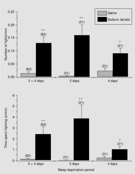
Increased fighting is an effect of desynchronized sleep deprivation (DSD) in rats, and recently this behavior has been suggested to be spontaneous panic and equivalent to panic disorder. In the present study we tested this hypothesis by evaluating the effect of sodium lactate on this aggressiveness, because this substance is recognized to induce spontaneous panic attacks in patients. A total of 186 male albino Wistar rats, 250-350 g, 90-120 days of age, were submitted to DSD (multiple platform method) for 0, 4, or 5 days. At the end of the deprivation period the rats were divided into subgroups respectively injected intraperitoneally with 1.86, 2.98 and 3.72 g/kg of 1 M sodium lactate, or 1.86 and 3.72 g/kg of 2 M sodium lactate. The control animals were submitted to the same procedures but received equiva-lent injections of sodium chloride. Regardless of DSD time, sleep-deprived animals that received sodium lactate presented a significant-ly higher mean number of fights (0.13 ± 0.02 fights/min) and a longer mean time spent in confrontation (2.43 ± 0.66 s/min) than the controls (0.01 ± 0.006 fights/min and 0.12 ± 0.07 s/min, respectively; P<0.01, Student t-test). For the sodium lactate group, concentration of the
solution and time of deprivation increased the number of fights, with the mean number of fights and mean duration of fighting episodes being greater with the 2.98 g/kg dose using 1 M lactate concentration. These results support the hypothesis that fighting induced by DSD is probably a spontaneous panic manifestation. However, additional investigations are necessary in order to accept this as a promising animal model for studies on panic disorder.
Co rre spo nde nce
F.A. Furlan
Faculdade de Medicina Universidade de Marília Av. Higyno Muzzi Filho, 1001 17525-902 Marília, SP Brasil
Fax: + 55-14-433-8961 E-mail: furlan@ laser.com.br
Research supported by CAPES. Publication supported by FAPESP.
Received March 9, 2000
Accepted January 17, 2001
Ke y wo rds
·Rat
·Sleep deprivation
·Panic
·Aggressiveness
·Sodium lactate
Intro ductio n
The homeotherms sleep is composed of two phases that cycle alternately (1-3): syn-chronized sleep, divided into 4 stages and characterized by electroencephalographic waves of high voltage and low frequency, and desynchronized sleep, also
character-ized by low voltage and high frequency waves, similar to the waves observed in the alert phase.
(5), a reduction in psychomotor performance (6) and mnemonic performance (7), hyper-phagia (8), and an increase in sexual activity (9). In rats, one of the effects of DSD is an increase in fighting behavior (10) which has been recognized as being of a defensive nature (11,12). Data from our laboratory (13,14) suggest that these agonistic behav-iors are panic manifestations.
Panic is an explosive reaction character-ized by a set of somatic and autonomic re-sponses associated with intense fear. It has been suggested that panic is a phenomenon related to the flight-or-fight behavior (15), and is manifested when a stimulus, recog-nized as dangerous, approaches or touches the individual (situational panic). Neverthe-less, panic-triggering mechanisms can suffer alterations and promote this behavior with-out justifiable environmental causes (spon-taneous panic). This phenomenon has been described for some non-human animal spe-cies in both natural (16) and experimental situations (17-19). Frequent recurrence of spontaneous panic attacks in humans char-acterizes the behavioral abnormality named panic disorder (PD). Since some episodes of fighting induced by DSD occur without the animal being actually threatened (normally conspecifics do not represent a threatening stimulus in groups of rats), they are pre-sumed to be a spontaneous panic manifesta-tion.
In the present study we evaluated the effects of sodium lactate administration on the fighting behavior of rats submitted to DSD in order to test the validity of the hypo-thesis that they are panic manifestations. Lactate was chosen because it is known, since the classic work of Pitts and McClure (20), that the infusion of sodium lactate can precipitate panic attacks in patients with PD. These panic attacks are symptomatically simi-lar to those that occur naturally (21), and the response to lactate is currently considered to be a sensitive and specific marker for PD (22).
Mate rial and Me tho ds
A total of 186 male albino Wistar rats weighing 250-350 g and aged 90-120 days were used for the study. After a minimum period of 7 days for adaptation to the labora-tory conditions, the animals were submitted to DSD for 0 (group 0), 4 (group DSD-4) or 5 (group DSD-5) days. Thirty minutes before the end of the DSD period, a large individual identification symbol was painted on the animals neck or trunk with a black dye for human hair (Henne) routinely used in our laboratory (23). Sodium lactate or sodium chloride was then injected ip, and the animals were kept in wire-mesh cages (25 x 30 x 25 cm) in groups of 4 animals per experimental treatment. Similar control groups of 5 animals each were also studied. The total number of animals in each sub-group is presented together with the results. The multiple platform method (23) was used for DSD. Ten rats were placed simulta-neously in a plastic box (105 x 55 x 27 cm) containing 15 cylindrical platforms (6 cm in height and 5 cm in diameter) fastened to the bottom of the box 15 cm apart from each other. The box was then filled with water up to 1 cm from the platform top. The animals kept on the platforms were able to sleep; however, when they reached the desynchro-nized sleep phase, the muscular atonia typi-cal of this phase would cause their nose to touch the water or the animal would fall into the water and be awakened. Natural temper-ature and light/dark cycle conditions were used. Water and food were provided ad libi-tum throughout the DSD period.
simi-larly injected with saline. The behavior of the rats was filmed during a period of 60 min after the end of the injections. Later, the number and duration of fighting episodes were quantified, without considering the data acquired during the first 10 min. Fighting behavior was considered to occur when two or more animals faced each other in a verti-cal position, noses elevated, front paws mu-tually touching (boxing) or when an ani-mal maintained the opponent immobilized (subjugation) in total or partial decubitus using its forepaws (on the top/on the back). The quantitative results are presented as percentages or as mean ± SEM number of fighting episodes. The number of fights dis-played by each animal was counted using the painted identification mark. When this was not possible, the video record was played back and reviewed as many times as neces-sary (generally in slow motion) for the iden-tification of the animal involved in the ago-nistic confrontation. A blind observer com-puted the fighting parameters. However, he was able to discriminate some lactate-treated groups due to the conspicuous differences displayed by them. The fighting behavior of animals that showed seizures and died dur-ing the observation period was not com-puted. Data were analyzed statistically by the unpaired Student t-test or, when this test could not be used, by the Mann-Whitney U-test. In both cases, the differences were con-sidered significant when P<0.05.
Re sults
The injection of hypertonic lactate or sodium chloride induced defensive reactions indicating pain. This was characterized by vocalization, loss of natural posture, agi-tated licking of the abdominal region, and biting of the cage bars or of other animals in different sequences. These manifestations ceased during the first 5 min. The animals that received sodium chloride progressively presented exploratory behavior intermittently
interrupted by cleaning behaviors, and sub-sequently culminating in sleep. Some of the rats stayed awake and eventually confronted or awakened those that slept, and the latter, for the most part, merely looked for a new place to sleep. The animals that received sodium lactate showed similar behavior up to the 30th min, with little grooming behav-ior, when they initiated locomotor activity that lasted approximately 30 min. Many ani-mals showed a freezing behavior during this period: they would stay apart, yet attentive to a fixed point or to movement in the cage, sometimes supporting themselves on their hind paws, maintaining an arched dorso and the front paws partially extended (freez-ing). These animals readily responded to another approaching rat or to a visual stimu-lus, or to external sounds, by rapidly stand-ing in a typical vertical defensive position and vocalizing, inducing a similar response in the animal nearby, and initiating a typical confrontation.
These agonistic behaviors, all character-istic of defense, were portrayed by vertical confrontations (82.8%) or on the top/on the back subjugation (18.2%). Vertical con-frontations involved 2 (71.1%), 3 (18.6%) or rarely more animals (0.3%). In some cases, both vertical confrontations and subjuga-tions occurred with one animal jumping on others (one or more).
The rats from the DSD-0 group did not fight. The number of rats that showed fighting behavior was greater in the groups treated with lactate (53.9 ± 13.51%) than in control groups (7.80 ± 5.75%) (P<0.01, Stu-dent t-test). The mean number of fights per minute and the mean time spent in confron-tations per minute were also significantly greater for the experimental animals (Fig-ures 1 and 2).
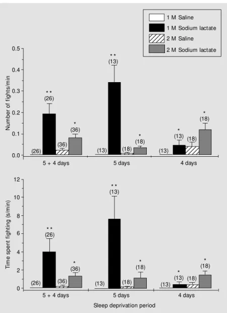
mean number of fights with the longest dura-tion was 2.98 g/kg. The highest doses of lactate and sodium chloride (3.72 g/kg of 2-M solutions) induced a behavioral depres-sion, followed by tremors, wild running and spontaneous convulsions in 20 rats, which eventually ended in death.
D iscussio n
The main results obtained in the present study show that the administration of so-dium lactate to rats significantly increases the number and duration of fights induced by sleep deprivation. This result supports the hypothesis that these fights might be mani-festations of panic, since lactate administra-tion, as mentioned before, precipitates panic attacks in PD patients. Such interpretation is supported by studies (13) addressing fight-ing induced by electrical shocks applied to the paws, which is quite similar to that ob-served in sleep-deprived rats, as well as panic. Ethological analysis to determine how fight-ing episodes start in DSD rats (13) has shown that the agonistic behavior is displayed only by those that quit self-cleaning and exhibit longer periods of behavioral freezing when removed from the deprivation cage, as seen in the present study. Freezing reactions, char-acterized by spontaneous movement arrest, hypertonic posture, and tremor, are indica-tive of extreme anxiety. Their occurrence in fighting DSD rats suggests a relationship between fighting and anxiety disorders. As seen before, an animal in a freezing posture shifts to the upright position in response to watchful stimuli (conspecific approach, acoustic stimuli, etc., although 20% start this behavior with no detectable environmental stimuli (13)), thereby inducing a similar re-sponse in nearby animals followed by con-frontation. Since no attempts to attack are seen during long-lasting confrontations, this may be interpreted as defensive behavior. Confrontation may also start when a behav-iorally frozen DSD animal, startled by a sudden conspecific shriek or other stimuli, triggers an episode of wild running flight. In this case, collision with mates generally induces confrontation simultaneously in two or more animals, often determining the on the top/on the back pattern of fighting, ei-ther because time is not enough to complete uprighting or because animals lose their
bal-N
u
m
b
e
r
o
f
fi
g
h
ts
/m
in
0.25
0.20
0.15
0.10
0.05
0.00
6
5
4
3
2
1
0
T
im
e
s
p
e
n
t
fi
g
h
ti
n
g
(
s
/m
in
)
(62) (62)
* *
(31)
* *
(31)*
(31)
(31)
5 + 4 days 5 days 4 days
Saline
Sodium lactate
(62)
(62) (31) (31)
(31) (31)
* *
* *
*
5 + 4 days 5 days 4 days
Sleep deprivation period
ance. The present classification of this con-frontation pattern - which accounts for al-most 20% of fighting episodes - as defensive seems to be correct since the characteristic bite threats aimed at the dorsal part of the opponent are absent, as is the characteristic tail rattling exhibited during offensive ag-gressiveness. The classification by Adams (24) is similar. In our previous study (12) of 139 DSD-induced fighting episodes, 105 proved to be defensive, whereas only three proved to be offensive using the criteria described in the literature (24-26). Defen-sive patterns of fighting in rats may be asso-ciated with panic (19,27,28), and hyperde-fensive fighting and flights induced in rats by intravenous administration of cocaine have been recently considered to be panic-like manifestations (29). On the basis of these considerations, it seems possible to interpret DSD-induced fighting as a manifestation of panic.
A parallel study in our laboratory (30) has shown that fighting after DSD is dis-played only by rats that respond with wild running flight to intense auditory stimula-tion in individual test chambers. This wild running flight response has also been shown to be facilitated by DSD and lactate adminis-tration, and impaired by imipramine admin-istration. Since this kind of flight is recog-nized as a manifestation of panic (28,29), the fighting behavior of DSD may be considered as the expression of the flight-or-fight re-sponse, which in turn is considered to be an expression of panic-related antipredatory defense (15,27).
Sleep disturbances in PD sufferers have been reported to be a consequence of anxiety disorders (31). The hypothesis that sleep deprivation may provoke panic manifesta-tions has been raised, with little emphasis, by Milasius et al. (32) for rabbits and by Labbate et al. (33) for humans, prior to ex-perimental evidence obtained by our group (13,14) showing a significant reduction of fighting in DSD rats by fluoxetine. In
agree-ment with these data, it has been docuagree-mented that sleep deprivation increases panic symp-toms in PD patients (34), who show in-creased partial pressure of CO2 (35), a con-dition recognized as being panicogenic (36). Although the above considerations
sup-Figure 2 - Fighting in sleep-deprived rats after lactate administration. M ean (± SEM ) number of fights per minute (upper graph) and time spent fighting per minute (low er graph) as a function of deprivation of desynchronized sleep time in rats that received sodium lactate solutions (black and gray columns) or NaCl (w hite and striped columns). The period of deprivation 5 + 4 (left columns) represents the combination of data obtained from sleep deprivation days 5 (center columns) and 4 (right columns). The number of animals is show n in parentheses above the bars of each group. * P<0.05 and * * P<0.01 for the comparison of sodium lactate vs saline w ith similar concentrations and w ithin the same deprivation period (M ann-Whitney U-test for the comparison of groups receiving 1-M solutions; Student t-test for the comparison of groups receiving 2-M solutions).
N
u
m
b
e
r
o
f
fi
g
h
ts
/m
in
0.5
0.4
0.3
0.2
0.1
0.0
12345 12345 12345 1234
1234 1234
1234
12345 12345 12345
1 M Saline
1 M Sodium lactate
2 M Sodium lactate 2 M Saline
(18)*
(13)* (18)
(13) (18)* (13)* *
(13) (18) (26)
(26)* *
(36) (36)*
1234 1234
1234 1234
T
im
e
s
p
e
n
t
fi
g
h
ti
n
g
(
s
/m
in
)
0 2 4 6 8 10 12
(26) (26)* *
(36) (36)
(13) (13)* *
(18) (18)*
(13)
(13)* (18) (18)*
5 + 4 days 5 days 4 days
5 + 4 days 5 days 4 days
Sleep deprivation period
port our conclusion that fighting after DSD possibly is a panic manifestation, sleep dep-rivation does not seem to be the main deter-minant cause, since not all sleep-deprived animals fight. Increased neuronal excitabil-ity resulting from DSD has been known since earlier investigations on the subject (37) and may be the facilitating factor in the main central panicogenic mechanism. What such mechanism is, however, is still unknown. Panic manifestations are concomitant with intense fear or terror and this emotional ex-perience is abolished after bilateral excision of the temporal lobes (Klüver-Bucy syn-drome). The amygdala nuclei, located in the temporal lobe, have been shown to be in-volved in the etiology of panic in humans (38). Fighting behavior is elicited in rats by experimental manipulations of the amygdala nuclei (15) and lactate and DSD may exert their facilitating action on these temporal lobe structures that permit a relation be-tween panic and fighting. Determination of the role played by such nuclei in DSD fight-ing seems to be the next important step for our line of investigation.
The possibility that lactate increases panic in sleep-deprived animals appears to have an important adaptive function under natural conditions since panic is an emotional re-sponse associated with defensive behavior patterns (15,27). The concentration of en-dogenous lactate - derived from the lactic acid of the anaerobic glycolysis - increases in high energy-demanding situations and fa-tigue. By triggering panic which enhances the defensive responses of fight or flight, lactate seems to provide the best conditions for the ultimate efforts to preserve life. Of all the numerous hypotheses proposed during the last 30 years for the mechanism of action of lactate (39), none has proved to be satis-factory (40). Nevertheless, lactate facilita-tion, along with the enhanced neural
excit-ability promoted by DSD, can explain the increase in the number and duration of panic fights when panic-sensitive animals are de-prived of sleep and given lactate. The occur-rence of seizures sometimes followed by death when higher doses are used seems to be a natural sequence of the increasing inten-sity of panic-inducing stimuli (30). Such manifestations are not seen clinically be-cause lactate is administered continuously and discontinued when the highest safe dose is reached. Since continuous administration was difficult in the conditions of the present experiment, a fixed dose was given in one or two injections.
As mentioned before, lactate administra-tion induces panic manifestaadministra-tions in PD pa-tients. The lactate-induced increase in fight-ing in DSD rats raises the question whether this behavioral manifestation in laboratory animals is equivalent to that seen in humans, and, by extension, whether it is possible to use it as an experimental model for PD. DSD-induced fighting fulfills some predic-tive and face validity criteria (many fighting episodes start without detectable stimuli, flu-oxetine reduces fighting, and lactate increases it); however, many neurochemical and neu-rophysiological parameters need to be inves-tigated in order to establish it as a reliable model of PD. In fact, the platform method used to promote DSD involves stress and fatigue (23). Panic fighting by DSD rats may be ascribed to stress or to demodulation resulting from excessive fatigue.
Re fe re nce s
1. Aserinsky E & Kleitman N (1953). Regu-larly occurring periods of eye motility, and concomitant phenomena, during sleep.
Science, 118: 273-274.
2. Dement W & Kleitman N (1957). Cyclic variations in EEG during sleep and their relation to eye movements, body motility and dreaming. Electroencephalography and Clinical Neurophysiology, 10: 291-296.
3. Timo-Iaria C, Negrão N, Schmidek WR, Hoshino K, M enezes CEL & Rocha CL (1970). Phases and states of sleep in the rat. Physiology and Behavior, 5: 1057-1062.
4. Hoshino K (1996). Food deprivation and hypothermia in desynchronized sleep-de-prived rats. Brazilian Journal of M edical and Biological Research, 29: 41-46. 5. Kubo C, Sogaw a H, Teshim a H &
Nakagaw a T (1992). The effects of starva-tion and sleep deprivastarva-tion on the immu-nological functions. Japanese Journal of Psychosomatic M edicine, 32: 13-20. 6. Kleitman N (1963). Sleep and
Wakeful-ness. The University of Chicago Press, Chicago.
7. Fishbein W & Gutw ein BM (1977). Para-doxical sleep and memory storage pro-cesses. Behavioral Biology, 19: 425-464. 8. Everson CA (1995). Functional
conse-quences of sustained sleep deprivation in the rat. Behavioural Brain Research, 69: 43-54.
9. M orden B, M ullens R, Levine S, Cohen H & Dement W (1968). Effect of REM sleep deprivation on the mating behavior of male rats. Psychophysiology, 5: 241 (Ab-stract).
10. Tufik S, Lindsey CJ & Carlini EA (1978). Does REM sleep deprivation induce a su-persensitivity of dopaminergic receptor in the rat brain? Pharmacology, 16: 98-105. 11. Sandrin M FN (1996). Sono de ratos
confi-nados em alta densidade populacional. M aster’s thesis, UNESP, Botucatu, SP, Brazil.
12. Sandrin M FN & Hoshino K (1999). Agres-sividade em ratos privados de sono: caracterização etológica dos confrontos agonísticos como padrões de comporta-mento defensivo. Revista de Etologia,1: 9-18.
13. M edeiros-Santille R (1998). Definição da agressividade induzida pela privação de sono na plataforma múltipla como mani-festação de pânico no rato albino macho. Doctoral thesis, UNESP, Botucatu, SP, Brazil.
14. Hoshino K, Sandrin M FN, M edeiros R, Lenneberg-Hoshino C & Tufik S (1998). Comportamento de pânico e generaliza-ção de brigas em ratos privados de sono.
Annals of the XIII Annual M eeting of the Federação de Sociedades de Biologia Ex-perimental, Caxambu, M G, Brazil, August 26-29, 58-59.
15. Graeff FG (1994). Neuroanatomy and neu-rotransmitter regulation of defensive be-haviors and related emotions in mam-mals. Brazilian Journal of M edical and Bio-logical Research, 27: 811-829.
16. Chertok L & Fontaine M (1965). Introduc-tion à une clinique psychosomatique vé-terinaire. In: Psychiatric Animale Deselee. Brouw er, Paris apud M edeiros-Santille R (1998). Definição da agressividade indu-zida pela privação de sono na plataforma múltipla como manifestação de pânico no rat o albino m acho. Doct oral t hesis, UNESP, Botucatu, SP, Brazil.
17. Jenck F, Broekkamp CLE & Van Delft AM L (1988). Serotonergic drugs on the panic response elicited by periaqueductal gray simulation in rats. Psychopharmacology, 96 (Suppl): 236 (Abstract).
18. Deakin JFW & Graeff FG (1991). 5-HT and mechanisms of defense. Journal of Psy-chopharmacology, 5: 305-315.
19. Graeff FG (1991). Neurotransmitters in the dorsal periaqueductal gray and animal models of panic anxiety. In: Briley M & File SE (Editors), New Concepts in Anxi-ety. CRC Press, New York, 288-307. 20. Pitts FM & M cClure JN (1967). Lactate
metabolism in anxiety neurosis. New Eng-land Journal of M edicine, 277: 1329-1336. 21. Liebow itz M R, Fyer AJ, Gorman JM , Dillon D, Appleby IL, Levy G, Anderson S, Levitt M , Palij M , Davies SO & Klein DF (1984). Lactate provocation of panic at-tacks. I. Clinical and behavioral findings.
Archives of General Psychiatry, 41: 764-770.
22. Cow ley DS & Arana GW (1990). The diag-nostic utility of lactate sensitivity in panic disorder. Archives of General Psychiatry, 47: 277-284.
23. M edeiros R, Lenneberg-Hoshino C, Hoshino K & Tufik S (1998). Neuroetho-logic differences in sleep deprivation in-duced by the single- and multiple-platform methods. Brazilian Journal of M edical and Biological Research, 31: 675-680. 24. Adams DB (1980). M otivational systems
of agonistic behavior in muroid rodents: comparative review and neural model.
Aggressive Behavior, 6: 295-346.
25. Eibl-Eibsfeldt I (1961). The fighting behav-ior of animals. Scientific American, 105: 112-122.
26. Blanchard RJ & Blanchard DC (1990). An ethoexperimental approach to the study of aggression. In: M orato S, Carobrez AP & Lima TCM (Editors), Neurosciences and Behavior. Faculdade de Filosofia, Ciências e Letras de Ribeirão Preto, Ribeirão Preto, SP, Brazil, 107-124.
27. Graeff FG, Silveira M CL, Nogueira RL, Audi EA & Oliveira RM W (1993). Role of the amygdala and periaqueductal gray in anxiety and panic. Behavioural Brain Re-search, 58: 123-131.
28. Griebel G, Blanchard DC & Blanchard RJ (1996). Predator-elicited flight responses in Sw iss-Webster mice: an experimental model of panic attacks. Progress in Neuro-Psychopharmacology and Biological Psy-chiatry, 20: 185-205.
29. Hebert M A, Blanchard DC & Blanchard RJ (1999). Intravenous cocaine precipitates panic-like flight responses and lasting hyperdefensiveness in laboratory rats.
Pharmacology, Biochemistry and Behav-ior,63: 349-360.
30. Paula HM G (1999). Comportamento de defesa antipredatória: possível partici-pação de mecanismos neurais convulsi-vos na etiologia das manifestações de pânico. M aster’s thesis, UNESP, Botu-catu, SP, Brazil.
31. Culebras A (1996). Clinical Handbook of Sleep Disorders. But t erw ort h-Heine-mann, Boston.
32. M ilasius AM , Grinevicius K-KA & Lapin IP (1990). Effect of quinolinic acid on w ake-fulness and sleep in the rabbit. Journal of Neural Transmission,82: 67-73. 33. Labbate LA, Johnson M R, Lydiard RB,
Braw manM intzer O, Emmanuel N, Craw -ford M , Kapp R & Ballenger JC (1997). Sleep deprivation in panic disorder and obsessive-compulsive disorder. Canadian Journal of Psychiatry, 42: 982-983. 34. Roy-Byrne PP, Uhde TW & Post RM
(1986). Effects of one night’s sleep depri-vation on mood and behavior in patients w ith panic disorder: comparison w ith de-pressed patients and normal controls. Ar-chives of General Psychiatry, 43: 895-899. 35. Craske M G & Barlow DH (1990). Noctur-nal panic: response to hyperventilation and carbon dioxide challenges. Journal of Abnormal Psychology, 99: 302-307. 36. Gorman JM , Fyer M R, Goetz R, Askanazi
patients w ith panic disorder. Archives of General Psychiatry, 45: 31-39.
37. Hoshino K (1972). Perturbações motoras induzidas pela lesão eletrolítica da forma-ção reticular m esencefálica em ratos privados de sono paradoxal. Doctoral the-sis, UNESP, Botucatu, SP, Brazil.
38. Shekhar A, Sajdyk TS, Keim SR, Yoder KK & Sanders SK (1999). Role of the basolat-eral amygdala in panic disorder. Annals of the New York Academy of Sciences, 877: 747-750.
39. Nutt D & Law son C (1992). Panic attacks: a neurochemical overview of models and
mechanisms. British Journal of Psychia-try, 160: 165-178.