Arterial blood gas analysis in two diferent
intra-hospital transport methods for postoperative
cardiac surgery patients
Gasometria arterial em dois diferentes métodos de transporte
intra-hospitalar no pós-operatório imediato de cirurgia cardíaca
INTRODUCTION
Extracorporeal circulation (ECC) might cause lung damage during the postoperative period of cardiac surgery, thus contributing to the mortality of patients due to physiological modiication of the acid-base and metabolic balance, increased inlammatory response, increased vascular permeability, increased pulmonary shunt or reduced lung compliance and gas exchange.(1-3) Most patients subjected to myocardial revascularization (MR) exhibit a reduction in lung compliance, whereas approximately one-third exhibit increased airway resistance. In half of patients presenting with comorbidities, the index of gas exchange decreases by 50%.(4) MR is associated with high rates (25 to 40%) of considerable postoperative complications.(5)
Disorders in the ventilation/perfusion ratio (V/Q ratio) cause a reduction in functional residual capacity (FRC) and hypoxemia. Age, body weight, left ventricular dysfunction and ECC are factors predictive of risk for hypoxemia; these factors could indicate the need for strategies such as positive end-expiratory pressure (PEEP) during transportation to reduce complications.(6-8) he state of a postoperative cardiac surgery patient upon his or her arrival at the coronary care unit (CCU) exerts a direct inluence on cost, the possibility of immediate ventilator weaning, and perhaps morbidity and mortally, especially when patients develop Newton Almeida Lima Junior1,6, Silvia Correa
Bacelar2,3, André Miguel Japiassú1,5, Samária Ali Cader2, Rosane Coelho Fernandes Lima4, Estélio Henrique Martin Dantas2, Alexandre Gomes Sancho6, Jefferson Braga Caldeira6
1. Instituto D’Or de Pesquisa e Ensino - IDOR - Rio de Janeiro (RJ), Brazil.
2. Laboratory of Human Mobility Biosciences, Universidade Federal do Estado do Rio de Janeiro - UNIRIO - Rio de Janeiro (RJ), Brazil. 3. Instituto Nacional do Câncer - INCA - Rio de Janeiro (RJ), Brazil.
4. Órtese, Prótese e Materiais Especiais - OPME - Unimed Rio - Rio de Janeiro (RJ), Brazil. 5.Instituto de Pesquisa Clínica Evandro Chagas, Fundação Oswaldo Cruz - FIOCRUZ - Rio de Janeiro (RJ), Brazil.
6. Universidade do Grande Rio - UNIGRANRIO - Rio de Janeiro (RJ), Brazil.
ABSTRACT
Objective: To evaluate the efects on blood gases by two methods of ventilation (with transport ventilation or self-inlating manual resuscitator) during intra-hospital transport of patients after cardiac surgery.
Methods: Observational, longitudinal, prospective, randomized study. Two samples of arterial blood were collected at the end of the surgery and another at the end of patient transport.
Results: We included 23 patients: 13 in the Group with transport ventilation and 10 in the Group with self-inlating manual resuscitator. Baseline characteristics were similar between both groups, except for
higher acute severity of illness in the Group with transport ventilation. We observed signiicant diferences in comparisons of percentage variations of gasometric data: pH (transport ventilation + 4% x MR -5%, p=0.007), PaCO2 (-8% x +13%, p=0.006), PaO2 (+47% x -34%, p=0.01) and SatO2 (+0.6% x -1.7%, p=0.001).
Conclusion: he use of mechanical ventilation results in fewer repercussions for blood gas analysis in the intra-hospital transport of cardiac surgery patients.
Keywords: Respiration, artiicial;
Transportation of patients; Blood gas analysis; Pulmonary gas exchange; Intensive care; Patient transfer
This study was conducted at the Hospital Quinta D’Or - Rio de Janeiro (RJ), Brazil.
Conflicts of interest: None.
Submitted on November 14, 2011 Accepted on March 28, 2012
Corresponding author:
Newton Almeida Lima Junior
Av. Prof. José de Souza Herdy, 1160 - Bairro 25 de Agosto - Duque de Caxias
more serious complications. Appropriate ventilation during and after surgery might improve the patient’s safety and could minimize metabolic and ventilatory repercussions that appear within a short period of time.(1,9,10)
Most of the studies that report physiological alterations during the transportation of severe patients were observational and associated such alterations with higher morbidity during stays in closed units.(11-13) he degree of arterial gas alteration during transportation of potentially severe patients using ventilatory methods (either by bag and mask ventilation or mechanical ventilator) has not been quantiied; this is a dynamic process that requires appropriate monitoring,
coordination, communication and equipment.(14,15) We
selected a population of patients in the postoperative period of cardiac surgery because it is a very homogeneous group that has been subjected to the inlammatory stimulus of ECC and is transported with tracheal intubation still in place to closed units after the end of the surgical procedure. Due to the scarcity of randomized controlled trials on this subject, we performed a systematic comparison of the blood gas alterations exhibited by patients transported using a transport ventilator (TV) and a self-inlating manual resuscitator (SMR).
METHODS
his was an prospective and randomized study (by random pickup of envelopes) conducted at a tertiary hospital. Intubated patients in the immediate postoperative period of cardiac surgery who were transported from the surgical center (SC) to the CCU were consecutively included. his study was approved by D’Or Network Research Ethics Committee (protocol nº 186/08). he participants and/or their caretakers were given all of the pertinent information and explanations of the aims and procedures of the study. hose that agreed to participate signed an informed consent form according to the Norms to Conduct Research on Human Beings, resolutions nº 196/96 and nº 404/2008 of the Brazilian National Health Council and the Declaration of Helsinki.
Two samples of arterial blood were obtained directly from the arterial line circuit using a 3 mL syringe previously lubricated with heparin (without any residual heparin) that was closed using the needle cap. he time between sample collection and obtaining the results of the blood gas analysis was no longer than 10 minutes.(16) A blood gas analyzer Radiometer ABL 5 (Reagentes/Eletrodos & Acessórios, São Paulo, SP, Brazil) was used for blood gas analysis. he irst sample for blood gas analysis was obtained before disconnection of the ventilator used during surgery, and the second one was obtained at the end of transportation before connection to the ventilator in the CCU. he allocation of
patients to the ventilatory methods to be investigated was performed randomly (by a simple envelope rale). Ventilation during transport was performed by the anesthesiologists. In the patients ventilated with SMR, the oxygen low varied between 5 and 6 L/min, which was transformed into the corresponding fraction of inspired oxygen (FiO2) according to a reference table.(17) In the patients ventilated with TV, FiO2 was 100%, and PEEP was maintained at 5 cm H2O according to the protocol established for this study.
he following data were collected from the clinical records: demographic information (age, gender, body mass index – BMI, Acute Physiology and Chronic Health Evaluation – APACHE II, European System for Cardiac Operative Risk Evaluation – EuroSCORE, length of hospitalization in the CCU and in the hospital) and surgical parameters (type, nature [emergency vs. elective] and length of surgery; length of ECC; and type of transport). he blood gas variables assessed in both groups were: arterial CO2 pressure (PaCO2), arterial O2 pressure (PaO2), base excess (BE), oxygen saturation (SaO2) and ratio of PaO2 to fraction of inspired O2 (PaO2/FiO2 ratio) before and after transport.
he main aim of this study was to establish whether the use of mechanical ventilation during the transport of patients after cardiac surgery could prevent the occurrence of blood gas alterations at the moment of arrival in the intensive care unit (ICU). Adopting a PaCO2 of 40 mmHg as a basis, we calculated a 20-patient sample to show 10% of alteration with 80% of study power, an error of 0.05 and 10% loss estimates. he numerical results were expressed as medians and interquartile intervals, whereas the categorical variables were expressed as absolute numbers and percentages. he statistical tests applied were selected as functions of the distribution of the data (Kolmogorov-Smirnov test). he Student’s t-test was used to compare numerical variables, and a chi-square test was used for categorical parameters. Statistical signiicance was established at p<0.05.
RESULTS
Although the patients subjected to TV exhibited greater severity, the length of surgery, ECC and transportation were similar in both groups. he TV group included a larger number of heart valve replacements (VRs) compared to the SMR group, although this diference was not statistically signiicant (6 versus 8 MRs and 7 versus 2 VRs, p=0.20). he nature of the surgery (elective versus emergency) was not signiicantly diferent between groups (7 versus 4 elective and 6 versus 6 emergency procedures, p=0.68) (Table 1).
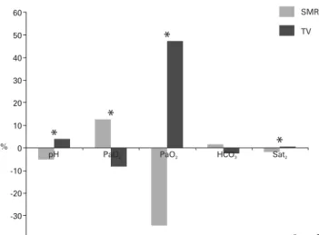
he patients in the TV group exhibited fewer blood gas alterations during transport compared to the SMR group, and their values were close to normal ranges upon arrival in the CCU. he arterial blood gas values prior to transportation were similar in both groups. he results of blood gas analysis after arrival in the CCU revealed signiicant diferences in regard to pH (TV 7.39 [7.36-7.43] versus SMR 7.29 [7.28 – 7.35], p=0.007), PaCO2 (TV 39 [36 - 44] versus SMR 49 [42 - 54] mmHg, p=0.006), PaO2 (TV 259 [224 - 349] versus SMR 173 [104 - 233] mmHg, p=0.01) and SaO2 (TV 96 [96 - 100] versus SMR 95 [94 - 95]%, p=0.001). Neither the bicarbonate levels nor the gas exchange ratios exhibited diferences (Table 2).
Comparisons between both groups in regard to the average blood gas values before and after transportation indicated that the pH, PaCO2, PaO2 and SaO2 levels were signiicantly diferent; the TV group presented better results (Figure 1). he percentages of variation in the SMR group before and after transportation were pH: -5%, paO2: -
34%, paCO2: +13%, HCO3: +1.6% and SaO2: -1.7%;
the corresponding values in the TV group were pH: +4%,
pO2: +47%, pCO2: -8%, HCO3: -2.5% and SatO2: +0.6%. Comparisons between the variations of the blood gas data
indicated signiicant diferences in pH (p=0.007), pO2
(p=0.01), pCO2 (p=0.006) and SatO2 (p=0.001) parameters, whereas the variations in bicarbonate levels were not diferent.
Figure 1 - Comparison between the averages of blood gas data before and after
transportation of the self-inflating manual resuscitator and transport ventilation groups.
SMR - self-inflating manual resuscitator group; TV - transport ventilator group. * - p<0.05.
DISCUSSION
Adverse efects of intra-hospital transportation causing impairment of respiratory function might occur more often among more severe patients or those exhibiting pre-transport lesions.(18) In this study, we were able to show that ventilation using a transport-speciic device caused less blood gas alterations
Table 2 – Comparison of blood gas analyses before and after transport of transport
ventilator and self-inflating manual resuscitator groups
Parameters Sample
collection time
TV (N=13) SMR (N=10) p
value
pH Pre-transport 7.36 (7.33-7.40) 7.37 (7.31-7.37) 0.17
Post-transport 7.39 (7.36-7.43) 7.29 (7.28-7.35) 0.007
PaCO2 Pre-transport 43.8 (38.3-47.2) 40.4 (39.4-45.0) 0.87 Post-transport 38.8 (35.7-44.0) 49.0 (42.4-54.1) 0.006
PaO2 Post-transport 184 (145.2-222.6) 213.5 (192-328.7) 0.06 Post-transport 259.3 (224.1-349.2) 173 (103.9-233.1) 0.01
HCO3 Pre-transport 23 (22.1-24.4) 22.2 (20.9-24.8) 0.35 Post-transport 22.4 (21.9-24) 22.7 (21-26.8) 0.71
BE Pre-transport -1.3 (-3-0) -2.9 (-3.7-0.3) 0.26
Post-transport -1 (-3--0.9) -3 (-4.4-0.3) 0.15
SaO2 Pre-transport 96.2 (94.5-99) 95 (94.9-95.3) 0.53
Post-transport 96 (95.7-100) 94.7 (94-94.9) 0.001
PaO2/FiO2 Pre-transport 188.6 (178-239.4) 332 (224.8-357.2) 0.08 Post-transport 259.3 (224.1-349.2) 432.5 (236.1-529.8) 0.11 TV – transport ventilator group; SMR – self-inflating manual resuscitator group; PaCO2 – arterial CO2 pressure; PaO2 – arterial O2 pressure; BE – base excess; SaO2 – O2 saturation; PaO2/FiO2 – ratio of PaO2 to fraction of inspired O2. Results are expressed as medians (minimum – maximum).
Table 1 – Demographic characteristics of the transport ventilator and self-inflating manual resuscitator groups
Characteristics TV (N=13) SMR (N=10) p value
Age 66 (55-72) 65 (59-69) 0.94
Male gender 8 7 0.98
BMI 29.3 (24.6-31.6) 25.9 (22.3-27.8) 0.08
LVEF (%) 60 (47-68) 60 (57-60) 0.96
APACHE II 16 (13-19) 12 (10-16) 0.03
EuroSCORE 7 (5-11) 3 (1-5) 0.02
∆t CCU 6 (4-10) 4 (4-6) 0.31
∆t H 12 (10-20) 9 (8-10) 0.09
Surgery type (MR/VR) 6/7 8/2 0.20
Nature (El/Em) 7/6 4/6 0.68
∆t surgery (min) 310 (290-355) 320 (295-355) 0.45
∆t ECC (min) 90 (75-100) 90 (80-106) 0.68
∆t transport (min) 10 (9-13) 8 (6-10) 0.62
compared to ventilation using SMR. Signiicant alterations in pH, oxygenation and retention of carbon dioxide were more frequent among patients transported with SMR.
he better results achieved in this study with the use of TV might be explained as a function of the advantages exhibited by the mechanical ventilator, which include continual monitoring of airway pressures, breathing rate, tidal volume given, PEEP and FiO2. Conversely, transportation using SMR with an oxygen reservoir involves variations in tidal volume and breathing rate and does not provide PEEP or safety parameters, such as peak pressure alarms or real FiO2 support. In addition, PEEP has the theoretical advantage of increasing oxygenation due to the expansion of collapsed alveoli, thus restoring the FRC and reducing the physiological shunt.(9)
Physiological alterations in severe patients during intra-hospital transport are a common occurrence. An observational study that assessed more than 3,000 clinical records identiied 59 adverse events (1.7%), most of which were related to hypoxia (25/59) or alterations in arterial blood pressure (25/59). Most of the interventions performed involved the adjustment of oxygen therapy (22/59) and the management of vasopressors (18/59). Because only 12 transport cases with adverse events (20%) needed to be aborted, one might conclude that the rate of clinically signiicant adverse events during transportation of patients is relatively low and needs little correction.(19) Although the sample in our study was small, no adverse events, such as accidental extubation or risk of cardiac arrest, were observed. he blood gas alterations identiied were reported to the team that received the patients in the CCU, and they were promptly treated by means of ventilation adjustment.
here are reports on respiratory impairment during and after transportation of severe patients.(20) Other studies observed that the use of SMR is acceptable when appropriate oxygen low is maintained.(21,22) Nevertheless, our results indicate that the use of a mechanical ventilator makes transportation safer, even when it is performed quickly, as it occurs after cardiac surgery. An observational study with 49 intubated patients with TV identiied ventilatory alterations in which 41 (84%) patients exhibited worse PO2/FiO2 ratios, and 21 (43%) experienced reductions of more than 20% compared to the baseline.(20) he efect on respiratory function lasted more than 24 hours in 10 patients (20%). Ventilation with positive pressure by means of PEEP exhibited signiicant correlation with fewer changes in PO2/FiO2 ratios after transportation. he authors further concluded that patients ventilated without PEEP are at higher risk for adverse events.(20) In turn, patients ventilated with high values of PEEP exhibit divergences between pulse oximetry and
arterial blood gas analyses.(23) Our study also shows this same efect in a randomized and controlled manner, along with fewer repercussions on gas exchange and CO2 retention when patients were ventilated with a mechanical ventilator.
During manual ventilation, the volume of air must be adjusted to the patient’s weight, the ventilation rate must be the same as that used during surgery, and the SMR reservoir must be able to supply FiO2 above 85%.(24) To guarantee further safety, the equipment should have a pressure manometer and a PEEP valve. However, these parameters are diicult to achieve, and hyper- or hypoventilation might occur even when ventilation is performed by experienced professionals.(25) Our results corroborate this possibility on the grounds of the diferences identiied, not only in regard to the PaCO2 level (as also reported in a pediatric study)(25) but also the oxygenation level (PaO2 and SaO2).
As a limitation of this study, we point to the fact that its design included sample size calculations to demonstrate diferences above 20% in the parameters directly related with blood gas analysis. he study power is not suicient to correlate our results with adverse events or outcomes, such as postoperative complications or mortality. In spite of randomization, the distribution of patients resulted in a greater level of severity in the TV group (a larger number of valve replacements and higher APACHE II and EuroSCORE assessments). his fact might reduce the power of our results. However, even with their theoretical disadvantage, the TV group exhibited a lower incidence of blood gas alterations. Finally, the methods used in this study could not be blinded because the anesthesiologists were informed as to the ventilatory method that had to be applied for transportation after the end of surgery.
CONCLUSION
Transportation using a mechanical ventilator causes fewer blood gas repercussions in patients after cardiac surgery. herefore, it should be the choice method for intra-hospital transportation of such types of patients.
RESUMO
Objetivo: Avaliar as repercussões gasométricas de dois mé-todos de ventilação (ventilador de transporte e ressuscitador manual autoinlável) durante o transporte intra-hospitalar de pacientes submetidos à cirurgia cardíaca.
Resultados: Foram incluídos 23 pacientes: 13 no Grupo ventilador de transporte e 10 no ressuscitador manual autoinlável. As características dos pacientes entre os grupos foram semelhantes, exceto pela maior gravidade no Grupo ventilador de transporte. Observaram-se diferenças signiicativas nas comparações das variações percentuais dos dados gasométricos: pH (VT: + 4% vs RMA: - 5%, p=0,007), PaCO2 (VT: - 8% vs RMA: + 13%, p=0,006), PaO2 (VT: + 47% vs RMA: - 34%, p=0,01) e SatO2
(VT: + 0,6% vs RMA: - 1,7%, p=0,001).
Conclusão: O uso de ventilador mecânico causa menor repercussão nos gases sanguíneos no transporte intra-hospitalar de pacientes após de cirurgia cardíaca.
Descritores: Respiração artiicial; Transporte de pacientes; Gasometria; Troca gasosa pulmonar; Terapia intensiva; Transferência de pacientes
REFERENCES
1. Luz HLM, Auler Júnior JOC. Temperatura e alterações no equilíbrio ácido-base de pacientes submetidos à cirurgia cardíaca com circulação extracorpórea, sob normotermia e hipotermia. Rev Bras Anestesiol. 2002;52(2):197-208.
2. Guizilini S, Gomes WJ, Faresin SM, Bolzan DW, Alves FA, Catani R, Buffolo E. Avaliação da função pulmonar em pacientes submetidos à cirurgia de revascularização do miocárdio com e sem circulação extracorpórea. Rev Bras Cir Cardiovasc. 2005;20(3):310-6.
3. Huang H, Yao T, Wang W, Zhu D, Zhang W, Chen H, Fu W. Continuous ultrafiltration attenuates the pulmonary injury that follows open heart surgery with cardiopulmonary bypass. Ann Thorac Surg. 2003;76(1):136-40. 4. Ambrozin ARP, Cataneo AJM. Aspectos da função pulmonar após
revascularização do miocárdio relacionados com risco pré-operatório. Rev Bras Cir Cardiovasc. 2005;20(4):408-15.
5. Almeida GF, Vegni R, Japiassú AM, Kurtz P, Drumond LE, Freitas M, et al. Complicações pós-operatórias de pacientes com dissecção de aorta ascendente tratados cirurgicamente. Rev Bras Ter Intensiva. 2011;23(3):304-11.
6. Szeles TF, Yoshinaga EM, Alencar W, Brudniewski M, Ferreira FS, Auler Júnior JOC, et al. Hypoxemia after myocardial revascularization: analysis of risk factors. Rev Bras Anestesiol. 2008;58(2):124-36.
7. Rock P, Rich PB. Postoperative pulmonary complications. Curr Opin Anaesthesiol. 2003;16(2):123-31.
8. Carvalho MRM, Silva NAS, Oliveira GMM, Klein CH. Complicações e tempo de internação na revascularização miocárdica em hospitais públicos no Rio de Janeiro. Rev Bras Ter Intensiva. 2011;23(3):312-20.
9. Scanlan CL, Wilkins RL, Stoller JK. Fundamentos da terapia respiratória de Egan. 9a ed. São Paulo: Manole; 2009.
10. Aoyagi T. Pulse oximetry: its invention, theory, and future. J Anesth. 2003;17(4):259-66.
11. Gervais HW, Eberle B, Konietzke D, Hennes HJ, Dick W. Comparison of blood gases of ventilated patients during transport. Crit Care Med. 1987;15(8):761-3.
12. Ashton CM, Petersen NJ, Wray NP, Kiefe CI, Dunn JK, Wu L, Thomas JM.
The incidence of perioperative myocardial infarction in men undergoing noncardiac surgical. Ann Intern Med. 1993;118(7):504-10.
13. Doherty GM, Way LW. Current surgical diagnosis and treatment. 12th ed. New York: McGraw-Hill; 2006.
14. Jensen LA, Onyskiw JE, Prasad NG. Meta-analysis of arterial oxygen saturation monitoring by pulse oximetry in adults. Heart Lung. 1998;27(6):387-408.
15. Warren J, Fromm RE Jr, Orr RA, Rotello LC, Horst HM; American College of Critical Care Medicine. Guidelines for the inter- and intrahospital transport of critically ill patients. Crit Care Med. 2004;32(1):256-62.
16. Viegas CAA. Gasometria arterial. J Pneumol. 2002;28(Supl 3):S233-8. 17. David CM. Ventilação mecânica: da fisiologia à prática clínica. 2a ed. Rio
de Janeiro: Revinter; 2011.
18. Kanter RK, Tompkins JM. Adverse events during interhospital transport: physiologic deterioration associated with pretransport severity of illness. Pediatrics. 1989;84(1):43-8.
19. Kue R, Brown P, Ness C, Scheulen J. Adverse clinical events during intrahospital transport by a specialized team: a preliminary report. Am J Crit Care. 2011;20(2):153-61; quiz 162.
20. Waydhas C, Schneck G, Duswald KH. Deterioration of respiratory function after intra-hospital transport of critically ill surgical patients. Intensive Care Med. 1995;21(10):784-9.
21. Shulman M, Schmidt G, Sadove MS. Evaluation of oxygen therapy devices by arterial oxygen tensions. Dis Chest. 1969;56(4):356-9.
22. Lahner D, Nikolic A, Marhofer P, Koinig H, Germann P, Weinstabl C, Krenn CG. Incidence of complications in intrahospital transport of critically ill patients--experience in a Austrian university hospital. Wien Klin Wochenschr. 2007;119(13-14):412-6.
23. Secker C, Spiers P. Accuracy of pulse oximetry in patients with low systemic vascular resistance. Anaesthesia. 1997;52(2):127-30.
24. Pereira Júnior GA, Carvalho JB, Ponte Filho AD, Malzone DA, Pedersoli CE. Transporte intra-hospitalar do paciente crítico. Medicina (Ribeirão Preto). 2007;40(4):500-8.