Abstract
Objective: To evaluate the peak inspiratory pressure, tidal volume and respiratory rate achieved during manual ventilation of premature lambs, using a self-inflating bag.
Methods: In this descriptive, experimental study, five pairs of physicians, selected at random among 35 neonatologists working at a neonatal intensive care unit and with experience in the resuscitation of newborn infants, ventilated five intubated premature lambs using a self-inflating bag. Pressure and flow monitor signals were passed through a transducer and digitized for recording and analysis. Tidal volume and pressure curves were obtained from the integral of flow rate, at peak, during the last 50 seconds of every fifth minute, and analyzed.
Results: Median pressure was 39.8 (IQ25-75% 30.2-47.2) cmH2O; being below 20 in 1.1% of cases and above 40 in 49.1%. Seven out of 10 physicians produced more than six pressure peaks of over 40 cmH2O. Median tidal volume/kg was 17.8 (IQ25-75% 14.1-22.4) mL, being below 5 mL in 0.1% of cases and greater than or equal to 20 mL in 37.7%. All of the physicians propelled five or more ventilation cycles with tidal volume/kg of 20 mL or more. Respiratory rate was between 30 and 60 cycles/minute in 65.9% of cases, being below 30 in 6.8% of cases and over 60 in 27.3% of cases.
Conclusions: There was major variation in peak inspiratory pressure and tidal volume/kg values, which were in many cases elevated, attaining levels that habitually cause biotrauma, while respiratory rates were adequate in the majority of cases.
J Pediatr (Rio J). 2006;82(4):279-83: Cardiopulmonary resuscitation, mechanical ventilation, neonate, neonatal asphyxia, tidal volume.
Evaluation of peak inspiratory pressure, tidal volume
and respiratory rate during ventilation
of premature lambs using a self-inflating bag
Jefferson G. Resende,1 Carlos A. M. Zaconeta,2 Antonio C. P. Ferreira,3 César A. M. Silva,4 Marcelo P. Rodrigues,5 Celso M. Rebello,6 Paulo Tavares7
O
RIGINALA
RTICLE1. Médico pediatra. Doutorando em Ciências Médicas, Universidade de Brasília (UnB), Brasília, DF, Brasil.
2. Médico neonatologista. Mestre, UnB, Brasília, DF, Brasil.
3. Médico intensivista pediátrico. Mestre, Universidade Federal de São Paulo (UNIFESP), São Paulo, SP, Brasil.
4. Fisioterapeuta. Mestre, UnB, Brasília, DF, Brasil. Fisioterapeuta, Hospital Universitário da UnB, Brasília, DF, Brasil.
5. Médico pneumologista. Mestre, UnB, Brasília, DF, Brasil. Professor, UnB, Brasília, DF, Brasil.
6. Médico. Doutor, Faculdade de Medicina, Universidade de São Paulo (USP), São Paulo, SP, Brasil. Coordenador, Unidade de Pesquisa Experimental, Instituto da Criança do Hospital das Clínicas, Faculdade de Medicina, USP, São Paulo, SP, Brasil.
7. Médico pneumologista. Doutor. Professor emérito, UnB, Brasília, DF, Brasil.
Financial support: This study was carried out at the Laboratory of Respiratory Apparatus Physiology, at the Universidade de Brasília (UnB), in Brasília, DF, with the cooperation of the Ovine Obstetrics Department, at the UnB Veterinary Medical Faculty. Radiant warmers were provided by Olidef CZ and resources belonging to the authors were also employed. This study is part of Jefferson Guimarães de Resende’s doctoral thesis, entitled “Evaluation of arterial gasses and lung injuries during ventilation of premature lambs, comparing two manual ventilation devices”.
Manuscript received Ago 25 2005, accepted for publication May 10 2006.
Suggested citation: Resende JG, Zaconeta CA, Ferreira AC, Silva CA, Rodrigues MP, Rebello CM, et al. Evaluation of peak inspiratory pressure, tidal volume and respiratory rate during ventilation of premature lambs using a self-inflating bag. J Pediatr (Rio J). 2006;82:279-83.
Introduction
Around 10 million newborn infants require some type of resuscitation every year globally and more than a million die from perinatal asphyxia complications.1
Self-inflating bags are the devices most commonly employed for manual mechanical ventilation, being recommended on the neonatal resuscitation course run by the Brazilian Society of Pediatrics.2 Research has demonstrated that
using bags can result in variations in tidal volume (Vt) and peak inspiratory pressure (PIP) during each inflation of the lungs.3 Mondolfi et al.4 observed large variations in Vt, PIP
and minute volume obtained by health professionals on a pediatric resuscitation dummy. Baskett et al.5 measured
the Vt considered adequate during resuscitation, based on thoracic expansion, and observed that it varied. Björklund et al.6 demonstrated that six large-volume breaths at the
start of ventilation of premature lambs reduced the response to surfactant given during the immediate neonatal period. Ingimarsson et al.7 demonstrated that when
premature lambs were subjected to five pulmonary inflations at a Vt of 20 mL/kg before receiving exogenous
Copyright © 2006 by Sociedade Brasileira de Pediatria doi:10.2223/JPED.1517
surfactant, this affected the intrapulmonary distribution of the surfactant, resulting in very uneven deposition, with almost all of the medication distributed to the dependent areas of the lungs. Wada et al.8 observed that the
application of large Vt to the lungs of premature lambs, soon after birth, can cause lung injuries and compromise response to exogenous surfactant.
The objective of this study is to evaluate the performance of physicians carrying out manual mechanical ventilation on premature lambs, using a self-inflating bag, through studying the PIP, Vt and respiratory rate (RR) they produced.
Materials and methods
This was a descriptive, experimental study, carried out at the Universitys Respiratory Physiology Laboratory.
Five premature lambs, of both sexes, carried by healthy ewes, were included on the study. Any major malformations observed in any of the animals at any phase of the study resulted in its exclusion.
Ten neonatologists were chosen from a list of 35, considered experienced professionals because they work at public and/or private neonatal intensive care units (ICU), by means of a list of random numbers. Neonatologists were defined as experienced if they were not recently-qualified, had completed residency in pediatrics and/or neonatology and were currently active in the field and familiar with neonatal resuscitation. All participating physicians signed free and informed consent forms.
Physicians were grouped into pairs at random and each pair ventilated one premature lamb for a 45-minute period. Pairs were used in order to allow ventilation to be performed in relays in order to reduce the risk of tiredness interfering with the results of the procedure. Thus, each physician ventilated a lamb for 20 or 25 minutes, alternating every 5 minutes.
The ewes and lambs were dealt with as described by Wada et al.8 Summing up, at 132 (±1) days gestational
age (full term is 150 days) each pregnant ewe was pre-anesthetized with 1 g of intramuscular ketamine, and given an epidural anesthetic with 10 mL of lidocaine at 2% and bupivacaine at 0.5% in a proportion of 1:1 (volume to volume). Each ewe was intubated using an endotracheal tube (EDT) in order to improve control over ventilation. The lamb was removed by caesarian, weighed and put under a radiant heat source (BA Matrix SC, Olidef CZ, Ribeirão Preto, SP, Brazil). The lamb was then dried and direct laryngoscopy was performed; the trachea was suctioned to remove excess alveolar liquid, then an EDT was inserted (number 4.0 or 4.5, depending on which was more appropriate for each lamb), and ventilation begun.
The EDT cuff was inflated in order to eliminate air leaks via the outer wall. Ketamine (10 mg/kg) and acepromazine (0.1 mg/kg) were given intramuscularly in order to allow total control over ventilation.
Each recently-delivered animal was catheterized via both the umbilical artery and the umbilical vein, with the tips of the catheters positioned within the inferior vena cava and thoracic aorta, respectively. These deep accesses allowed 5% glucose to be infused at a rate equivalent to 100 mL/kg/day, saline to be infused when necessary to correct arterial hypotension and for gasometry (arterial) to be performed and systolic and diastolic arterial pressure, in addition to central venous pressure, to be controlled. Control and recording of pressures was performed by means of a polygraph (Model 7C, Grass® Instrument Co.,
Quincy, MA, USA), via two transducers (P23Db Gould Statham®, USA).
Between the bag and the tracheal cannula there was a lateral connection and a pneumotachograph (Fleisch nº 0). The pneumotachograph was coupled to a differential transducer (PT5A, Grass, Quincy, MA, USA), which allowed the airflow to be measured and recorded, which in turn allowed volume to be calculated. The lateral connection was coupled to an absolute pressure transducer (Statham, Gould, USA), which allowed tracheal pressure (PIP) to be measured and recorded.
All of the transducers were connected to the polygraph, where signals were filtered and amplified. From the polygraph the signals were passed to a biological signal conditioning module, designed for the measurement of ventilatory mechanics (Emgsystem do Brazil, São José dos Campos, São Paulo, Brazil), and from there they were sent to a microcomputer where, by means of an analogue to digital converter (CAD-1232, Lynx Tecnologia Eletrônica, São Paulo, SP, Brazil), the signals were digitized for storage and later analysis. The software employed for storage and analysis of the signals was AqDados 5 for Windows®. A sample rate of 200 Hz was used throughout
the experiment.
The self-inflating bag was new, Lifesaver® brand
(Hudson RCI®, Temecula, CA, USA), neonatal size, 280
mL capacity, with a low-complacency reserve bag to allow 100% FiO2. The oxygen supply had an output pressure of 3.5 kgf/cm2, and measured oxygen flow was 10 L/min,
always maintaining gasses in the low-complacency reserve bag. The pressure release valve was stopped for purposes of the study.
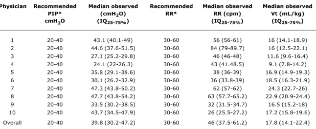
Table 1 - Peak pressure, respiratory rate and tidal volume for each participant and for the whole group
PIP = peak inspiratory pressure; RR = respiratory rate, in cycles/minute; Vt = tidal volume (mL/kg). * ILCOR9
Figures are expressed in medians with their respective 25-75% interquartile ranges.
Physician Recommended Median observed Recommended Median observed Median observed
PIP* (cmH2O) RR* RR (cpm) Vt (mL/kg)
cmH2O (IQ25-75%) (IQ25-75%) (IQ25-75%)
1 20-40 43.1 (40.1-49) 30-60 56 (56-61) 16 (14.1-18.9)
2 20-40 44.6 (37.6-51.5) 30-60 84 (79-89.7) 16 (12.5-22.1)
3 20-40 27.1 (25.2-29.8) 30-60 46 (46-48) 11.6 (9.6-16.4)
4 20-40 24.1 (22-26.3) 30-60 43 (41.48.5) 9.1 (7.8-14.2)
5 20-40 35.8 (29.1-38.6) 30-60 38 (36-39) 16.9 (14.9-19.3)
6 20-40 30.1 (26.2-32.9) 30-60 36 (33.8-39) 18.5 (16.3-21.9)
7 20-40 47.3 (43.8-50.2) 30-60 62 (57-62) 24.3 (22.7-26)
8 20-40 47.7 (43.8-54.2) 30-60 63 (57.7-65.2) 22.9 (20.9-24.4)
9 20-40 33.5 (30.2-38.5) 30-60 32 (31.5-34.7) 16.5 (15.2-18)
10 20-40 43.7 (34.5-47.9) 30-60 26 (25.5-27.2) 17.2 (15.8-19.6)
Overall 20-40 39.8 (30.2-47.2) 30-60 46 (37.5-61.2) 17.8 (14.1-22.4)
Data were collected and stored continuously, with traces of pressure and airflow against time being recorded. The exact last 50 seconds of every fifth minute were used for data analysis. The RR per minute was obtained by multiplying the number of cycles analyzed during 50 seconds by 1.2. The Vt/kg was calculated by dividing the Vt on each flow curve by the weight of the animal.
Excel® (Microsoft®) and SPSS® were used to evaluate
the data.
The study protocol was submitted to the Animal Research Ethics Committee at the University, which gave its approval.
Results
Median PIP was 39.8 cmH2O (IQ25-75% 30.2-47.2). In 1.1% of cases PIP below 20 cmH2O was recorded and in 49.1% it was above 40. Seven out of 10 physicians produced more than six PIP above 40 cmH2O. Median Vt/kg was 17.8 mL (IQ25-75% 14.1-22.4), being below 5 mL in 0.1% of cases and greater than or equal to 10 mL in 90.2% and greater than or equal to 20 mL in 37.7%. All of the physicians propelled five or more ventilation cycles with Vt/kg at 20 mL or more. The RR was between 30 and 60 cycles/minute in 65.9% of cases, being fewer than 30 in 6.8% and more than 60 in 27.3%. Table 1 presents the medians and interquartile (IQ) ranges for each physician, together with overall results.
Discussion
In this experiment 1,872 PIP and Vt curves were analyzed, demonstrating that there was major variation in the parameters investigated. Both pressure and Vt/kg reached elevated levels compared with those suggested in manual ventilation guides. In 49.1% of cases the physicians ventilated the lambs at PIP of over 40 cmH2O. Both the ILCOR9 and the Brazilian Society of Pediatrics neonatal
resuscitation course2 suggest that maximum PIP should
be 40 cmH2O. Varying ventilatory pressures with bags have been reported before; Mondolfi et al.4 demonstrated
mean PIP of 35±19 cmH2O, using a pediatric resuscitation dummy. Our study shows, as can be observed in the tabulated results, that all of the physicians produced variations in pressure and Vt/kg.
The limitations of self-inflating bags as ventilation instruments are already well documented.10-15 For
example, a study carried out by Barnes et al.14 investigated
the performance and safety of 10 disposable manual resuscitators, four of which were for pediatric use, and concluded that just one of the pediatric systems and three of the adult ones met all of the requirements of the F920-93 ASTM standard (American Society for Testing and Materials).16
For this experiment the decision was taken to stop the pressure-release valve. This type of valve is preset by the manufacturer, according to F920-93,16 so that pressure is
model. These valves sometimes fail however,10,15 and,
even when they have been tested they can still release pressure at levels other than the recommended ones. Work done by Finer et al.,15 for example, studying three
different brands of neonatal bag, found that the valves opening pressures varied from 41 to 72 cmH2O for one bag, from 51 to 97 cmH2O for another and, from 38 to 106 cmH2O for the third bag. These pressures are very different from those that the manufacturers had stated that the valves had been calibrated to (35 cmH2O, 43-60 cmH2O and 40 cmH2O, respectively).
Even with the bags pressure-release valve stopped, in 49% of cases the physicians were unable to avoid passing the maximum pressure limit recommended by ILCOR,9
and 70% ventilated more than six times with pressure over 40 cmH2O, taking into account only 50 seconds of every 5 minutes. It is possible to speculate that the presence of a functioning pressure release valve would have made the physicians pass the 40 cmH2O PIP mark more often, if the valve was set to 45 cmH2O (still within the limits set by ASTM), since they would have had the safety valve as a security factor, alleviating them from the responsibility to concern themselves with limiting pressure.
Median Vt/kg was 17.8 mL (IQ25-75% 14.1-22.4), being greater than or equal to 10 mL in 90.2% and greater than or equal to 20 mL in 37.7%. Mondolfi et al.4 also
identified large variations in volume: mean of 25±10 mL (limits from 3 to 103 mL). The reasons for this variability are dependent not only on ventilation pressure, but also on the complacency of the respiratory system.
The potential for damage of levels like those observed here has already been demonstrated in the literature. Wada et al.8 found that, in premature lambs, Vt of
10 mL/kg during the first 30 minutes after birth resulted in compromise to ventilatory mechanics and gaseous exchange during the first 6 hours of pulmonary ventilation even when given surfactant, when compared with lambs that were ventilated for the same period but had been ventilated at 30 minutes with Vt of 5 mL/kg. In the present experiment, in 90.2% of cases, the physicians produced a Vt/kg greater than or equal to 10 mL. Research by Ingimarsson et al.7 has demonstrated that five pulmonary
inflations with high tidal volumes (20 mL/kg) given to premature lambs affects the intrapulmonary distribution of exogenous surfactant given afterwards. In the current study, all of the physicians produced at least five ventilation cycles with Vt/kg of 20 mL or more.
The appropriateness of Vt is habitually monitored by means of chest expansion observation. However, as Baskett et al.5 have demonstrated, the observation of
thoracic expansion does not appear to be sufficient to achieve adequate Vt, at least in adults. Hird et al.17 applied
pressure until premature newborn infants achieved what they considered to be adequate thoracic expansion, finding
that this varied from 14 to 30 cmH2O, and was not correlated with the weight of the preterms nor with their gestational age. The PIP and Vt/kg levels observed in the current experiment demonstrate that the physicians had different points of view as to what equated to adequate thoracic expansion.
There is consensus that, Physical expansion of the lungs, with establishment of functional residual capacity, and increase in alveolar oxygen tension both mediate the critical decrease in pulmonary vascular resistance and result in an increase in pulmonary blood flow after birth (...) failure to adequately expand alveolar spaces may result in intrapulmonary shunting of blood with resultant hypoxemia.9 Published data6-8 indicate that there is a
limit, possibly a very fine one, at which adequacy is defined, since this must be very exact so as to avoid passing the exact point at which the lungs are able to act as media for gaseous exchange, without compromising their function in the near future. The aim is to achieve permanently adequate gaseous exchange. The application of elevated pressures over prolonged periods has been suggested18 as a guarantee that increased residual
functional capacity is acquired rapidly, improving oxygenation, being rapidly incorporated into clinical practice as the ideal. Nowadays, this practice is no longer universally recommended,9 and research by Björklund et al.,6
Ingimarsson et al.7 and Wada et al.8 warns of the dangers.
The literature demonstrates concern with the fact that bronchopulmonary dysplasia could be a disease that has its origin in the delivery room, caused by extremely aggressive ventilation, causing hypocapnia and volutrauma.1,19,20
The physicians performed best at maintaining RR, remaining within the recommended range in 65.9% of cases.
This experiment has its limitations. The conditions under which the physicians performed did not have the same stress as when treating a human neonate, and the figures observed here could even be better than those that would be found in a real situation. The number of animals and the number of physicians involved could be considered small. Since this was an animal experiment, two issues are raised: the animal most often employed as a model of the human neonate is the lamb,6-8,21,22 and research with this
delineation could not be carried out with humans due to ethical limitations. The number of animals per group varies from four to eight in several studies of recognized quality.6-8,21,22 In the current experiment, the number of
References
1. Wiswell TE. Neonatal resuscitation. Respir Care. 2003;48:288-94. 2. Sociedade Brasileira de Pediatria. Manual do curso de reanimação neonatal. São Paulo: Universidade Federal de São Paulo Escola Paulista de Medicina; 1996. [Traduzido do inglês. Copyright by American Heart Association.]
3. Barnes TA. Emergency ventilation techniques and related equipment. Respir Care. 1992;37:673-90; discussion 690-4. 4. Mondolfi AA, Grenier BM, Thompson JE, Bachur RG. Comparison
of self-inflating bags with anesthesia bags for bag-mask ventilation in the pediatric emergency department. Pediatr Emerg Care. 1997;13:312-6.
5. Baskett P, Nolan J, Parr M. Tidal volumes which are perceived to be adequate for resuscitation. Resuscitation. 1996;31:231-4. 6. Björklund LJ, Ingimarsson J, Curstedt T, John J, Robertson B,
Werner O, et al. Manual ventilation with a few large breaths at birth compromises the therapeutic effect of subsequent surfactant replacement in immature lambs. Pediatr Res. 1997;42:348-55. 7. Ingimarsson J, Björklund LJ, Curstedt T, Jonson B, Larsson A, Robertson B, et al. Uneven distribution of exogenous surfactant after hyperinflation of the lungs at birth in immature lambs.
Correspondence:
Jefferson Guimarães de Resende
SHIN QL 10, conjunto 1, casa 14, Lago Norte CEP 71525-015 Brasília, DF Brazil Tel.: +55 (61) 3368.4665
Fax: +55 (61) 3386.7481 E-mail: [email protected] neonatologists do not represent the totality of experienced
neonatologists. We believe that the technology employed in the experiment was of a sufficient level of sophistication to ensure reliability of data; however, the data published here, together with the conclusions drawn, should be treated with the reservations appropriate to experimental studies with animals.
The results of this study indicate that physicians, even experienced and well-trained ones, are not necessarily capable of successfully ventilating premature lambs employing a self-inflating bag in accordance with internationally established standards. There was great variation in PIP and Vt/kg, both of which were often elevated and which reached levels that habitually cause biotrauma. In the majority of cases, the RR achieved was adequate.
Acknowledgements
The authors would like to thank Hercilia Maria Nogueira de Resende and Patrícia Carvalho Baião Câmara for revising the texts.
Conflict of interest
Jefferson Guimarães de Resende is the inventor and patent holder of a mechanical pulmonary ventilation system.
2001 Pediatric Academic Societies Annual Meeting; 2001 Apr 28-May 1; Baltimore, Maryland: US. Abstract 2200.
8. Wada K, Jobe AH, Ikegami M. Tidal Volume effects on surfactant treatment responses with the initiation of ventilation in preterm lambs. J Appl Physiol. 1997;83:1054-61.
9. Kattwinkel J, Niermeyer S, Nadkarni V, Tibballs J, Phillips B, Zideman D, et al. An advisory statement from the Pediatric Working Group of the International Liaison Committee on Resuscitation Pediatrics. 1999;103:e56. http://www.pediatrics. org/cgi/content/full/103/4/e56.
10. Connors R, Kisson N, Tiffin N, Frewer TC. An evaluation of the physical and functional characteristics of infant resuscitators. Pediatr Emerg Care. 1993;9:104-7.
11. Mills PJ, Baptiste J, Preston J, Barnas GM. Manual resuscitators and spontaneous ventilation an evaluation. Crit Care Med. 1991;19:1425-31.
12. Martell RJ, Soder CM. Laerdal infant resuscitators are unreliable as free-flow oxygen delivery devices. Am J Perinatol. 1997;14:347-51.
13. Hess D, Hirsch C, Marquis-DAmico C, Kacmarek RM. Imposed work and oxygen delivery during spontaneous breathing with adult disposable manual ventilators. Anesthesiology. 1994;81: 1256-63.
14. Barnes TA, McGarry WP 3rd. Evaluation of ten disposable manual resuscitators. Respir Care. 1990;35:960-8.
15. Finer NN, Barrington KJ, Al-Fadley F, Peters KL. Limitations of self-inflating resuscitators. Pediatrics 1986;77:417-20. 16. American Society for Testing and Materials. Standard specification
for minimum performance and safety requirements for resuscitators intended for use with humans. ASTM International. 1993;F 920-93.
17. Hird MF, Greenough A, Gamsu HR. Inflating pressures for effective resuscitation of preterm infants. Early Hum Dev. 1991;26:69-72.
18. Vyas H, Milner AD, Hopkin IE, Boon AW. Physiologic responses to prolonged and slow-rise inflation in the resuscitation of the asphyxiated newborn infant. J Pediatr. 1981;99:635-9. 19. Sharek PJ, Baker R, Litman F, Kaempf J, Burch K, Schwarz E, et
al. Evaluation and development of potentially better practices to prevent chronic lung disease and reduce lung injury in neonates. Pediatrics. 2003;111:e426-31. http://pediatrics.aappublications. org/cgi/reprint/111/4/SE1/e426.
20. Burch K, Rhine W, Baker R, Litman F, Kaempf JW, Schwarz E, et al. Implementing potentially better practices to reduce lung injury in neonates. Pediatrics. 2003;111:e432-6. http:// pediatrics.aappublications.org/cgi/reprint/111/4/SE1/e432. 21. Naik AS, Kallapur SG, Bachurski, Jobe AH, Michna J, Kramer BW,
et al. Effects of ventilation with different positive end-expiratory pressures on cytokine expression in the preterm lamb lung. Am J Respir Crit Care Med. 2001;164:494-8.