Ventilation strategy and its influence on the functional
performance of lung grafts in an experimental model of
single lung transplantation using non-heart-beating donors*
Influência da estratégia ventilatória no desempenho funcional de enxertos pulmonares em um modelo experimental de transplante pulmonar
unilateral de doadores após parada cardiocirculatória
Elaine Aparecida Felix, Cristiano Feijó Andrade, Paulo Francisco Guerreiro Cardoso, Gabriela Cury Thiesen,
Ana Carolina Peçanha Antonio, Lucas Krieger Martins, Tiago Antonio Tonietto
Abstract
Objective: To compare the influence of two different ventilation strategies—volume-controlled ventilation (VCV) and pressure-controlled ventilation (PCV)—on the functional performance of lung grafts in a canine model of unilateral left lung transplantation using donor lungs harvested after three hours of normothermic cardiocirculatory arrest under mechanical ventilation. Methods: The study comprised 40 mongrel dogs, randomized into two groups: VCV and PCV. Of the 20 recipients, 5 did not survive the transplant, and 5 died before the end of the post-transplant assessment period. The remaining 10 survivors (5 in each group) were evaluated for 360 min after lung transplantation. The functional performance of the grafts was evaluated regarding respiratory mechanics, gas exchange, and lung graft histology. Results: There were no significant differences between the groups regarding respiratory mechanics (peak inspiratory pressure, plateau pressure, mean airway pressure, dynamic compliance, and static compliance) or gas exchange variables (PaO2, venous oxygen tension, PaCO2, venous carbon dioxide tension,
and the arterial-venous oxygen content difference). The histopathological findings were consistent with nonspecific acute lung injury and did not differ between the groups. Conclusions: This model of lung transplantation showed that the functional performance of lung grafts was not influenced by the ventilation strategy employed during the first six hours after reperfusion.
Keywords: Pulmonary ventilation; Respiration, artificial; Lung transplantation; Dogs; Organ preservation.
Resumo
Objetivo: Comparar a influência de duas estratégias ventilatórias — ventilação controlada a volume (VCV) e ventilação controlada a pressão (VCP) — no desempenho funcional de enxertos pulmonares em um modelo canino de transplante pulmonar unilateral esquerdo, utilizando-se doadores cujos pulmões foram captados após três horas de parada cardiocirculatória em temperatura ambiente e sob ventilação mecânica. Métodos: O estudo incluiu 40 cães mestiços randomizados nos grupos VCV e VCP. Dos 20 receptores, 5 não sobreviveram ao transplante, e 5 não sobreviveram ao período de avaliação pós-transplante. Os 10 receptores sobreviventes (5 em cada grupo) foram avaliados durante 360 min após o término do transplante pulmonar. O desempenho funcional dos enxertos foi estudado através da avaliação da mecânica respiratória, trocas gasosas e histologia do enxerto. Resultados: Não houve diferenças significativas entre os grupos quanto às variáveis de mecânica respiratória (pressão de pico inspiratória, pressão de platô, pressão média de vias aéreas, complacência dinâmica e complacência estática) e de trocas gasosas (PaO2, pressão venosa mista de oxigênio, PaCO2, pressão venosa mista de CO2 e diferença arteriovenosa de oxigênio). As alterações histopatológicas foram compatíveis com o padrão de lesão pulmonar aguda não específica e não diferiram entre os grupos. Conclusões: Este modelo de transplante pulmonar mostrou que o desempenho funcional do enxerto não foi influenciado pela estratégia ventilatória utilizada até seis horas após a reperfusão.
Descritores: Ventilação pulmonar; Respiração artificial; Transplante de pulmão; Cães; Preservação de órgãos.
* Study carried out in the Jean Carlo Kohmann Laboratory of Experimental Surgery, Postgraduate Program in Medical Sciences, Federal University of Health Sciences of Porto Alegre, Porto Alegre, Brazil, and as part of the Postgraduate Program in Pulmonology, Federal University of Rio Grande do Sul, Porto Alegre, Brazil.
Correspondence to: Paulo F. Guerreiro Cardoso. Instituto do Coração/HCFMUSP, Disciplina de Cirurgia Torácica, Rua Dr. Enéas de Carvalho Aguiar, 44, bloco 2, sala 9, Cerqueira Cesar, CEP 05403-000, São Paulo, SP, Brasil.
Tel 55 11 3069-5248. E-mail: [email protected]
Financial support: This study received financial support from Vitrolife, Sweden, in the form of the donation of lung preservation solutions and from Siemens, Brazil, in the form of the donation of a ventilator and a hemodynamic monitor.
pneumatic electronic ventilator (Narcosul Ltda, Porto Alegre, Brazil) under VCV: FiO2 = 1.0; tidal volume (VT) = 15 mL/kg; RR = 20 breaths/min; inspiratory/expiratory ratio = 1:2; and positive end-expiratory pressure (PEEP) = 5 cmH2O. These settings were defined as the standard strategy for VCV. Subsequently, arterial blood samples were collected for baseline blood gas analysis. The dogs received anticoagulation therapy with heparin (5 mg/kg i.v.) and were injected with a lethal dose of thiopental sodium (65 mg/kg i.v.). After cardiac arrest, the lower lobe of the right lung was biopsied, and the animals were maintained on mechanical ventilation for 180 min at room temperature. By the end of the period of normothermic ischemia, the cardiopulmonary block was removed after retrograde pulmonary perfusion with 50 mL/kg of Perfadex® solution (Vitrolife, Kungsbacka, Sweden).(8,9) After two hours of hypothermic ischemia (at 4°C), the left lung was prepared for transplantation into the recipient. The recipient animals were submitted to anesthesia similar to that employed for the donors. The recipients were then intubated and ventilated with a Servo 900-C ventilator (Siemens Elema, Solna, Sweden), the standard VCV strategy settings being used. Using a Tracer 5 graphic ventilation monitor (Intermed Ltda, São Paulo, Brazil), we registered the following curves: pressure-time; flow-time; volume-time; pressure-volume; and flow-volume. Hemodynamic monitoring included mean arterial pressure (MAP), pulmonary artery pressure (PAP), heart rhythm, and HR. The parameters were maintained at physiological levels for the species, that is, MAP = 60-110 mmHg, PAP = 15-20 mmHg, HR = 60-120 bpm, and sinus heart rhythm, with hemodynamic management and the use of vasoactive drugs when necessary. Esophageal temperature was monitored, as was blood volume, which was estimated at 80 mL/kg.(10) The ventilation variables included were as follows: expiratory VT; expiratory minute volume; airway peak inspiratory pressure (PIP); plateau pressure (PPLAT); mean airway pressure (Paw); dynamic compliance (Cdyn) of the respiratory system; and static compliance (Cstat) of the respiratory system. The PPLAT readings were taken after a five second end-inspiratory pause. Oxygen saturation was studied using the following parameters: oxygen saturation index (PaO2/
Introduction
The use of lungs obtained from non-heart-beating donors has become a clinical necessity due to the increasing demand for organs. However, it is anticipated that grafts from such donors will show ischemic changes that can result in graft dysfunction in the immediate post-transplant period. Ischemic injury can cause complications resulting from graft reperfusion, such as ischemia-reperfusion (I/R) lung injury, acute graft failure, and hyperacute rejection.(1,2) The major cause of morbidity and mortality in the first weeks after lung transplantation is I/R injury.(3) In 15% of cases, there is severe lung injury, the pattern of which is similar to that seen in ARDS.(4) Greater I/R injury severity increases the chance of need for prolonged mechanical ventilatory support.(5) Pulmonary dysfunction associated with acute lung injury and I/R injury have clinical and pathophysiological aspects that are similar to those of ARDS. Therefore, ventilation strategies that are effective in patients with ARDS might be applicable to patients submitted to lung transplantation.(6) The objective of optimizing ventilation is to reduce barotrauma, volutrauma, and biotrauma (the inflammatory response), all of which can have systemic repercussions that might contribute to morbidity and mortality.(7) There have been few studies focusing on mechanical ventilation in the immediate post-operative period and its potential influence on the performance of the lung graft. The objective of this study was to compare the influence of two different ventilation strategies—volume-controlled ventilation (VCV) and pressure-controlled ventilation (PCV)—in a canine model of unilateral left lung transplantation using donor lungs harvested after three hours of cardiac arrest.
Methods
pathologist, who was blinded to the protocol, as well as to the region from which the sample was obtained, and who used a histological scoring system.(12)
The statistical analysis was performed with the Statistical Package for the Social Sciences, version 10.0 (SPSS Inc., Chicago, IL, USA). The parametric Student’s t-test for independent samples was used in order to compare the groups with each other, and the values obtained are expressed as means and standard deviations. In order to compare the VCV and PCV groups regarding hemodynamic, respiratory, and gas exchange measurements, taken 30-360 min after lung transplantation, we used ANOVA with repeated measures, followed by the least significant difference multiple comparison test. The results are expressed as mean ± SE. The histopathological characteristics of the initial and final samples were compared by Fisher’s exact test, and the results obtained are expressed as absolute and relative frequencies. The level of statistical significance was set at p < 0.05. FiO2); mixed venous oxygen pressure (PvO2);
and arterial-venous oxygen content difference, that is, ΔSO2 = SaO2 − mixed venous oxygen saturation (SvO2). Ventilation was assessed by measurement of PaCO2 levels and mixed venous carbon dioxide pressure (PvCO2). It was established that the PaO2/FiO2 ratio should be maintained at 200 mmHg or less and that PaCO2 should be maintained between 35 and 50 mmHg. Anesthesia was maintained with a continuous infusion of midazolam
(0.005‑0.010 µg • kg−1 • min−1), fentanyl
hydrochloride (0.1‑0.2 µg • kg−1 • min−1), and
pancuronium (1-2 mg/h). Unilateral left lung transplantation was performed in accordance with the technique previously described by Kohmann et al.(9) Hemodynamic, respiratory, and gas exchange variables were assessed at the following time points: baseline; 15 min after clamping of the left pulmonary artery, followed by a second biopsy of the left lung; 15 min after reperfusion; and 15 min after right lung exclusion by clamping of the right main bronchus and right pulmonary artery. After chest wall closure, the animals were placed in the supine position and were randomized into two groups (VCV and PCV). Thereafter, measurements were taken every 30 min for 360 min. For the VCV group, the ventilator settings were maintained as the standard strategy. For the PCV group, PIP was adjusted to obtain a VT of 15 mL/kg. All remaining parameters were adjusted to settings identical to those employed for the VCV group. Over the course of the experiment, RR was adjusted (20-30 breaths/min), aiming at a minute volume
of 300 mL • kg−1 • min−1—with a PIP limit of
50 cmH2O, assuming a PPLAT < 35 cmH2O and a PaO2 > 200 mmHg—and tolerating hypercapnia of 50-80 mmHg.(11) In the presence of hypoxemia, alveolar recruitment maneuvers were performed using an expiratory VT that was 50% higher than that initially established and maintaining PIP at 35 cmH2O for eight seconds. At the end of the assessment period, the lung graft was again biopsied, and the animals were injected with a lethal dose of thiopental sodium (65 mg/kg i.v.) The wet weight/dry weight ratio of the lung grafts was determined.(8) The lung tissue samples were fixed, embedded in paraffin, sectioned, and stained with H&E. The inflammatory changes, as well as the severity and location of the histopathological lesions, were evaluated by a
aAssessed at four time points—at baseline; 15 min after
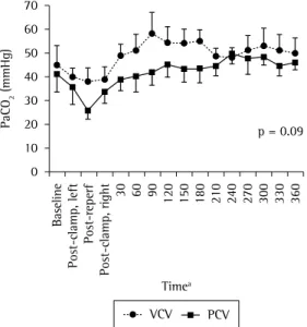
values for the PaO2/FiO2 ratio, PvO2, PaCO2, and PvCO2 were similar in the two groups, despite the progressive and persistent increase in PaCO2 (Figure 3). The PaO2/FiO2 ratio tended to decrease progressively over time (p = 0.09). There was a progressive increase in ΔSO2 over the course of the assessment period (p = 0.04; Figure 4). The lung biopsies revealed diffuse alveolar damage that was similar in the two groups. Alveolar septal rupture was found in 2 animals in the VCV group and in 1 animal in the PCV group (Figure 5).
Discussion
After extensive experimental investiga-tion,(9,13,14) the use of lungs obtained from non-heart-beating donors has become a clinical reality. Recently, the survival of recipients of lungs obtained from non-heart-beating donors was found to be somewhat lower than that of recipients of lungs from brain-dead donors, although within acceptable levels, the one-year and two-year survival rates being 78% and 69% in the former group, respectively, compared with
Results
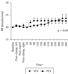
Of the 20 recipient animals, 5 were excluded because they did not survive the transplant. Another 5 (3 in the VCV group and 2 in the PCV group) died before the end of the 360-min assessment period, due to excessive bleeding and hemodynamic instability. Of the remaining 10 survivors, 5 belonged to the VCV group and 5 belonged to the PCV group. There were no significant differences between the groups regarding body weight, total ischemic time, anastomosis time, wet weight/dry weight ratio, total anesthetic consumption, volume of electrolyte solutions administered, or urinary output. There were also no statistically significant differences between the groups regarding MAP, PAP, HR, or esophageal temperature. In addition, volumetric measurements taken in both groups revealed no significant differences regarding VT values (p = 0.49). There was a tendency toward higher RR values in the VCV group (p = 0.09; Figure 1). Over time, RR increased, being significantly higher by the end of the experiment (p = 0.01). There were no significant differences between the groups regarding PIP, PPLAT (Figure 2), mean Paw, Cdyn, or Cstat. The
Figure 3 - PaCO2 over time in the pressure-controlled ventilation (PCV) and volume-controlled ventilation (VCV) groups. Despite a progressive increase in PaCO2, there were no significant differences between the groups.
Figure 2 - Plateau pressure over time in the pressure-controlled ventilation (PCV) and volume-pressure-controlled ventilation (VCV) groups.
aAssessed at four time points—at baseline; 15 min after clamping of the left pulmonary artery (post-clamp, left); 15 min after
indicate altered tissue oxygenation. There was a gradual and variable increase in ΔSO2 (ΔSO2 > 30%), reflecting significant changes in SvO2, which is an indicator of oxygen transfer through the alveolar-capillary membrane, as well as of cardiac output and peripheral utilization of oxygen. A SvO2 value < 60 mmHg in the presence of adequate gas exchange indicates decreased cardiac output and high oxygen consumption, or it might reflect a change in the ventilation/ perfusion ratio. The increase in ΔSO2 can result from reduced oxygen transfer in the lungs, decreased oxygen transport to the tissues, or increased tissue oxygen consumption, probably due to different responses to vasoactive drugs and hemodynamic management.(16) Regarding the variables used to monitor ventilation, there were considerable increases in PaCO2 and PvCO2 levels from the end of the transplantation procedure onward. In the VCV group, PaCO2 and PvCO2 values always remained higher. Therefore, with the use of similar minute volumes, minor adjustments in RR, and higher mean Paw, the animals in the PCV group presented with lower PaCO2 values, providing evidence of an improved capacity to eliminate CO2. The changes found are likely due to the increase in the physiological dead space, resulting from the major changes in the ventilation/perfusion ratio. A review of transplant recipients(17) revealed that decreased diffusing capacity of the lung for CO2 is common in lung grafts. The PCV strategy is theoretically more likely to promote lower PaCO2 levels and a lower dead space volume/VT ratio due to its propensity to promote a better intra-alveolar gas distribution.(18) One group of authors(19) stated that, over time, the PCV strategy seems to be accompanied by a tendency toward a more rapid normalization of CO2 elimination. This would be the advantage to be considered, particularly in cases in which there is a tendency toward hypercapnia.
The ventilation strategies were comparable in terms of VT, RR, expiratory minute volume, and mean inspiratory flow. Those values confirmed the effect that the settings had on RR, since there was a tendency toward higher levels over time. Other studies have compared the efficacy of ventilation strategies by assessing patients or animals with the use of different minute volumes.(20,21) In the two groups, PIP, P
PLAT, and mean Paw were similar. The PIP varies due to 94% and 87% in the latter.(15) It is anticipated
that, in those non-heart-beating donor lungs, the post-arrest ischemic changes will be severe and will lead to a greater number of complications in the recipients. Few studies involving experimental models of lung transplantation have specifically addressed the ventilation strategy used. To our knowledge, the present study is the first to test two different conventional controlled ventilation strategies (VCV and PCV) in an experimental model of lung transplantation in dogs using donor lungs harvested after three hours of normothermic ischemia following cardiac arrest. Regarding respiratory mechanics, gas exchange, and histopathological findings, we found no significant differences to suggest an unequivocal benefit of one strategy over the other.
Despite the progressive functional impairment found, the lung grafts maintained their ability to perform gas exchanges, despite the observed reduction in PaO2/FiO2 and PvO2. The changes in PvO2 (values below 40 mmHg) Figure 4 - Difference in oxygen-hemoglobin saturation (ΔSO2) between the pressure-controlled ventilation (PCV) and volume-controlled ventilation (VCV) groups. There was a progressive increase over the course of the assessment period, revealing a difference between the groups (p = 0.04).
aAssessed at four time points—at baseline; 15 min after
damage in the groups. Diffuse alveolar damage is a limited and stereotypical characteristic of the pulmonary reaction to acute injury.(24) Our results confirm the expected pattern of earlier changes in acute lung injury. However, several factors in combination, such as oxygen toxicity, mechanical ventilation, and the I/R injury itself, can lead to this pattern of injury.(25,26)
Three experimental studies have addressed ventilation strategies after lung graft reperfusion. Two of those studies used partial liquid ventilation with perfluorocarbons in models of lung transplantation.(8,27) The third evaluated a gas ventilation strategy in a model of I/R injury after lung transplantation in rats.(28) A conventional ventilation strategy was compared with a minimal stress mechanical ventilation strategy After three hours of reperfusion, the group submitted to the protective strategy showed better results in terms of oxygen saturation, cytokine levels, and compliance, as well as in terms of the morphological signs of lung injury. The study proved that the ventilation strategy used in the initial phases of lung graft reperfusion can influence I/R injury. Therefore, the results are not comparable, since the choice of the controlled ventilation strategy alone did not necessarily provide a minimal stress strategy. In this scenario, the ventilation strategy in isolation was not sufficient to change the performance of the grafts during the first six hours after reperfusion. A larger sample could reveal whether the tendencies observed for the PCV strategy—greater elimination of carbon dioxide and lower PIP values—can reduce stress in the lung tissues and their consequent inflammatory responses.
The principal limitation of our experimental model is the exclusion of the contralateral native lung. Although this makes it possible to evaluate the function of the transplanted organ exclusively, it creates a non-physiological situation in which the newly transplanted lung receives all of the cardiac output. Since the early days of this experimental model,(29) this strategy has been used in order to simulate the clinical condition in which the native lung does not participate in gas exchange. Therefore, it is possible that there will be effects other than those of I/R injury, probably aggravated by the increase in intrapulmonary hydrostatic pressure. The cumulative effect of these factors certainly contributed to the high mortality rate parameters inherent to the patient and the
ventilator, and among such parameters are lung compliance, airway resistance, and inspiratory flow. No barotrauma occurred, although PIP reached values above 35 cmH2O in the VCV group. During the assessment period, there was a gradual increase in PIP, especially in the VCV group, coincident with the need for major adjustments in RR. Inspiratory flow increased in direct proportion with the need for adjustments in RR, and this led to higher PIP values. In accordance with the tendency shown in our results, one group of authors,(22) comparing the VCV and PCV strategies in patients submitted to single-lung ventilation, observed that PIP, PPLAT, and pulmonary shunt were significantly higher during VCV, whereas PaCO2 was higher during PCV. The authors concluded that PCV is an alternative to VCV in patients requiring single-lung ventilation and can be superior to VCV in patients with respiratory disease. The low PEEP levels used in this experiment (5 cmH2O) might have contributed to the low PaO2 levels and facilitated the occurrence of atelectasis, as well as the occurrence of alveolar recruitment and derecruitment, factors that negatively affect gas exchange. Although there have been few studies reporting the PEEP levels used during the postoperative period in lung transplant recipients, one group of authors(23) stated a preference for moderate PEEP levels (5 to 8 cmH2O), used in combination with the PCV strategy.
5. Khan SU, Salloum J, O’Donovan PB, Mascha EJ, Mehta AC, Matthay MA, et al.Acute pulmonary edema after lung transplantation: the pulmonary reimplantation response. Chest. 1999;116(1):187-94.
6. Buchanan SA, DeLima NF, Binns OA, Mauney MC, Cope JT, Langenburg SE, et al.Pulmonary function after non-heart-beating lung donation in a survival model. Ann Thorac Surg. 1995;60(1):38-44; discussion 44-6. 7. Hudson LD. Progress in understanding
ventilator-induced lung injury. JAMA. 1999;282(1):77-8. 8. Andrade CF, Martins LK, Tonietto TA, Koefender C,
Anflor LC Jr, da Silva NB, et al.Partial liquid ventilation with perfluorodecalin following unilateral canine lung allotransplantation in non-heart-beating donors. J Heart Lung Transplant. 2004;23(2):242-51.
9. Kohmann JC, Lima e Silva U, Madke G, Pilla ES, Felicetti JC, Camargo JJ, et al.Perfusão pulmonar anterógrada versus retrógada na preservação pulmonar para transplante em modelo canino de viabilidade pulmonar pós-mortem. J Pneumol. 1999;25(2):78-83.
10. Flecknell PA. Anaesthesia of animals for biomedical research. Br J Anaesth. 1993;71(6):885-94.
11. Della Rocca G, Costa GM, Coccia C, Pompei L, Di Marco P, Pietropaoli P. Preload index: pulmonary artery occlusion pressure versus intrathoracic blood volume monitoring during lung transplantation. Anesth Analg. 2002;95(4):835-43.
12. Fujino Y, Goddon S, Chiche JD, Hromi J, Kacmarek RM. Partial liquid ventilation ventilates better than gas ventilation. Am J Respir Crit Care Med. 2000;162(2 Pt 1):650-7.
13. Steen S, Kimblad PO, Sjöberg T, Lindberg L, Ingemansson R, Massa G. Safe lung preservation for twenty-four hours with Perfadex. Ann Thorac Surg. 1994;57(2):450-7. 14. Steen S, Ingemansson R, Budrikis A, Bolys R, Roscher R,
Sjöberg T. Successful transplantation of lungs topically cooled in the non-heart-beating donor for 6 hours. Ann Thorac Surg. 1997;63(2):345-51.
15. Mason DP, Thuita L, Alster JM, Murthy SC, Budev MM, Mehta AC, et al.Should lung transplantation be performed using donation after cardiac death? The United States experience. J Thorac Cardiovasc Surg. 2008 Oct;136(4):1061-6.
16. Russell WJ, James MF. The effects on increasing cardiac output with adrenaline or isoprenaline on arterial haemoglobin oxygen saturation and shunt during one-lung ventilation. Anaesth Intensive Care. 2000;28(6):636-41.
17. Toivonen HJ. Anaesthesia for patients with a transplanted organ. Acta Anaesthesiol Scand. 2000;44(7):812-33. 18. Lessard MR, Guérot E, Lorino H, Lemaire F, Brochard L.
Effects of pressure-controlled with different I:E ratios versus volume-controlled ventilation on respiratory mechanics, gas exchange, and hemodynamics in patients with adult respiratory distress syndrome. Anesthesiology. 1994;80(5):983-91.
19. Rappaport SH, Shpiner R, Yoshihara G, Wright J, Chang P, Abraham E. Randomized, prospective trial of pressure-limited versus volume-controlled ventilation in severe respiratory failure. Crit Care Med. 1994;22(1):22-32. 20. Valiatti JL. Ventilação controlada a volume versus
ventilação controlada a pressão. Efeitos sobre trocas gasosas e mecânica pulmonar [thesis]. São Paulo: Universidade Federal de São Paulo; 1999.
(34%) observed in this experimental model. Although this is a model in which the organ is harvested after severe ischemic lung injury following cardiac arrest, it allows the analysis of various options of hemodynamic management and ventilation management, providing the possibility of identifying any strategy that can have a significant impact and can affect the overall outcomes. Knowledge of the risks and consequences of ventilator-induced lung injury has changed the philosophy of respiratory therapy and influenced the recommendations and standardizations of the use of mechanical ventilation. It is no longer sufficient to achieve adequate physiological values of gas exchange, but rather it is necessary to promote less traumatic ventilation in order to minimize ventilator-associated lung injury and the side effects in other organs.(30)
We conclude that, in this experimental model of single lung transplantation using donor lungs harvested after three hours of normothermic cardiac arrest and employing two different ventilation strategies, the performance of lung grafts during the first six hours after reperfusion did not differ substantially in relation to the ventilation strategy employed. Further studies, involving alternative methods of lung preservation, lung reconditioning, and protective ventilation, should be carried out in order to determine the best strategies to be used in extreme situations, such as transplantation with non-heart-beating donor lungs.
Acknowledgments
We would like to thank the engineer Leria R. Holsbach, from the Department of Clinical Engineering of the Santa Casa Hospital Complex in Porto Alegre, for the technical support provided.
References
1. DeMeo DL, Ginns LC. Lung transplantation at the turn of the century. Annu Rev Med. 2001;52:185-201. 2. de Perrot M, Liu M, Waddell TK, Keshavjee S.
Ischemia-reperfusion-induced lung injury. Am J Respir Crit Care Med. 2003;167(4):490-511.
3. King RC, Binns OA, Rodriguez F, Kanithanon RC, Daniel TM, Spotnitz WD, et al.Reperfusion injury significantly impacts clinical outcome after pulmonary transplantation. Ann Thorac Surg. 2000;69(6):1681-5. 4. Christie JD, Bavaria JE, Palevsky HI, Litzky L, Blumenthal
26. Dos Santos CC, Slutsky AS. Invited review: mechanisms of ventilator-induced lung injury: a perspective. J Appl Physiol. 2000;89(4):1645-55.
27. Itano H, Aoe M, Ichiba S, Yamashita M, Date H, Andou A, et al.Partial liquid ventilation for acute allograft dysfunction after canine lung transplantation. Ann Thorac Surg. 1999;67(2):332-9.
28. de Perrot M, Imai Y, Volgyesi GA, Waddell TK, Liu M, Mullen JB, et al.Effect of ventilator-induced lung injury on the development of reperfusion injury in a rat lung transplant model. J Thorac Cardiovasc Surg. 2002;124(6):1137-44.
29. Jones MT, Hsieh C, Yoshikawa K, Patterson GA, Cooper JD. A new model for assessment of lung preservation. J Thorac Cardiovasc Surg. 1988;96(4):608-14.
30. Tremblay LN, Slutsky AS. Ventilator-induced injury: from barotrauma to biotrauma. Proc Assoc Am Physicians. 1998;110(6):482-8.
21. Davis K Jr, Branson RD, Campbell RS, Porembka DT. Comparison of volume control and pressure control ventilation: is flow waveform the difference? J Trauma. 1996;41(5):808-14.
22. Tuğrul M, Camci E, Karadeniz H, Sentürk M, Pembeci K, Akpir K. Comparison of volume controlled with pressure controlled ventilation during one-lung anaesthesia. Br J Anaesth. 1997;79(3):306-10.
23. Briegel J, Groh J, Haller M. Perioperative management of patients undergoing lung transplantation. Curr Opin Anaesthesiol. 1998;11(1):51-9.
24. Travis WD, Colby TV, Koss MN, Rosado-de-Christenson ML, Müller NL, King Jr TE. Non-Neoplastic Disorders of the Lower Respiratory Tract. Washington: Armed Forces Institute of Pathology/American Registry of Pathology; 2002. p. 747-65.
25. Adoumie R, Serrick C, Giaid A, Shennib H. Early cellular events in the lung allograft. Ann Thorac Surg. 1992;54(6):1071-6; discussion 1076-7.
About the authors
Elaine Aparecida Felix
Adjunct Professor. Department of Surgery, Federal University of Rio Grande do Sul, Porto Alegre, Brazil.
Cristiano Feijó Andrade
Thoracic Surgeon. Porto Alegre Hospital de Clínicas, Federal University of Rio Grande do Sul, Porto Alegre, Brazil, and Santo Antônio Children’s Hospital, Santa Casa Hospital Complex in Porto Alegre, Porto Alegre, Brazil.
Paulo Francisco Guerreiro Cardoso
Associate Professor. Department of Surgery, Thoracic Surgery Section, Federal University of Health Sciences of Porto Alegre, Porto Alegre, Brazil; Thoracic Surgeon. Pereira Filho Ward, Santa Casa Hospital Complex in Porto Alegre, Porto Alegre, Brazil; Visiting Professor. Department of Thoracic Surgery, University of São Paulo, São Paulo, Brazil.
Gabriela Cury Thiesen
Anesthesiologist. Padre Jeremias Hospital, Cachoeirinha, Brazil.
Ana Carolina Peçanha Antonio
Internist. Porto Alegre Hospital de Clínicas, Federal University of Rio Grande do Sul, Porto Alegre, Brazil.
Lucas Krieger Martins
Physician. Uruguaiana Institute of Cardiology, Uruguaiana, Brazil.
Tiago Antonio Tonietto