*Correspondência: Serviço de Endocrinologia do Hospital de Clínicas de Porto Alegre
Ramiro Barcelos 2350, Prédio 12, 4° andar, CEP: 90035-003, Porto Alegre, RS, Brasil. Fone: 51 21018127 Fax: 55 51 21018777 [email protected]
Summary
Prevalence of diabetic retinopathy in patients with type 1 diabetes mellitus
Objectives. Diabetic retinopathy (DR) is the leading cause of legal blindness in young adults. Scarce data from Brazilian subjects with type 1 diabetes mellitus (DM) are available. Aims: The objectives of this study were to determine the prevalence of DR and its risk factors in type 1 diabetes mellitus (DM) outpatients from a general hospital.
MethOds. A cross-sectional study of 437 type 1 DM (50.3% males, 82.4% whites) was conducted. DR was graded as absent, mild and moderate non-proliferative DR (mild/moderate NPDR) or severe non-proliferative and proliferative DR (advanced DR). Presence of clinically signiicant macular edema (CSME) was also recorded.
Results. Any DR was present in 44.4% of subjects. In multivariate analysis, DM duration, systolic blood pressure (SBP) and A1C test were associated with mild/moderate NPDR (P<0.005). Advanced DR, was associated with DM duration, SBP, smoking [odds ratio (OR) 2.75, 95%CI 1.15-6.60] and micro-or macroalbuminuria (OR 8.53, 95%CI 3.81-18.05). CSME was present in 21 (9.4%) patients and was associated with smoking (OR 3.19, 95%CI 1.24-8.2). Its frequency increased with the severity of DR (16.4% in advanced DR, 9.6% in mild/moderate NPDR, and 4.7% in the group without DR; P = 0.020).
cOnclusiOn. Patients with type 1 DM attending an endocrine out-patient clinic at a general hospital had a high prevalence of DR associated with traditional risk-factors and smoking.
Keywords: Type 1 diabetes mellitus. Diabetic retinopathy. Risk factors.
pRevalence
Of
diabetic
RetinOpathy
in
patients
with
type
1
diabetes
Mellitus
jORge f. esteves1, caROline K. KRaMeR2, MiRela jObiMde azevedO3, andRessa p. stOlz4, MuRilO f. ROggia4, andRéia laRangeiRa5, suellen a. MiOzzO5, caROlina ROsa5, jOse huMbeRtO laMbeRt5, MiRiaM pecis6, ticiana c. ROdRigues7, luis henRique santOs canani*8
Trabalho realizado no Serviço de Oftalmologia, Hospital de Clínicas de Porto Alegre, Porto Alegre, Brasil, Serviço de Endocrinologia, Hospital de Clínicas de Porto Alegre, Porto Alegre, Brasil Universidade Federal do Rio Grande do Sul, Porto Alegre, RS
1. Professor assistente do Departamento de Oftalmologia da Universidade Federal do Rio Grande do Sul, Porto Alegre, RS
2. Aluna de doutorado do Programa de Pós Graduação Ciências Médicas: Endocrinologia da Universidade Federal do Rio Grande do Sul, Porto Alegre, RS 3. Professora adjunta do Departamento de Medicina Interna da Universidade Federal do Rio Grande do Sul, Porto Alegre, RS
4. Médica residente do Serviço de Oftalmologia do Hospital de Clinicas de Porto Alegre, Porto Alegre, RS 5. Estudante de medicina - Estudante de Medicina da Universidade Federal do Rio Grande do Sul, Porto Alegre, RS 6. Bolsista de Pós-doutorado do CNPq
7. Médica do Serviço de Endocrinologia do Hospital de Clínicas de Porto Alegre, Porto Alegre, RS
8. Professor adjunto do Departamento de Medicina Interna da Universidade Federal do Rio Grande do Sul, Porto Alegre, RS
intROductiOn
Diabetic retinopathy (DR) is the most frequent microvascular complication of diabetes mellitus (DM), resulting in blindness for over 10,000 people with DM every year1 and is the leading
cause of legal blindness2. In type 1 DM, the overall prevalence
of DR after eleven years of follow-up is 66.6%3, and almost all
patients have some degree of DR after 20 years of DM 4, 5. Further,
severe forms of the disease leading to visual impairment occur in 50% of type 1 DM patients2.
The main risk factors for the development and progress of DR are persistent hyperglycemia, DM duration and high blood pressure levels6-11. However, there is an important individual
variability in incidence of DR among diabetic patients. The
question often asked is why some patients under good metabolic control develop DR while others remain free of this complication, despite poorly controlled DM12. This may be due to different
genetic backgrounds.
The aims of the present study were to describe prevalence of DR and its risk factors in type 1 DM outpatients from a general hospital in Southern Brazil.
MethOds
research design
Subjects
Patients with type 1 DM attending the Hospital de Clínicas de Porto Alegre, Brazil, in the Endocrine Clinic and referred to the Ophthalmology Clinic for routine eye examination were included. The criteria for referral were patients more than 18 years of age with a diagnosis of type 1 DM for ive years or more. Deinition of type 1 DM was based on the presence of DM, diagnosed before 30 years of age, at least one episode of diabetic cetoacidosis and/or cetonemia and need for insulin therapy within 1 year of DM diagnosis13.
Eye examination and classiication of retinopathy
Eye examination included, in addition to fundoscopy, visual acuity test (logMAR notation), refraction, tonometry and biomi-croscopy of the anterior segment.
DR was graded at the time of ophthalmologic assessment by fundoscopy through dilated pupils by the same researcher (JFE) and severity was established using the scale developed by the Global Diabetic Retinopathy Group 14. The irst level was “absent DR”, with no fundus abnormalities; the second was “mild non proliferative diabetic retinopathy (NPDR)”, microaneurysms only; the third, “moderate NPDR”, included more than just microaneu -rysms, but less than severe NPDR; the fourth, “severe NPDR”, included any of the following: >20 intra-retinal hemorrhages in each of the 4 quadrants, deinite venous beading in 2+ quadrants, prominent intra-retinal microvascular abnormalities in 1+ quadrant, and no signs of proliferative DR; and the ifth level, “proliferative DR” (PDR), which includes eyes with one or more of the following: deinite neovascularization or vitreous/ pre-retinal hemorrhage15.
Classification of patient DR was based on the most severe degree of retinopathy in the worst affected eye. We have previously described an excellent agreement of DR classifica-tion (95.3%) carried out by different trained ophthalmologists from our group16. Therefore, in the present study only a single
observer, not aware of the patients’ clinical data, classified all the subjects
According to the DR classiication, three groups were deined for further analysis: 1- absent DR; 2- mild and moderate NPDR (mild/moderate NPDR) and 3- severe non proliferative and proli-ferative DR (advanced DR group).
Macular edema was evaluated upon dilated eyes, using slit-lamp biomicroscopy in a subset of patients. Clinically signiicant macular edema (CSME) was deined as one or more of the following: any retinal thickening within 500mm of the center of the macula, with or without loss of retinal transparency; hard exudates associated with retinal thickening within 500mm of the center of the macula; or one disc area of thickening within one disc diameter of the center of the macula17, 18.
Clinical evaluation
Risk factors for DR were recorded at the time of ophthalmo-logic examination and included age, age at onset of DM, DM duration, ethnicity (self reported), smoking habit, body mass index (BMI), systolic blood pressure (SBP) and diastolic blood pressure (DBP). All patients answered a brief standard ques-tionnaire and underwent physical examination and laboratory
tests. They were weighed wearing light outdoor clothes without shoes and height was recorded. BMI was calculated as weight (kilograms)/height2 (meters). Waist circumference was
measured on a horizontal plane, midway between the inferior margin of the ribs and the superior border of the iliac crest. Sitting blood pressure was measured twice on the right arm to the nearest 2mm Hg after a 10 minute rest using a standard mercury sphygmomanometer (phases I and V of Korotkoff sounds). Subjects who smoked one or more cigarettes daily were classified as current smokers. Those who had smoked in the past and stopped for more than one year were classified as former smokers.
Laboratory methods
Laboratory evaluations consisted of measuring A1C test, lipid proile, serum creatinine and urinary albumin excretion (UAE). FPG was determined by enzymatic colorimetric assay (through glucose oxidase enzyme). A1C test was measured by high-performance liquid chromatography system (reference range 4.7 - 6.0%; Merck-Hitachi 9100, Merck, Darmstadt, Germany). Fasting plasma glucose was measured by the glucose-peroxidase colorimetric enzymatic method (Biodiagnostica). Creatinine was measured by the Jaffé method and serum total choles-terol, triglycerides were measured by enzymatic-colorimetric methods (Merck Diagnostica, Darmstadt, Germany; Boeringher Mannheim, Buenos Aires, Argentina) and HDL cholesterol was measured by homogeneous direct method (autoanalyzer, ADVIA 1650). LDL cholesterol was calculated using the Frie-dewald formula. Albuminuria was measured in a sterile spot urine sample by turbidimetric immunoassay on at least two occasions in patients without end-stage renal disease (ESRD); values below 17 mg/l were considered as normoalbuminúria (n = 190), between 17-174 mg/l microalbuminuria (n = 56) and >174 mg/dl, as macroalbuminuria (n = 33) [19]. Patients with ESRD (n = 10, dialysis) were included in the macroalbuminuric group. Glomerular iltration rate (GFR) was estimated using the Modiication of Diet in Renal Disease (MDRD) formula: 186 x [plasma creatinine (mg/dl)-1.154 x age (yr)-0.203 x (1.212 if black)
x (0.742 if female)][20].
The study protocol was approved by the Hospital Ethics Committees and an informed consent was obtained from all patients.
Statistical analysis
Results
Sample description
A total of 437 patients were evaluated (50.3% males, 82.4% whites). Mean age at ophthalmologic examination was 26.8 ± 7.8 years and at diagnosis of DM was 12.9 ± 7.1 years. Duration of DM was 14.4 ± 7.3 years. Overall prevalence of any DR was 44.4% (n = 194). Sixty-six patients (15.1%) had mild NPDR, 18 patients (4.2%) moderate NPDR, 13 (3.0%) patients severe NPDR, and 97 patients (22.2%) were diagnosed with PDR.
Patients with mild and moderate NDPR were grouped as mild/ moderate (n=84), and patients with severe NPDR and PDR were grouped as advanced DR (n = 110).
Demographic, anthropometric and smoking habit data
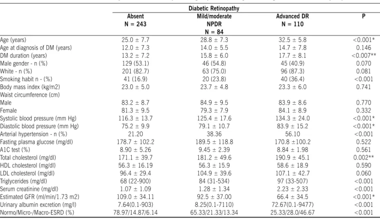
Clinical and laboratory features of type 1 DM patients grouped according to the degree of DR are shown in Table 1.
Patients with absent DR had a shorter duration of DM and were younger than the patients with mild/moderate NPDR and advanced DR. Duration of DM was not different between patients with mild/moderate NPDR and advanced DR. Onset of DM occurred earlier for patients without DR when compared to patients with mild/moderate NPDR and advanced DR. Gender proportion, ethnic group and anthropometric indices did not differ among groups. Neither general obesity (BMI) nor central obesity (waist circumference) was linked to DR.
Current or past smoking history was associated with DR (P <0.001). There was a progressive increase in the frequency of smokers/former smokers among those with absent DR, mild/ moderate DR and advanced DR (P for trend <0.001).
Blood pressure and glycemic control
Patients with mild/moderate NPDR and advanced DR had higher SBP than patients without DR (Table 1). The DBP levels were higher in the group with advanced DR than in patients without DR, but DBP was not different between those without DR and with mild/moderate NPDR. There was a progressive increase in the prevalence of arterial hypertension from those without DR to mild/moderate NPDR and advanced DR (21.0% vs. 38.1 vs. 56.4%: P <0.001).
There was no difference in FPG values among the three groups (178.7 ± 102.2 vs. 189.5 ± 118.8 vs. 170.8 ± 100.2 mg/dl, P = 0.520). The A1C test was higher among those with mild/moderate NPDR and advanced DR when compared to those without DR.
Figure 1 shows the frequency of advanced DR according to SBP and A1C test quartiles. Prevalence of advanced DR was 8.2% in patients in the lower A1C test and SBP quartiles. Even in patients with the best metabolic control (A1C test <7.2%), prevalence of advanced DR increased with the increase of blood pressure quartiles, reaching 28% in those of the upper SBP quar-tile (>130 mm Hg). The same pattern was observed for the A1C quartiles in those with low SBP (<110 mm Hg). Those in the
Table 1 - Clinical and laboratory characteristics of patients grouped according to the stages of diabetic retinopathy
Diabetic retinopathy absent
N = 243
mild/moderate NPDr N = 84
advanced Dr N = 110
P
Age (years) 25.0 ± 7.7 28.8 ± 7.3 32.5 ± 5.8 <0.001*
Age at diagnosis of DM (years) 12.0 ± 7.3 14.0 ± 5.5 14.7 ± 7.8 0.146
DM duration (years) 13.2 ± 7.2 15.8 ± 6.0 17.7 ± 8.1 <0.007**
Male gender - n (%) 129 (53.1) 46 (54.8) 45 (40.9) 0.070
White - n (%) 201 (82.7) 63 (75.0) 96 (87.3) 0.081
Smoking habit n - (%) 41 (16.9) 20 (23.8) 40 (36.4) <0.001
Body mass index (kg/m2) 23.0 ± 5.0 23.7 ± 4.8 23.3 ± 6.0 0.741
Waist circumference (cm)
Male 83.2 ± 8.7 84.9 ± 9.5 83.9 ± 8.6 0.770
Female 81.3 ± 9.5 79.3 ± 7.9 84.1 ± 8.9 0.332
Systolic blood pressure (mm Hg) 116.3 ± 13.7 125.4 ± 17.6 134.3 ± 24.0 <0.001*
Diastolic blood pressure (mm Hg) 75.2 ± 9.9 79.1 ± 10.7 83.9 ± 15.2 <0.001*
Arterial hypertension - n (%) 21.20 38.36 56.10 <0.001
Fasting plasma glucose (mg/dl) 178.7 ± 102.2 189.5 ± 118.8 170.8 ±100.2 0.522
A1C test (%) 8.90 ± 5.26 9.45 ± 2.39 8.84 ± 1.98 0.561
Total cholesterol (mg/dl) 171.1 ± 39.7 181.2 ± 49.6 190.9 ± 45.1 0.002**
HDL cholesterol (mg/dl) 56.3 ± 16.19 56.3 ± 15.9 58.6 ± 18.9 0.590
LDL cholesterol (mg/dl) 96.4 ± 29.4 104.9 ± 39.6 107.1 ± 42.7 0.060
Triglycerides (mg/dl) 68 (22-900) 84 (31-534) 97 (33-507) <0.001
Serum creatinine (mg/dl) 1.07 ± 1.09 1.28 ± 1.34 2.23 ± 2.33 <0.001
Estimated GFR (ml/min/1.73 m2) 109.0 ± 34.11 92.5 ± 37.00 66.4 ± 34.5 <0.001*
Urinary albumin excretion (mg/l) 7.64(0.1-903) 8.25(0.1-7110) 72.67(0.1-9477) <0.001
Normo/Micro-/Macro-ESRD (%) 78.97/14.87/6.14 65.33/21.33/13.34 25.33/28.0/46.67 <0.001
Data expressed as mean (± standard deviation), median [range] or number of cases (%). NPDR= non proliferative diabetic retinopathy
upper quartiles for A1C test and SBP had the highest prevalence of severe DR (41%).
Lipid proile
Patients with advanced DR had higher values of total choles-terol, LDL cholesterol and triglycerides than patients without DR. There were no differences in the levels of HDL cholesterol values among groups.
Renal function
Patients with advanced-DR had higher serum creatinine values than patients with mild/moderate NPDR and without DR. Estimated GFR was also lower among patients with advanced DR than among those with mild/moderate NPDR or without DR. There was a progressive increase in UAE according to the degree of retinal involvement, lower among those patients without DR and higher among those with advanced DR. To establish an index of magnitude, subjects were divided according to the UAE into normo, micro or macroalbuminurics. Subjects with ESRD were included in the macroalbuminuric group. Microalbuminuria increases the chance of advanced DR by 4.8 times (95% CI 2.5-9.4, P <0.001), but not the mild/moderate forms (OR 1.75, 95%CI 0.9 - 3.5, P = 0.320). Macroalbuminuria was associated with both, mild/moderate NPDR (OR 2.6, 95%CI 1.1-6.3, P =0.020) and advanced DR (OR 23.3, 95%CI 11.0 - 50. 1, P <0.001).
macular edema
In the subset of 223 patients in whom the presence of CSME was evaluated, 21 patients (9.4%) presented CSME, and this frequency increased with the severity of DR: 16.4% in advanced DR, 9.6% in mild/moderate NPDR, and 4.7% in the group without DR (P = 0.020). Current smoking was also associated with CSME (OR 3.19, 95%CI 1.24-8.2, P = 0.012). There was a progressive increase in the frequency of CSME according to renal status: normo 5.4%, micro 11.4%, and macroalbu-minuria 22.2% (P for trend 0.005). CSME was not associated with gender, ethnicity, blood pressure levels, lipid proile, serum creatinine or metabolic control.
MultivaRiate analysis
Mild/moderate non proliferative diabetic retinopathy
Mild/moderate NPDR was associated with most variables, except for total cholesterol, and smoking. SBP, A1C test, micro-albuminuria (log transformed), DM duration, total cholesterol and smoking (current or past) were included in the initial multivariate logistic regression model. For each increase in one year of DM duration, in one mmHg in SBP or in one point in A1C test, there was an increased chance of presenting mild/moderate NPDR of 6%, 2% and 2% (P <0.005), respectively.
Other models were constructed substituting SBP for DBP or arterial hypertension, or substituting total cholesterol for triglyc-erides (log transformed), or degree of albuminuria for stages of diabetic nephropathy (norn, micro or macroalbuminuria) or serum creatinine. The inclusion of arterial hypertension instead of SBP showed an OR of 3.12 (95%CI 1.06-9.40). Neither DBP nor triglycerides were associated with mild/moderate NPDR. When microalbuminuria was replaced by serum creatinine, the OR for mild/moderate NPDR was 1.76 (95% CI 1.03-3.48).
advanced diabetic retinopathy
Advanced DR was associated to all variables with the excep-tion of total cholesterol and A1C test. Each increase in one year of DM duration or in one mmHg in SBP was associated with an increase in the odds of advanced DR of 4% (95% CI 1.3-7.8, P <0.05). Smoking increased chances for advanced DR by 2.75 times (OR 95%CI 1.15-6.60). Hypertension was associated with an OR of 2.48 (95% CI 1.13-5.40) for advanced DR. Presence of diabetic nephropathy (DN) (micro- or macroalbuminuria) was associated with an OR of 8.53 (95% CI 3.81-18.05). When serum creatinine was used in the model instead of microalbumin-uria it was also associated with advanced DR (OR 2.64 - 95% CI 1.40-5.01).
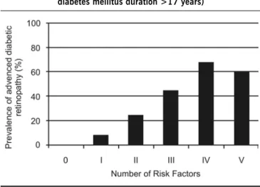
The ive major independent risk factors for advanced DR were dichotomized into present or absent (arterial hypertension, DN and smoking) or above or below the median value (A1C test - 8.7% and DM duration - 17 years). Forty-two of the patients (9.6%) had no risk factors, 131 patients (30%) had one, 139 (31.8%) two, 49 (11.4%) three, 50 (11.4%) four, and only 7 patients (1.6%) had all ive risk factors. The prevalence of advanced DR increased with the number of risk factors (Figure 2). However, even in the presence of four or ive risk factors, about 40% of the subjects were free of the most severe degree of DR (proliferative form).
discussiOn
In this study, DR was present in a high percentage of this sample of type 1 DM patients and it was associated with the main traditional risk factors, namely glycemic control, blood pressure and DM duration. On the other hand, glycemic control was not associated with advanced DR in multivariate analyses. This may suggest that for more severe forms of DR, glycemic control does not play a major role as observed for systolic blood pressure. An alternative explanation is that absence of association could relect improvement of glycemic control that results from medical advice, once diagnosis of this severe microvascular chronic
Figure 1 - Frequency of advanced-DR diabetic retinopathy
complication is established. However, the cross-sectional design adopted precludes conirmation of this hypothesis.
Another aspect that must be highlighted is the association of smoking with any degree of DR found in this study. Smoking habits and DR are controversially related 11 and no association
has been described by some authors 21. In patients with type 2
DM we, as others, had previously described a protective asso-ciation of smoking and DR 16, 22.
Overt DN is well known to be associated with DR 23. Also,
association of incipient DN and DR was previously described in type 1 DM patients 24. In the present study, microalbuminuria was
associated only with more severe forms of DN. We have shown a similar association in patients with type 2 DM 16. Concordance
of DN and DR could be due to common risk factors or could be a marker of general vascular damage, leading to leakage of protein from retinal vessels7.
Association of total cholesterol levels with DR had been clearly demonstrated, especially in type 2 DM patients 25, 26. However,
this was not observed in the present study for any DR. This could be explained by low mean levels of total cholesterol (<200 mg/ dl) of our patients studied, probably related to their young age. Finally, unmarked inluence of A1C test levels in patients with severe forms of DR could relect the major role of genetic factors in advanced DR stages of diabetic eye disease. This aspect is reinforced by a high magnitude of OR for almost all DR risk factors evaluated in this study.
Diabetic maculopathy is the most common cause of visual impairment in diabetic patients. 27 CSME must be considered for
treatment with laser photocoagulation irrespective of the level of visual acuity or DR stage, because treatment reduces the risk of visual loss by 50%27. Risk factors for CSME are not conclusively
established. A positive relation of CSME with DN, severity of DR and smoking history was found in our patients. Association of albuminuria and CSME is an interesting inding from a clinical point of view. Probably, presence of even early DN stages should alert the physician to the need of an ophthalmologic evaluation.
High values of LDL cholesterol and total cholesterol-HDL choles-terol ratio have been suggested to increase by two and fourfold the risk of CSME, respectively28. In type 1 DM patients, old age
at diagnosis of DM, male gender and higher A1C test levels signiicantly increase the risk of clinically signiicant macular edema29. We did not ind an association between demographic data, blood pressure, lipid proile or metabolic control indices and CSME. This could be due to the low power to detect these associations since there were only 22 patients with CSME. This would also preclude performing a multivariate analysis to identify the independence of the associations found.
cOnclusiOn
In conclusion, prevalence of 44.4% of any DR in type 1 DM patients attending a general hospital shows that this condition continues to be a major public health problem despite current knowledge about advanced DR. Furthermore, prevalence of 24% of advanced DR stages is a warning sign. Those with a long DM duration, positive smoking, elevated blood pressure, poor meta-bolic control and albuminuria are at highest risk of presenting advanced DR forms. Finally, CSME should be suspected in pres-ence of smoking or any degree of DN.
acKnOwledgeMents
Grant Support: Research supported by grants from Conselho Nacional de Desenvolvimento Cientíico e Tecnológico (CNPq) and Fundo de Incentivo a Pesquisa e Eventos (FIPE)- Hospital de Clínicas de Porto Alegre. LHC was the recipient of a postdoctoral (ProDoc) grant from Fundação de Coordenação de Aperfeiçoamento de Pessoal de Ensino Superior (Fundação CAPES) and MP was the recipient of a posdoctoral grant from CNPq.
Conlict of interest: none
ResuMO
pRevalênciadeRetinOpatiadiabéticaeMpacientescOM diabetes MellitustipO 1
ObjetivOs. Determinar a prevalência de RD e seus fatores de
risco em pacientes com DM tipo 1 atendidos em um hospital geral.
MétOdOs. Foi realizado um estudo transversal com 437
pacientes (50,3% homens, 82,4% brancos). RD foi agrupada em: 1) ausente; 2) não proliferativa leve e moderada (RDNP leve/moderada); 3) não prolifetiva grave e RD proliferativa (RD avançada). Edema de mácula clinicamente significativo (EMCS) também foi registrado.
ResultadOs. Qualquer grau de RD esteve presente em
44,4% dos pacientes. Na análise multivariada, duração do DM, pressão arterial sistólica e teste A1C foram associados com a RD leve/moderada (P<0,005). RD avançada foi asso-ciada com duração do DM, pressão arterial sistólica (PAS), fumo [razão de chances (RC) 2,75, IC 95% 1,15-6,60] e micro- ou macroalbuminúria (RC 8,53, CI 95% 3,81-18,05). EMCS esteve presente em 21 (9,4%) dos pacientes associado ao fumo, aumentando com a gravidade da RD (16,4% RD avançada; 9,6% RD leve/modera, e 4,7% no grupo sem RD; P = 0,020).
Figure 2 - Prevalence of advanced diabetic retinopathy and number of risk factors present (hypertension, diabetic
COnClusãO. Pacientes com DM tipo 1 vistos em um hospital
geral têm uma alta prevalência de RD, a qual foi associada aos fatores de risco tradicionais e fumo. [Rev Assoc Med Bras 2009; 55(3): 268-73]
unitermos: Diabetes mellitus tipo 1. Retinopatia diabética.
Fa-tores de risco.
RefeRences
1. Fong DS, Aiello LP, Ferris FL, 3rd, Klein R. Diabetic retinopathy. Diabetes Care. 2004; 27:2540-53.
2. Roy MS, Klein R, OColmain BJ, Klein BE, Moss SE, Kempen JH. The prevalence of diabetic retinopathy among adult type 1 diabetic persons in the United States. Arch Ophthalmol. 2004; 122:546-51.
3. Bryden KS, Dunger DB, Mayou RA, Peveler RC, Neil HA. Poor prognosis of young adults with type 1 diabetes: a longitudinal study. Diabetes Care. 2003;26:1052-7.
4. Lovestam-Adrian M, Agardh CD, Torffvit O, Agardh E. Diabetic retinopathy, visual acuity, and medical risk indicators: a continuous 10-year follow-up study in Type 1 diabetic patients under routine care. J Diabetes Complica-tions. 2001;15:287-294.
5. Krolewski AS, Warram JH, Rand LI, Christlieb AR, Busick EJ, Kahn CR. Risk of proliferative diabetic retinopathy in juvenile-onset type I diabetes: a 40-yr follow-up study. Diabetes Care. 1986; 9:443-52.
6. Agardh E, Torffvit O, Agardh CD. The prevalence of retinopathy and associated medical risk factors in type I (insulin-dependent) diabetes mellitus. J Intern Med. 1989;226:47-52.
7. Ciulla TA, Amador AG, Zinman B. Diabetic retinopathy and diabetic macular edema: pathophysiology, screening, and novel therapies. Diabetes Care. 2003;26:2653-64.
8. Van Leiden HA, Dekker JM, Moll AC, Nijpels G, Heine RJ, Bouter LM, et al. Risk factors for incident retinopathy in a diabetic and nondiabetic population: the Hoorn study. Arch Ophthalmol. 2003;121:245-51.
9. Klein R, Klein BE, Moss SE, Davis MD, DeMets DL. Glycosylated hemoglobin predicts the incidence and progression of diabetic retinopathy. JAMA. 1988;260:2864-71.
10. Janka HU, Warram JH, Rand LI, Krolewski AS. Risk factors for progression of background retinopathy in long-standing IDDM. Diabetes. 1989; 38:460-4. 11. Esteves J, Laranjeira AF, Roggia MF, Dalpizol M, Scocco C, Kramer CK, et
al. [Diabetic retinopathy risk factors]. Arq Bras Endocrinol Metabol. 2008; 52:431-441.
12. Zhang L, Krzentowski G, Albert A, Lefebvre PJ. Risk of developing retinopathy in Diabetes Control and Complications Trial type 1 diabetic patients with good or poor metabolic control. Diabetes Care. 2001; 24:1275-1279.
13. Diabetes Mellitus: Report of a WHO Study Group. Geneva: World Health Org; 1985.
14. Wilkinson CP, Ferris FL, 3rd, Klein RE, Lee PP, Agardh CD, Davis M, et al. Proposed international clinical diabetic retinopathy and diabetic macular edema disease severity scales. Ophthalmology. 2003; 110:1677-1682.
15. The Diabetic Retinopathy Study Research Group: a modiication of the Arlie House classiiation of Diabetic retinopathy (DRS report no. 7). Invest Ophthalmol Vis Sci. 1981; 21:210-226.
16. Boelter MC, Azevedo MJ, Gross JL, Lavinsky J. Fatores de risco para retinopatia diabética. Arq Bras Oftalmol. 2003;66:239-47.
17. Photocoagulation for diabetic macular edema. Early Treatment Diabetic Reti-nopathy Study report number 1. Early Treatment Diabetic RetiReti-nopathy Study research group. Arch Ophthalmol. 1985;103:1796-806.
18. Grading diabetic retinopathy from stereoscopic color fundus photographs--an extension of the modiied Airlie House classiication. ETDRS report number 10. Early Treatment Diabetic Retinopathy Study Research Group. Ophthalmology. 1991;98:786-806.
19. Gross JL, de Azevedo MJ, Silveiro SP, Canani LH, Caramori ML, Zelmanovitz T. Diabetic nephropathy: diagnosis, prevention, and treatment. Diabetes Care. 2005;28:164-76.
20. Levey AS, Bosch JP, Lewis JB, Greene T, Rogers N, Roth D. A more accurate method to estimate glomerular iltration rate from serum creatinine: a new prediction equation. Modiication of Diet in Renal Disease Study Group. Ann Intern Med. 1999;130:461-70.
21. Cantoni A CM, Congeti I, Carrerasi G, Castell C, Tresserras R. Type 1 Diabetes Mellitus in Catalonia: Chronic complications and Metabolic control ten years after onset. Med Sci Monit. 2005;10:185-90.
22. Stratton IM, Kohner EM, Aldington SJ, Turner RC, Holman RR, Manley SE, et al. UKPDS 50: risk factors for incidence and progression of retinopathy in Type II diabetes over 6 years from diagnosis. Diabetologia. 2001;44:156-63. 23. Klein R, Moss SE, Klein BE. Is gross proteinuria a risk factor for the incidence
of proliferative diabetic retinopathy? Ophthalmology. 1993;100:1140-6. 24. Parving HH, Hommel E, Mathiesen E, Skott P, Edsberg B, Bahnsen M, et
al. Prevalence of microalbuminuria, arterial hypertension, retinopathy and neuropathy in patients with insulin dependent diabetes. Br Med J (Clin Res Ed). 1988;296:156-60.
25. Klein BE, Moss SE, Klein R, Surawicz TS. The Wisconsin Epidemiologic Study of Diabetic Retinopathy. XIII. Relationship of serum cholesterol to retinopathy and hard exudate. Ophthalmology. 1991;98:1261-5.
26. Chew EY, Klein ML, Ferris FL, 3rd, Remaley NA, Murphy RP, Chantry K, et al. Association of elevated serum lipid levels with retinal hard exudate in diabetic retinopathy. Early Treatment Diabetic Retinopathy Study (ETDRS) Report 22. Arch Ophthalmol. 1996;114:1079-84.
27. Kanski JJ. Diabetic retinopathy--a preventable cause of blindness. Practitioner. 1985;229:343-8.
28. Miljanovic B, Glynn RJ, Nathan DM, Manson JE, Schaumberg DA. A pros-pective study of serum lipids and risk of diabetic macular edema in type 1 diabetes. Diabetes. 2004;53:2883-92.
29. Vitale S, Maguire MG, Murphy RP, Hiner CJ, Rourke L, Sackett C, et al. Clini-cally signiicant macular edema in type I diabetes. Incidence and risk factors. Ophthalmology. 1995;102:1170-6.