Universidade Nova de Lisboa
Instituto de Higiene e Medicina Tropical
Neutrophil-Leishmania interaction during the initial phase
of Leishmania infantum infection in dogs
Maria de Aires Machado Pereira
DISSERTATION IN SUPPORT OF A CANDIDATURE FOR A DOCTORATE DEGREE IN BIOMEDICAL SCIENCES, SPECIALIZATION IN CELLULAR AND MOLECULAR BIOLOGY,
BY UNIVERSIDADE NOVA DE LISBOA, INSTITUTO DE HIGIENE E MEDICINA TROPICAL
Universidade Nova de Lisboa
Instituto de Higiene e Medicina Tropical
Neutrophil-Leishmania interaction during the initial phase
of Leishmania infantum infection in dogs
Author: Maria de Aires Machado Pereira
Degree in Veterinary Medicine (UTAD) Master in Medical Parasitology (IHMT-UNL)
Supervisor: Prof. Dr. Gabriela Santos-Gomes (IHMT-UNL)
Dissertation in support of a candidature for a Doctorate degree in Biomedical Sciences, specialization in Cellular and Molecular Biology by Universidade Nova de Lisboa, Instituto de Higiene e Medicina Tropical, under supervision of Prof. Dr. Gabriela Santos-Gomes. Finantial support by PhD scholarship SFRH/BD/2011 and projects PTDC/CVT/113121/2009 and PTDC/CVT/118566/2010 funded by the Portuguese Foundation for Science and Technology (FCT) with the participation of European Union Fund (FEDER).
COMMUNICATIONS AND PUBLICATIONS
National scientific meetings
Poster presentation
Rodrigues, MA, Santos-Mateus D, Alexandre-Pires G, Valério-Bolas A, Santos M, Fernandes MR, Ligeiro D, Pereira M, Nunes T, Pereira da Fonseca I, Santos-Gomes G. 2016. 3D-hepatocyte culture for parasitological studies: the example of Leishmania infantum infection. Ciência 2016- Encontro com a Ciência e Tecnologia em Portugal. Centro de Congressos de Lisboa 4 a 6 Julho.
Gabriel M, Armada A, Pereira M, Santos-Gomes G, Viveiros M. 2015. Inflammatory immune response of macrophages in the presence of efflux inhibitors. Microbiotec’15,
10-12 de Dezembro, Évora, Portugal. Abstract book, pp 217.
Pereira M, Alexandre-Pires G, Margarida C, Rodrigues A, Martins C, Valério-Bolas A,
Tomas A, Santos M, Pereira da Fonseca I, Santos-Gomes G. 2015. Os neutrófilos caninos asseguram a transferência de Leishmania infantum para os macrófagos. 6as Jornadas Científicas do IHMT, Lisbon, Portugal, 11th December.
http://www.ihmt.unl.pt/wp-content/uploads/2015/11/Indice-geral-de-resumos.pdf
Rodrigues A, Alexandre-Pires G, Santos-Mateus D, Aires-Pereira M, Valério-Bolas A, Rafael-Fernandes M, Silva-Pedrosa R, Pereira da Fonseca I, Santos-Gomes G. 2014. A exposição a Leishmania infantum causa alterações metabólicas nos hepatócitos caninos. 5as Jornadas Científicas do IHMT, Lisbon, Portugal, 12th December.
http://www.ihmt.unl.pt/images/uploaded/news/Resumo_MariaArmanda.pdf
Pereira MA, Santos-Mateus D, Valério-Bolas A, Rodrigues A, Marques C, Fonseca IP,
Santos-Gomes G. Exocitose enzimática por células polimorfonucleares expostas a Leishmania infantum. 4as Jornadas Científicas do IHMT, Lisbon, Portugal, 13th
December 2013.
Valério-Bolas A, Pereira MA, Santos M, Rafael-Fernandes M, Rodrigues A, Santos-Mateus D, Claro M, Pereira da Fonseca I, Santos-Gomes GM. 2013. Importance of neutrophils and macrophages in the immune response to Leishmania spp. infection. iMed 5.0 Conference, 11-13th October, Lisboa.
Alves I, Pereira M, Veloso L, Santos-Mateus D, Santos-Gomes G, Semião-Santos S, Martins L. 2012. Rastreio preliminar da susceptibilidade/resistência do Rafeiro do Alentejo à leishmaniose Canina. VIII Congresso do Hospital Veterinário Montenegro. Santa Maria da Feira, Portugal, 11-12th February.
Oral presentation
Pereira M, Valério-Bolas A, Santos-Mateus D, Rodrigues A, Santos M, Rocha H, Santos
A, Alexandre-Pires G, Martins C, Pereira da Fonseca I, Santos-Gomes G. 2014. Leishmania infantum regula a ativação dos mecanismos efetores de neutrófilos caninos. 5as Jornadas Científicas do IHMT, Lisbon, Portugal, 12th December.
http://www.ihmt.unl.pt/images/uploaded/news/Resumo_MariaAiresPereira.pdf
Santos-Gomes G, Santos-Mateus D, Valério-Bolas A, Rafael-Fernandes M, Silva-Pedrosa R, Pereira M, Rodrigues A. 2014. Human leishmaniasis. Portuguese Institute of Blood and Transplant, Lisbon, Portugal. 17-18th December.
Santos-Gomes G, Santos-Mateus D, Valério-Bolas A, Rafael-Fernandes M, Silva-Pedrosa AR, Pereira M, Rodrigues A. 2014. Leishmanioses. Infeção emergente nos migrantes & transplantados. OMV, 29-30th November, Lisboa.
Pereira MA, Santos-Mateus D, Valério-Bolas A, Rodrigues A, Santos-Gomes G. 2014.
Terapêutica e controlo da leishmaniose canina. Ciclo de palestras sobre leishmaniose canina, GAAF-AEFMV, Faculdade de Medicina Veterinária de Lisboa, Lisbon, Portugal, 21st March (by invitation).
Rodrigues A, Alexandre-Pires G, Santos-Mateus D, Aires-Pereira M, Valério-Bolas A, Rafael-Fernandes M, Marques C, Pereira da Fonseca I, Santos-Gomes G. 2013. Impacto na função metabólica e na resposta imunitária de hepatócitos de cão expostos a
Leishmania infantum. 4as Jornadas Científicas do IHMT, Lisbon, Portugal, 13th
December.
http://www.ihmt.unl.pt/images/uploaded/news/ResumosemLogo_MariaArmandaRodrig ues.pdf
Pereira MA, Santos-Mateus D, Valério-Bolas A, Rodrigues A, Santos-Gomes G. 2013.
Terapêutica e controlo da leishmaniose Canina. VII Congresso da OMV, IV Encontro de Formação, Lisbon, Portugal, 30th November and 1st December (by invitation).
Santos-Gomes G, Santos-Mateus D, Valério-Bolas A, Rafael-Fernandes M, Claro M. Rodrigues A, Pereira MA. 2013. Imunidade e resposta imunitária na leishmaniose canina. IV Encontro de Formação e VIII Congresso da Ordem dos Médicos Veterinários 30th November and 1st December, Lisboa.
Santos-Gomes G, Santos-Mateus D, Valério-Bolas A, Rodrigues MA, Pereira MA. 2012. Leishmaniose canina: a resposta imunitária e o diagnóstico sorológico. Jornadas de Parasitologia II. Escola Superior de Tecnologia da Saúde do Porto. Vila Nova de Gaia, Portugal, 24th May.
Pereira MA, Santos-Mateus D, Valério-Bolas A, Rodrigues MA, Santos-Gomes G.
2012.Tratamento e monitorização da leishmaniose canina. Jornadas de Parasitologia II. Escola Superior de Tecnologia da Saúde do Porto. Vila Nova de Gaia, Portugal 24th May
(by invitation).
International scientific meetings
Poster presentation
Pereira M; Alexandre-Pires G; Câmara M; Gabriel ÁM; Ruas P; Manjate GC; Rodrigues
A; Santos M; Pereira da Fonseca I, Santos-Gomes G. 2016. A high resolution image technique for the study of recently discovered macrophage effector functions. XXXI Congress of the European Association of Veterinary Anatomists. 27-30th July, Vienna,
Pereira M, Valério-Bolas A, Santos-Mateus D, Rodrigues A, Santos M, Rocha H, Santos
A, Câmara M, Martins C, Alexandre-Pires G, Pereira da Fonseca I, Santos-Gomes G. 2015. The balance between activation and parasite modulation of dog neutrophils is crucial for Leishmania infantum infection. 4th European Congress of Immunology,
Vienna, Austria, 6-9th September. Abstract book pp 140.
Rodrigues A, Alexandre-Pires G, Santos-Mateus D, Ligeiro D, Valério-Bolas A, Martins C, Rafael-Fernandes M, Pereira M, Pereira da Fonseca I, Santos-Gomes G. 2015. Innate immune receptors of canine kupffer cells sense Leishmania infantum. 4th European Congress of Immunology, Vienna, Austria, 6-9th September. Abstract book pp 528.
Pereira M, Alexandre-Pires G, Valério-Bolas A, Câmara M, Santos-Mateus D,
Rodrigues A, Santos M, Pereira da Fonseca I, Santos-Gomes G. 2015. Leishmania infantum does not induce dog`s neutrophil extracellular traps. 24th International Symposium on Morphological Sciences, Turkey, Istanbul, 2-6th September. Anatomy,
Vol 9(2): S133(P-12).
Rodrigues A, Pires GA, Mateus DS, Bolas AV, Fernandes MF, Pereira M, Da Fonseca IP, Gomes, GS. 2015. 3D canine hepatocyte culture system modeling the Leishmania infantum liver infection. 24th International Symposium on Morphological Sciences, Turkey, Istanbul, 2-6th September. Anatomy, Vol 9 (2): S132(P-11).
Rodrigues A, Alexandre-Pires G, Santos-Mateus D, Valério-Bolas A, Rafael-Fernandes M, Aires-Pereira M, Pereira-da-Fonseca I, Santos-Gomes G. 2014. Impact on cell metabolism of dog hepatocytes when exposed to Leishmania infantum. Congress of the European Association of Veterinary Anatomists, Cluj-Napoca, Romania, 23-26th July,
Journal of Veterinary Medicne C Anatomia Histologia Embriologia Vol 43, Issue S1: pp. 80-81.
Oral presentation
Silva-Pedrosa R, Baptista-Fernandes T, Rodrigues A, Santos-Mateus D,Alexandre-Pires G, Pereira M, Toscano C, Semião-Santos S, Nery Costa CH, Passero LFD, Santos-Gomes G. 2016. Host immunocompetence influences the activation of neutrophils when
exposed to Leishmania spp. 2ndInternational Conference on Parasitology. August 01-03rd,
Manchester, UK.
Pereira MA. Alexandre-Pires G, Câmara M, Rodrigues A, Armada A, Martins C, Tomas
A, Santos M, de Jesus JA, Passero FD, Pereira da Fonseca I, Santos-Gomes G. 2015. Leishmania infantum: the double face of dog’s neutrophils. Global Challenges in Neglected Diseases. León, Spain, 13-15th July. Abstract book OC11, p53.
Rodrigues A, Santos-Mateus D, Alexandre-Pires G, Valério-Bolas A, Rafael-Fernandes M, Alves-Azevedo R, Ligeiro D, Pereira M, Nunes T, Pereira da Fonseca I, Santos-Gomes G. 2015. Leishmania infantum modulates the metabolism of dog hepatocyte. XIX Congreso de la Sociedad Española de Parasitología. Vitoria-Gasteiz, Spain, 23-25th July.
http://www.congresoparasitologia.org/info/H.pdf
Pereira, MA. 2013. Novas abordagens na vacinação de cães e gatos. III Congresso
Internacional de Enfermagem Veterinária, Elvas, Portugal, 25-27th October (by
invitation).
Santos-Gomes GM, Rodrigues O, Marques C, Santos-Mateus D, Valério-Bolas A,
Pereira MA, Rodrigues A, Pereira da Fonseca I. 2013. Role of Toll-like receptors during L. infantum infection. Worlsleish5. Porto Galinhas, Pernambuco, Brasil, 13-17th May.
Abstract book O331.
Scientific Articles
Santos-Mateus D, Passero F, Rodrigues A, Valério-Bolas A, Silva-Pedrosa R, Pereira
M, Laurenti MD, Santos-Gomes G. 2016. The battle between Leishmania and the host
immune system at a glance. International Trends in Immunity 4: 28-34. DOI: 10.18281/iti.2016.1.3.
Rodrigues A, Claro M, Alexandre-Pires G, Santos-Mateus D, Martins C, Valério-Bolas A, Rafael-Fernandes M, Pereira MA, Pereira da Fonseca I, Tomás AM, Santos-Gomes G. Leishmania infantum antigens modulate memory cell subsets of liver resident T lymphocyte. Immunobiology (in press). DOI: 10.1016/j.imbio.2016.08.009.
To all of you who helped me remove rubble and build bridges
Acknowledgements
First of all I would like to express my sincere gratitude to Professor Doutor Gabriela Santos-Gomes, my supervisor for her continuous support. Her close guidance helped me throughout my research and writing stages and her level of demand was essential for my growth as far as in research is concerned. I chose my supervisor before choosing the topic for my thesis and I am certain that I made the right choice. Thank you Professor Gabriela Santos-Gomes, you are the best supervisor I could have ever had!
I would also like to thank the members of my tutorial committee Professor Doutor Isabel Pereira da Fonseca (FMV-UL) and Professor Doutor Henrique Silveira (IHMT-UNL) for their insightful comments and for all their critical discussions and suggestions.
My sincere thanks to Grupo de Intervenção Cinotécnico da Guarda Nacional Republicana, especially to Major Veterinarian Vitor Agostinho Martins de Oliveira and to the Captain Veterinarians Hugo Manuel Bernardo Rocha, Ana Margarida Moniz Pereira dos Santos and João Carlos Agostinho Alves and also to Canil Municipal de Évora, in particular to Veterinarian Margarida Câmara, Mrs. Inácia Moreirinho and Mr. Eduardo Santos for allowing me to use some of their animals as blood donors and helping me during blood collection. Their cooperation was crucial for this study.
I would like to acknowledge the precious collaboration of Professor Doutor Graça Alexandre-Pires (FMV-UL) whose dedication, commitment, effort and enthusiasm were essential for this work. It was a privilege to have met Professor Graça and worked with her. I also thank Doutor Catarina Martins (CEDOC) for her time and help with flow cytometry and Dr. Telmo Nunes (FCUL) for his aid with SEM and fluorescence.
I must thank once again to Professor Doutor Isabel Pereira da Fonseca (FMV-UL) and also Marcos Santos (FMV-UNL) and Cátia Marques (FMV-UNL) for their collaboration in testing the dogs.
I thank Professor Doutor Filipe Passero, Professor Doutor Marcia Dalastra Laurenti and Jessica Adriana de Jesus from Universidade Federal de São Paulo for their precious collaboration in TEM analysis.
I would also like to express my gratitude to Doutor MaríaVictoria Alarcón Sánchez from Centro de Investigaciones Científicas y Tecnológicas de Extremadura (CICYTEX), Laboratorio de Biología Celular y Microscopía, and Professor Doutor Ludovina Padre and Maria João Vila-Viçosa from Laboratório de Parasitologia Victor Caeiro, Departamento de Medicina Veterinária, Universidade de Évora for allowing me to use their fluorescent and optical microscope, respectively.
My most sincere thanks to David Mateus, Ana Bolas and Armanda Rodrigues who taught me how to work in a research lab. Without their generous help I would not have been able to present these findings. I also thank Geraldina Manjate, Pedro Ruas and Aurea Gabriel for their help in the lab work.
To all staff members of IHMT, in particular to Professor Doutor Celso Cunha, Doutor João Tabanez, Dr. Ana Armada, Professor Doutor Lenea Campino, Professor Doutor Henrique Silveira, Professor Doutor Fátima Nogueira, Professor Doutor Ana Domingues, Doutor Sandra Antunes and Professor Doutor Marcelo Sousa Silva for providing access to the laboratory facilities and equipment and also Professor Doutor Lúzia Gonçalves for the important statistical teachings. I am also grateful to Dr. Carla Santos for English review.
I would also like to thank my colleagues from Escola Superior Agrária de Elvas, Dr. Luisa Raimundo, Professor Doutor Rute Santos, Professor Doutor Graça Pacheco and Professor Doutor Ana Cordeiro the encouragement they all gave me.
To all my friends and colleagues for the strong support and incentive, namely Susana Carrega, Margarida Câmara, Rita Velez, Ana Almeida and Maria João Tavares. My sincere thanks to Laura Nicolau, Leonor Pinho and Amélia Fernandes for their precious assistance.
Resumo
Interação Neutrófilo-Leishmania na fase inicial da infeção por
Leishmania infantum em cães
Maria de Aires Machado Pereira
PALAVRAS-CHAVE: Leishmaniose canina; neutrófilos, interação neutrófilo-macrófago
A leishmaniose canina (LCan) é uma doença zoonótica causada pelo protozoário Leishmania infantum e transmitida por insetos do género Phlebotomus e Lutzomyia, respetivamente no Velho e Novo Mundos. Trata-se de uma doença grave, sistémica e potencialmente fatal. Embora estejam disponíveis algumas medidas profiláticas e terapêuticas, a sua eficácia é questionável. Apesar dos neutrófilos (PMN) serem a primeira linha de defesa do organismo, fagocitando rapidamente o parasita após inoculação, os macrófagos (MØ) são as células hospedeiras definitivas, na medida em que suportam a sua multiplicação e disseminação. Este trabalho teve como objetivos identificar os mecanismos efetores ativados pelos PMN em resposta à exposição ao parasita e o impacto da interação PMN-MØ na fase inicial da infeção canina, determinando a sua contribuição no controlo da infeção ou, pelo contrário, no estabelecimento da doença clínica. Foram selecionados animais clinica e analiticamente saudáveis que testaram negativamente para as principais patologias infeciosas e parasitárias e estabeleceram-se culturas PMN-Leishmania. Verificou-se por citometria e observação microscópica que promastigotas de L. infantum foram eficazmente fagocitados por PMN na maioria dos canídeos avaliados, activando os mecanismos oxidativos (produção de superóxido) e não oxidativos (exocitose de elastase neutrofílica), mas prevenindo a libertação de armadilhas extracelulares neutrofílicas (neutrophil extracelular traps, NET). Para além disso, promastigotas e sobrenadantes de cultura induziram a migração de PMN, mas o contacto prévio com Leishmania inibiu a quimiotaxia, o que contribui para a retenção dos PMN no local da inoculação. A interação com o parasita teve um impacto negativo na viabilidade dos PMN, tendo induzido a necrose secundária. A interação PMN-parasita resultou numa diminuição da viabilidade parasitária, embora alguns promastigotas intracelulares tenham sobrevivido e mantido a capacidade de proliferar, assegurando o estabelecimento da infeção. MØ diferenciados a partir de monócitos sanguíneos sofreram alterações morfológicas importantes com o intuito de conter o parasita e também libertaram Histona H1, cujo efeito leishmanicida não foi ainda provado em promastigotas de L. infantum. A transferência do parasita para os MØ foi confirmada por citometria em co-culturas de MØ+PMN infetados aos quais foram removidos os parasitas extracelulares. A observação microscópica destas co-culturas mostrou que a eferocitose de PMN infetados e provavelmente a fagocitose de parasitas libertados por PMN apoptóticos são importantes na transferência do parasita para a célula hospedeira definitiva. Inesperadamente, a interação PMN-MØ ativou a produção de óxido nítrico e induziu a libertação de NET, o que pode contribuir para a contenção do parasita e para o controlo da infeção numa fase precoce. Os resultados obtidos ampliam o conhecimento da infeção por L. infantum no cão e podem contribuir para o desenvolvimento de novas ferramentas terapêuticas e profiláticas que conduzam à redução acentuada da LCan.
Abstract
Neutrophil-Leishmania interaction during the initial phase of
Leishmania infantum infection in dogs
Maria de Aires Machado Pereira
KEYWORDS: Canine leishmaniasis; neutrophils; interaction neutrophil-macrophage
Canine leishmaniosis (CanL) is a zoonotic disease caused by the protozoan Leishmania infantum transmitted by insects of the genus Phlebotomus and Lutzomyia in the Old and New Worlds, respectively. It is a severe, systemic and potentially fatal disease. Despite the availability of some prophylactic and therapeutic tools, their effectiveness is questionable. Although neutrophils (PMN) are the first line of defense of the organism, rapidly phagocytizing the parasite after inoculation, macrophages (MØ) are the definitive host cells, supporting their multiplication and spread. This study aimed to identify the effector mechanisms activated by PMN in response to L. infantum promastigotes and the impact of PMN-MØ interaction in the initial phase of canine infection by determining its contribution to infection control or, on the contrary, the establishment of clinical disease. Clinically and analytically healthy animals who tested negative for the main infectious and parasitic diseases were selected and PMN-Leishmania cultures were established. It was found by flow cytometry and microscopic observation that L. infantum promastigotes are efficiently phagocytized by PMN in the majority of the dogs, activating oxidative (superoxide production) and non-oxidative (neutrophil elastase exocytosis) mechanisms, but preventing the release of neutrophil extracellular traps (NET). Furthermore, promastigotes and culture supernatants induced PMN migration, but the prior contact with Leishmania inhibited chemotaxis, which contributes to PMN retention at the inoculation site. The interaction with the parasite had a negative impact on PMN viability, promoting secondary necrosis. PMN-parasite interaction resulted in a decrease in parasite viability, although some intracellular promastigotes survived and maintained their proliferative capacity, contributing to the establishment of the infection. MØ differentiated from blood monocytes underwent major morphological changes in order to contain the parasite and also released Histone H1, but its leishmanicidal effect was not yet proven in L. infantum promastigotes. Parasite transfer to MØ was confirmed by flow cytometry in co-cultures of MØ-infected PMN whose extracellular parasites were removed. The microscopic observation of these co-cultures showed that infected PMN efferocytosis and probably phagocytosis of parasites released from apoptotic PMN are crucial for parasite transfer to the definitive host cell. Unexpectedly, PMN-MØ interaction activated nitric oxide production and induced NET release, which can contribute to parasite containment and to the early control of the infection. These findings broaden the knowledge of L. infantum infection in the dog and can shed light in the design of new therapeutic and prophylactic tools, leading to a marked reduction in CanL.
Table of Contents
COMMUNICATIONS AND PUBLICATIONS ... I NATIONAL SCIENTIFIC MEETINGS ... I
Poster presentation ... i
Oral presentation ... ii
INTERNATIONAL SCIENTIFIC MEETINGS ... III Poster presentation ... iii
Oral presentation ... iv SCIENTIFIC ARTICLES ... V ACKNOWLEDGEMENTS ...IX RESUMO ...XI ABSTRACT ...XIII TABLE OF CONTENTS ... XV LIST OF FIGURES ... XIX LIST OF TABLES... XXI ABBREVIATION LIST ... XXIII 1 GENERAL INTRODUCTION ... 1
1.1 CANINELEISHMANIOSIS ... 3
1.1.1 Etiology and geographical distribution ... 3
1.1.2 Leishmania infantum infection ... 4
1.1.3 Infection by “exotic” Leishmania species ... 7
1.1.4 Dog as Leishmania host and reservoir ... 8
1.1.5 Dynamics of dog infection ... 9
1.1.6 Pathology and clinical manifestations ... 10
1.1.7 Predisposing factors... 13 1.1.8 Treatment ... 15 1.1.9 Prevention ... 17 1.2 THEPARASITE ... 19 1.2.1 Morphology ... 19 1.2.2 Virulence factors... 20 1.3 PARASITE-HOST-VECTORTRIANGLE ... 23
1.3.1 Parasite-Vector interface ... 24
1.3.2 Vector-Host Interaction ... 28
1.3.3 Host-Parasite interaction ... 31
2 EFFECTOR FUNCTIONS OF NEUTROPHILS ... 41
2.1 INTRODUCTION ... 43
2.1.1 General characteristics of neutrophils ... 43
2.1.2 Neutrophil granules ... 44
2.1.3 Neutrophil recruitment, priming and activation ... 46
2.1.4 Phagocytosis and intracellular killing ... 49
2.1.5 Extracellular killing ... 51
2.1.6 Neutrophil apoptosis ... 52
2.1.7 Leishmania-neutrophils interaction ... 54
2.2 OBJECTIVES... 56
2.3 MATERIALANDMETHODS ... 57
2.3.1 Animals ... 57
2.3.2 Parasites ... 58
2.3.3 Isolation and purification of dog PMN ... 58
2.3.4 PMN cultures and controls ... 59
2.3.5 PMN purity and viability... 60
2.3.6 Chemotaxis assay ... 61
2.3.7 Interaction of L. infantum promastigotes with PMN ... 61
2.3.8 Superoxide production ... 62
2.3.9 Exocytosis of NE and catG ... 62
2.3.10 Watching PMN-parasite relationship through different microscopy techniques ... 63
2.3.11 PMN cell death ... 64
2.3.12 Viability of L. infantum promastigotes after PMN contact ... 65
2.3.13 Statistical analysis ... 65
2.4 RESULTS ... 66
2.4.1 Double-density gradient protocol ensures higher cell purity ... 66
2.4.2 L. infantum promastigotes bind to dog PMN in a orientated manner and are engulfed via funnel-like pseudopods ... 67
2.4.3 Dog PMN efficiently bind L. infantum promastigotes ... 67
2.4.4 Dog neutrophils efficiently internalize L. infantum promastigotes ... 71
2.4.5 Previous contact with L. infantum decreases PMN migration... 72
2.4.6 L. infantum stimulates dog PMN oxidative burst ... 73
2.4.7 L. infantum regulates NE and catG exocytosis ... 74
2.4.9 L. infantum impacts PMN cell death ... 79
2.4.10 L. infantum delays apoptosis of parasitized neutrophils ... 86
2.4.11 PMN reduce L. infantum promastigote viability by extracellular and intracellular parasite killing ……….86
2.5 DISCUSSION ... 91
3 EFFECTOR FUNCTIONS OF MACROPHAGES ... 101
3.1 INTRODUCTION ... 103
3.1.1 Monocytes and macrophages ... 103
3.1.2 Leishmania recognition and attachment by macrophages ... 104
3.1.3 Phagosome maturation ... 107
3.1.4 Modulation of phagosome fusion machinery by Leishmania... 109
3.1.5 Macrophage activation ... 113
3.1.6 Leishmania modulation of macrophage intracellular signaling pathways ... 117
3.1.7 Superoxide production ... 119
3.1.8 Nitric oxide synthesis ... 120
3.1.9 Modulation of the arginase pathway by Leishmania ... 122
3.1.10 Macrophage extracellular traps ... 125
3.1.11 Antigen presentation ... 126
3.1.12 Efferocytosis ... 127
3.2 OBJECTIVES... 130
3.3 MATERIALANDMETHODS ... 131
3.3.1 Animals and parasites ... 131
3.3.2 Isolation of dog PMN and monocytes ... 131
3.3.3 Differentiation of blood monocyte-derived macrophages and purity assessment ... 133
3.3.4 Macrophage in vitro infection ... 133
3.3.5 Binding and internalization of L. infantum promastigotes by macrophages ... 133
3.3.6 Morphological features of macrophage activation... 134
3.3.7 Evaluation of macrophage activation by immunostaining ... 134
3.3.8 PMN in vitro infection and removing of free parasites ... 135
3.3.9 Viability of PMN-phagocytized parasites ... 135
3.3.10 Macrophage-PMN co-culture ... 136
3.3.11 Parasite transference from PMN to macrophages ... 136
3.3.12 Morphological characterization of parasite transference ... 136
3.3.13 Urea production ... 137
3.3.14 Nitrate plus nitrite measurement ... 137
3.3.15 Statistical analysis ... 137
3.4.1 Dog blood monocytes take 96 h to differentiate into macrophages ... 138 3.4.2 The differentiation process ensures a high purity cell population ... 138 3.4.3 Macrophages efficiently bind and internalize the parasite ... 140 3.4.4 L. infantum promastigotes adhere with the tip of the flagellum to dog macrophages ... 143 3.4.5 L. infantum induces morphological changes in dog macrophages ... 144 3.4.6 L. infantum induces the release of nuclear histone ... 149 3.4.7 Dog macrophages activate killing effector mechanisms in response to L. infantum infection ……….152 3.4.8 The parasite maintains the viability and the capacity of multiplication after neutrophil phagocytosis ... 154 3.4.9 In vitro efferocytosis of apoptotic neutrophils ... 155 3.4.10 The parasite is transferred from infected PMN to macrophages ... 159 3.4.11 Efferocytosis of apoptotic neutrophils ensures parasite transference to macrophages . 161 3.4.12 Extracellular traps are released in co-cultures ... 163 3.4.13 Urea production is suppressed in infected co-cultures... 166 3.4.14 Nitric oxide production is induced in co-cultures ... 166 3.5 DISCUSSION ... 168 4 CONCLUDING REMARKS ... 179 5 REFERENCE LIST ... 185
List of Figures
FIGURE 1:CLINICAL SIGNS PRODUCED BY L. INFANTUM INFECTION IN THE DOG ... 14
FIGURE 2:SIDE EFFECTS OF LONG TERM ALLOPURINOL ADMINISTRATION... 17
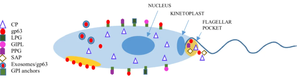
FIGURE 3:LEISHMANIA VIRULENCE FACTORS.. ... 23
FIGURE 4:PARASITE-HOST-VECTOR TRIANGLE ... 24
FIGURE 5:INTRACELLULAR MORPHOLOGY OF QUIESCENT DOG NEUTROPHIL ... 46
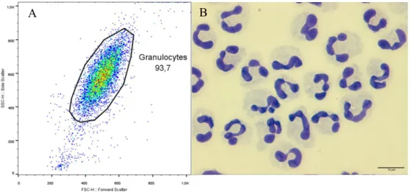
FIGURE 6:PURITY OF NEUTROPHILS ISOLATED FROM DOG PERIPHERAL BLOOD.. ... 66
FIGURE 7:ATTACHMENT AND PHAGOCYTOSIS OF L. INFANTUM PROMASTIGOTES BY DOG PMN. ... 69
FIGURE 8:LEVELS OF L. INFANTUM ASSOCIATED (INTERNALIZED OR BOUND) TO DOG PMN ... 70
FIGURE 9:LEVELS OF L. INFANTUM INTERNALIZATION BY DOG NEUTROPHILS. ... 71 FIGURE 10:PROMASTIGOTE UPTAKE BY NEUTROPHILS ... 72
FIGURE 11:ABILITY OF L. INFANTUM TO INDUCE DOG PMN CHEMOTAXIS. ... 73
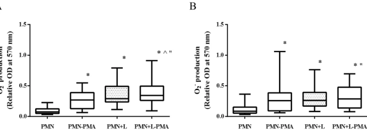
FIGURE 12:SUPEROXIDE (O2-) PRODUCTION BY PMN EXPOSED TO L. INFANTUM PROMASTIGOTES .. 74
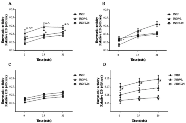
FIGURE 13:ENZYMATIC ACTIVITY OF DOG PMN PRIMARY GRANULES. ... 75
FIGURE 14:NET RELEASE EVALUATED BY IMMUNOLABLING. ... 77 FIGURE 15:EXTRACELLULAR INTERACTION BETWEEN DOG PMN AND L. INFANTUM
PROMASTIGOTES ... 78
FIGURE 16:NEUTROPHIL EXTRACELLULAR TRAPS (NET) LIMIT PARASITE SPREAD ... 79
FIGURE 17:GATING STRATEGY USED TO SELECT VIABLE, APOPTOTIC AND NECROTIC CELLS.. ... 80
FIGURE 18:LEVELS OF VIABLE AND APOPTOTIC DOG PMN AFTER EXPOSURE TO L. INFANTUM
PROMASTIGOTES ... 82
FIGURE 19:LEVELS OF NECROTIC DOG PMN... 83 FIGURE 20:LEVELS OF NECROTIC PMN EXPRESSING DIFFERENT AMOUNTS OF PROPIDIUM IODINE
(PI). ... 85 FIGURE 21:L. INFANTUM DELAYS APOPTOSIS OF PARASITIZED NEUTROPHILS………...86
FIGURE 22:VIABILITY OF L. INFANTUM PROMASTIGOTES AFTER EXPOSURE TO DOG PMN ... 87
FIGURE 23:LARGE PHAGOLYSOSOMES ENSURE PARASITE KILLING.. ... 88 FIGURE 24:PARASITE KILLING BY DOG NEUTROPHILS. ... 90
FIGURE 25:SIMPLIFIED OVERVIEW OF THE MAMMALIAN ARGININE PATHWAY ... 125
FIGURE 26:TIMELINE OF BLOOD COLLECTION, CELL ISOLATION AND CELL INFECTION ... 132
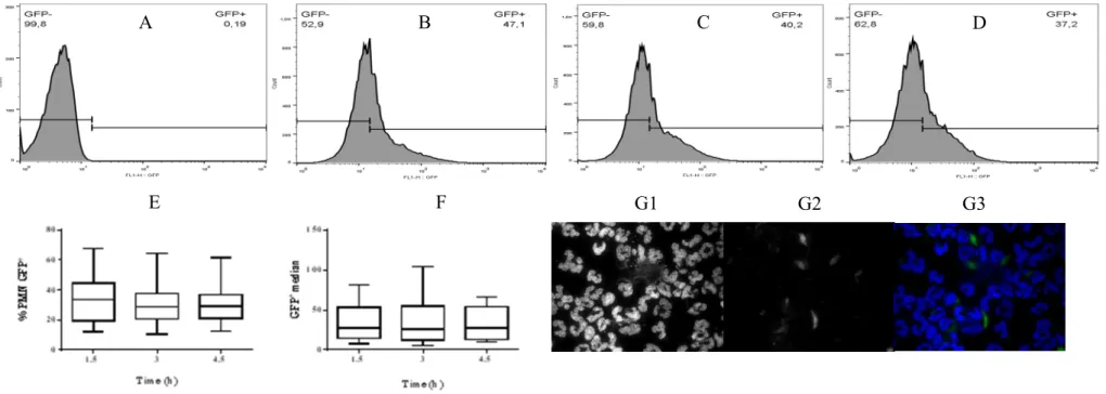
FIGURE 27:FLOW CYTOMETRY ANALYSIS OF MACROPHAGE DIFFERENTIATION ... 139
FIGURE 28:MACROPHAGE PURITY ASSESSED BY FLOW CYTOMETRY. ... 140
FIGURE 29:L. INFANTUM BIND OR ARE INTERNALIZED BY DOG MACROPHAGES... ... 142
FIGURE 30:DOG MACROPHAGE INTERNALIZING THE PARASITE AND ENSURING PARASITE
DIFFERENTIATION INTO THE AMASTIGOTE-LIKE FORM. ... 143
FIGURE 31:L. INFANTUM PROMASTIGOTES ADHERE BY THE TIP OF THE FLAGELLUM TO DOG
MACROPHAGES. ... 146
FIGURE 32:MORPHOLOGICAL CHANGES OF DOG MACROPHAGES. ... 148
FIGURE 33:STIMULATED DOG MACROPHAGES RELEASE HISTONE 1. ... 150 FIGURE 34:LEISHMANIA INFANTUM EXPOSED-MACROPHAGES RELEASE HISTONE H1 INTO THE
EXTRACELLULAR MEDIUM ... 151
FIGURE 35:DOG MACROPHAGES PROMOTE INTRACELLULAR PARASITE KILLING. ... 152
FIGURE 36:INTRACELLULAR KILLING OF L. INFANTUM PARASITES BY DOG MACROPHAGES. ... 153
FIGURE 37:NEUTROPHIL PHAGOCYTIZED PARASITES MAINTAIN THE VIABILITY AND THE
CAPACITY OF MULTIPLICATION. ... 154
FIGURE 38:APOPTOTIC NEUTROPHIL EFFEROCYTOSIS... 156 FIGURE 39:NEUTROPHIL EFFEROCYTOSIS DOCUMENTED BY TRANSMISSION ELECTRON
MICROSCOPY (TEM). ... 157
FIGURE 40:EFFEROCYTOSIS DOCUMENTED BY SCANNING ELECTRON MICROSCOPY (SEM). ... 158
FIGURE 41:DOG NEUTROPHIL ENSURE PARASITE TRANSFERENCE TO MACROPHAGES.. ... 160
FIGURE 42:TRANSFERENCE OF INTRACELLULAR L. INFANTUM PARASITES FROM NEUTROPHILS TO
MACROPHAGES. ... 162
FIGURE 43:PARASITE TRANSFERENCE DOCUMENTED BY SCAN ELECTRON MICROSCOPY (SEM). .. 163
FIGURE 44:NEUTROPHILS CO-CULTURED WITH MACROPHAGES RELEASE NEUTROPHIL
EXTRACELLULAR TRAPS.. ... 164
FIGURE 45:NEUTROPHIL EXTRACELLULAR TRAP RELEASE BY INFECTED PMN IN CO-CULTURE WITH MACROPHAGES. ... 165
FIGURE 47:INTERACTION BETWEEN DOG NEUTROPHILS AND MACROPHAGES AT THE EARLY PHASE OF L. INFANTUM INFECTION. ... 183
List of Tables
TABLE 1:TAXONOMIC CLASSIFICATION OF LEISHMANIA SPECIES THAT INFECT DOGS BASED ON
MULTILOCUS ENZYME ELECTROPHORESIS, ITS GEOGRAPHICAL DISTRIBUTION AND
VECTORS INVOLVED IN TRANSMISSION. ... 4
TABLE 2:CURRENT THERAPEUTIC PROTOCOLS FOR CANINE LEISHMANIOSIS. ... 17
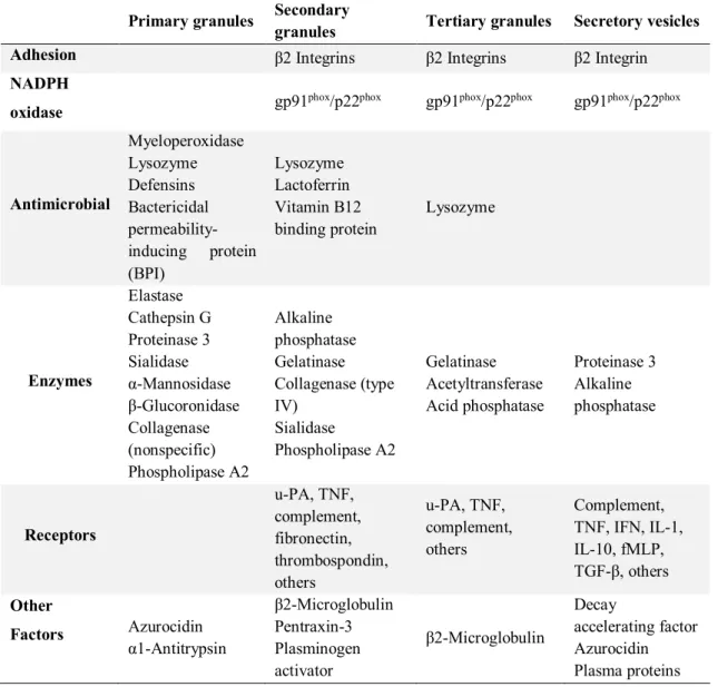
TABLE 3:CONTENT OF SECRETORY GRANULES AND OF SECRETORY VESICLES. ... 45
TABLE 4:FORWARD (FW) AND REVERSE (RV) PRIMERS USED TO DETECT DIFFERENT
MICROORGANISMS BY Q-PCR, THE RESPECTIVE PROBES AND BASE PAIR NUMBER (BP) OF THE AMPLIFIED FRAGMENTS. ... 58
Abbreviation List
< Less than
×g Times gravity
≈ Approximately equal to
2P-IVM Two-photon intra-vital microscopy
3’NT/NU 3’-nucleotidase/nuclease
Ag Antigen
AP-1 Activator protein-1
APC Antigen presenting cells
Arp2/3 Actin-related protein-2/3
Bcl-2 B-cell lymphoma 2
BH4 Tetrahydrobiopterin
BID Twice a day
bp Base pair
BPI Bactericidal permeability-inducing protein
C1q Complement component 3 subunit q
C3 Complement component 3
Ca2+ Calcium
CaM Calmodulin
Campto (S)-(+)-camptothecin
CanL Canine Leishmaniosis
CARD; NLRC Caspase activation and recruitment domain
catG cathepsin G
CCL2; MCP-1 C-C motif ligand 2 chemokine; monocyte chemotactic protein 1
CCR2+ Chemokine receptor 2 positive monocytes
CD Cluster of differentiation
Cdc42 Cell division control protein 42 homolog
CEDOC Chronic Diseases Research Center
CIE Counterimmunoelectrophoresis
CO2 Carbon dioxide
CP Cysteine protease
CPB Cysteine protease B
CPDA-1 Citrate Phosphate Dextrose Adenine-1
CpG Cytosine-phosphate-guanosine
CR Complement receptor
CSF Colony-stimulating factor
CSF-1R Colony-stimulating factor-1 receptor
CXCL8; IL-8 C-X-C motif chemokine 8; Interleukine-8
Cyt D Cytochalasin D
DAMP Damage-associated molecular patterns
DAPI Fluorescent nuclear dye 4’, 6-diamidino-2-phenylindole
DAT Direct agglutination test
DC Dendritic cells
DFMO Difluoromethyl ornithine
DLA-DRB1 Dog leukocyte antigen-DRB1
DNA Deoxyribonucleic acid
DTH Delayed-type hypersensitivity
e.g Exempli gratia
EDTA Ethylenediamine tetraacetic acid
EEA1 Early endosome antigen 1
eGFP Expressed green fluorescent protein
ELISA Enzyme-Linked Immunosorbent Assay
ER Endoplasmic reticulum
ESL-1 E-selectin ligand-1
ET Extracellular traps
Fas-FasL FAS-Fas ligand
FasR FAS receptor
FBS Fetal bovine serum
FcR Fc receptor
FcγR Fc gamma receptor
Fe2+ Iron ion
Fe-SODe Iron superoxide dismutase excreted
FITC Fluorescein isothiocyanate
FL1-H FL1 height channel
FL2-H FL2 height channel
FML Fucose mannose ligand
fMLP N-formyl-methionyl-leucyl-phenylalanine
FMN Flavin mononucleotide
FMV-UL Faculdade de Medicina Veterinária-Universidade de Lisboa
FnR Fibronectin receptors
FOXP3 Forkhead box P3 or scurfin
fPPG Filamentous proteophosphoglycans
FSC Forward-scattered light
FSC-A Forward scatter area
FSC-H Forward scatter height
Fw Forward
G-CSF Granulocyte colony-stimulating factor
GFP Green Fluorescent Protein
GIPL Glycosylinositol phospholipids
GM-CSF Granulocyte/macrophage colony-stimulating factor
gp63 Glycoprotein of 63kDa
gp91phox (Nox2) gp91 phagocytic oxidase
GPcR G-protein-coupled receptor
GTP Guanosine-5’-triphosphate
GTPases Guanosine triphosphatase
h Hour
H2A Histone H2A
H2O Water
H2O2 Hydrogen peroxide
HBSS Hanks’ Balanced Salt Solution
HCl Hydrochloric acid
HOCl Hypochlorous acid
HOP Heat-shock proteins organizer protein
HSC Haematopoietic stem cell
HSP Heat-shock proteins
HSP100 Heat-shock proteins 100 kDa
HVL Human visceral leishmaniosis
IAP Inhibitor of apoptosis protein
iC3b Inactivated C3b
ICAM Intercellular adhesion molecule
IFAT Indirect immuno-fluorescent antibody test
IFN Interferon
IFN-γR Interferon-gamma receptor
IgG Immunoglobulin G
IL Interleukin
IL-10R Interleukin 10 receptor
IL-12p40 Interleukin-12 subunit p40
IL-2R-α chain (CD25)
Interleukin 2 receptor alpha chain
iNOS Inducible nitric oxide synthase
IP-10; CXCL10 Interferon gamma-induced protein 10; C-X-C motif chemokine 10
IRF Interferon regulatory factor
JAK Janus kinase 1
JNK Jun N-terminal kinase
kDNA Kinetoplast deoxyribonucleic acid
LAMP Lysosome-associated membrane protein
L Leukotriene
LC Langerhans cells
LCF Leishmania chemotactic factor
LdAAP3 Leishmania donovani amino acid permease 3
LJL Recombinant Lutzomya longipalpus salivary protein
LPG Lipophosphoglycan
LPS Lipopolysaccharide
LTA Lipoteichoic acid
M1 Classically activated macrophages
M2 Alternatively activated macrophages
M6PR Mannose-6-phosphate receptor
MAC Membrane attack complex
MAP Mitogen-activated protein
MAPK; ERK Mitogen-activated protein kinase; extracellular signal-regulated kinase
MARCKS Myristoylated alanine-rich C kinase substrate
MARCO Macrophage receptor with collagenous structure
MBL Mannose-binding lectin
mCAT2B Mouse cationic amino acid transporter 2B
Mcl-1 Myeloid cell leukemia-1
M-CSF Macrophage colony-stimulating factor
MET Macrophage extracellular traps
MHCI Class I molecules of major of histocompatibility complex
MIP Macrophage inflammatory proteins
mM Milimolar
Mn2+ Manganese ion
MØ Macrophages
MPO Myeloperoxidase
mPPG Membrane bound proteophosphoglycans
MR Mannose receptor
mRNA Messenger ribonucleic acid
MRP MARCKS-related proteins
MT-ZVL Mountain-type-zoonotic visceral leishmaniosis
MyD88 Myeloid differentiation factor 88
N Normality
n1 Necrotic 1
n2 Necrotic 2
n3 Necrotic 3
NaCl Sodium chloride
NADPH; PHOX Nicotinamide adenine dinucleotide phosphate
NBT Nitroblue tetrazolium
NE Neutrophilic elastase
NET Neutrophil extracellular traps
NF-kB Nuclear factor-κB
NK Natural killer cells
NLR Nucleotide-binding and oligomerization domain (NOD)-like
receptors
NLRA Nucleotide-binding and oligomerization domain (NOD)-like
receptors acidic transactivation domain
NLRB Nucleotide-binding and oligomerization domain (NOD)-like
receptors baculoviral inhibitory repeat-like domain
NLRP Nucleotide-binding and oligomerization domain (NOD)-like
receptors pyrin domain
NOD Nucleotide-binding and oligomerization domain
NOD1 Nucleotide-binding and oligomerization domain-containing
protein 1
NOD2 Nucleotide-binding and oligomerization domain-containing
protein 2
nor-NOHA Nώ-hydroxy-nor-L-arginine
O2- Superoxide anion
OAT Ornithine aminotransferase
ºC degree Celsius
ODC Ornithine decarboxylaseTh2
OH-arg Nω-hydroxy-arginine
OM Optical microscopy
ONOO- Peroxynitrite
p Probability
p22phox (CYBA) p22 phagocytic oxidase
p38 MAPK p38 Mitogen-activated protein kinase
p38MAP p38 mitogen-activated protein
p40phox p40 phagocytic oxidase
p47phox p47 phagocytic oxidase
p67phox p67 phagocytic oxidase
PAD4 Peptidylarginine deiminase 4
PAMP Pathogen-associated molecular patterns
PBMC Peripheral blood mononuclear cells
PBS Phosphate-buffered saline
PCR Polymerase chain reaction
PFR Paraflagellar rod
PG Prostaglandin
pH Potential of hydrogen
PI Propidium iodide
PICD Phagocytosis-induced cell death
PIK Phosphatidyl inositol kinases
PK Protein kinase
PM Peritrophic matrix
PMA Phorbol myristate acetate
PMN Polymorphonuclear leukocytes
PO Per os, by mouth
PPARγ Peroxisome proliferator-activated receptor-γ
PPG Proteophosphoglycans
PpGalec Phlebotomus papatasi galactose binding protein
PpSP15 Phlebotomus papatasi salivary protein 15
PRR Pattern recognition receptors
PS Phosphatidylserine
PSG Promastigote secretory gel
PSGL-1 P-selectin glycoprotein ligand-1
PTX3 Pentraxin 3
q-PCR Quantitative- polymerase chain reaction
QuilA Quillaja saponin
Rac1 Ras-related C3 botulinum toxin substrate 1
RANTES Regulated on activation, normal T cell expressed and secreted
RFP Red fluorescent protein
Rho Ras homolog family
RhoA Ras homolog gene family, member A
RNA Ribonucleic acid
RNS Reactive nitrogen species
ROS Reactive oxigen species
RPMI Roswell Park Memorial Institute medium
RV Reverse
SBE STAT-binding elements
SC Subcutaneous
SCHN Schneider medium
SDS Sodium dodecyl sulfate
SEM Scanning electron microscopy
SID Once a day
Slc11a1; N-RAMP1 Solute carrier family 11 member 1
SNAP Semi-HMM-based Nucleic Acid Parser
SNARE Soluble-N-ethylmaleimide sensitive-factor accessory-protein (SNAP) receptor
SNP Single nucleotide polymorphism
sPPG Secreted proteophosphoglycans
SSC Side scatter
SSC-A Side scatter-area
SSC-H Side scatter-height
STAT Signal transducer and activator of transcription
STI1 Stress-inducible protein 1
Syt Synaptotagmin
T CD8+ Cytotoxic T cell
T-bet Type 1 associated transcription factor
TEM Transmission electron microscopy
TGF Transforming growth factor
Th1 T helper cells type 1
Th2 T helper cells type 2
TLR Toll-like receptors
TNF Tumor necrosis factor
TNT Tunneling nanotubes
TRAIL TNF-related apoptosis-inducing ligand
TRIF Toll-interleukin 1 receptor domain-containing adaptor inducing interferon-β
Tris-HCl Tris hydrochloride
Tyk2 Tyrosine kinase 2
U Unit
u-PA Urokinase-type plasminogen activator
USA United States of America
v/v; w/v Volume/volume; weight/volume
VAMP8 Vesicle-associated membrane protein 8
v-ATPase vesicular ATPase
VCAM Vascular cell adhesion molecule
VL Visceral leishmaniosis
vs Versus
WASP Wiskott-Aldrich syndrome protein
WHO World Health Organization
Y-NBT Yellow-colored nitroblue tetrazolium
1.1 CANINE LEISHMANIOSIS
1.1.1 Etiology and geographical distribution
Canine leishmaniosis (CanL) is a parasitic disease caused by a protozoan of the genus Leishmania transmitted to the domestic dogs (Canis familiaris) through the bite of a phlebotomine sand fly (Alvar et al, 2004; Dantas-Torres, 2009).
Leishmania belongs to the order Kinetoplastida, family Trypanosomatidae and genus Leishmania, which comprises the subgenera Leishmania and Viannia (Lainson et al, 1987) and several complexes. With some exceptions, all species of the subgenus Viannia are found in the New World, while those belonging to the subgenus Leishmania are isolated in the Old World. Species of L. mexicana complex are located in the New World and L. infantum (syn. L. chagasi) is present in both the New and Old Worlds (Maurício et al, 2000; Alvar et al, 2004; Bañuls et al, 2007; Baneth et al, 2008). Recent studies based on multilocus microsatellite typing and genomic studies support the hypothesis that L. infantum was introduced into the Americas by infected dogs of European settlers during the colonization of South America in the 16th century and that L. infantum and L. chagasi
are indistinguishable (Maurício et al, 2000; Kuhls et al, 2011; Leblois et al, 2011).
CanL is endemic in more than 70 countries from South and Central America, the Mediterranean region, Africa and Asia (Baneth et al, 2008; Solano-Gallego et al, 2009, 2011; Dantas-Torres et al, 2009, 2012).
At least 14 Leishmania species infect the dog (Saridomichelakis, 2009; Solano-Gallego et al, 2009; Cantacessi et al 2015) (Table 1). Presenting a wide geographical distribution in both the Old and New World, high prevalence of infection and disease in endemic areas, increased severity and life-threatening character associated with a zoonotic nature, L. infantum is probably the most representative species (Gramiccia and Gradoni, 2005; Saridomichelakis, 2009; Solano-Gallego et al, 2009; Saridomichelakis and Koutinas, 2014). However, at least 11 isoenzymatic variants (zymodemes) of L. infantum have been isolated in the dog (Gramiccia et al, 1992; Dereure et al, 1999; Gaskin et al, 2002).
Table 1: Taxonomic classification of Leishmania species that infect dogs based on multilocus enzyme electrophoresis, its geographical distribution and vectors involved in transmission. All species listed belong to the section Euleishmania, except L. colombiensis that belongs to Paraleishmania section. Adapted from Solano-Gallego et al (2009) and Cantacessi et al (2015).
1.1.2 Leishmania infantum infection
In the Mediterranean, seroprevalence of L. infantum infection varies from 5% to 30%, depending on the region and its ecological aspects (Solano-Gallego et al, 2009). It has been estimated that at least 2.5 million dogs are infected in Southwest Europe (Moreno and Alvar, 2002). However, surveys employing other detection methods, such as amplification of Leishmania deoxyribonucleic acid (DNA) or the detection of specific anti-Leishmania cellular immunity have revealed even higher infection rates, reaching 70% in some foci (Baneth et al, 2008).
Subgenus Species complex Species Geographical distribution Proven or suspected sand fly vectors
L. infantum Europe, America, Asia, Africa L. longipalpus, L. evansi, P. neglectus, P.
perniciosus, others
L. donovani East Africa P. orientalis , P. martini , P. rodhaini
L. archibaldi Sudan
L. tropica L. tropica India, Iran, Israel, Morocco, Syria P. sargenti
L. major Egypt, Saudi Arabia P. papatasi
L. arabica Saudi Arabia P. papatasi
L. amazonensis Brazil L. flaviscutellata , L. nociva , L. whitmani
L. mexicana Ecuador, USA L. ayacuchensis , L. olmeca
L. pifanoi Ecuador L. flaviscutellata , L. youngi
L. braziliensis Argentine, Bolivia, Brazil, Colombia, Peru, Venezuela L. intermedia , L. migonei , L. wellcomei , L. whitmani , others
L. peruviana Peru L. peruensis , L. verrucarum
L. panamensis Colombia, Ecuador, Panama L. hartmanni , L. gomezi , L. panamensis,
L. trapidoi
L. guyanensis Colombia L. anduzei , L. umbratilis , L. whitmani
L. colombiensis Venezuela L. hartmanni
Leishmania L. donovani L. major L. mexicana Viannia L. braziliensis L. guyanensis
The endemicity of the infection in Portugal was confirmed by a national survey carried out in January 2009 with the screening of 3974 dogs from all the 18 districts of mainland Portugal. Using the direct agglutination test (DAT), an overall prevalence of 6.31%, ranging from 0.88% to 16.16% was observed and the emergency of Beja, Castelo Branco and Portalegre districts as new foci was noticed. According to this study, the oldest foci of Trás-os-Montes and Alto Douro, Lisbon and Algarve regions lost their importance (Cortes et al, 2012). A seroprevalence of 21.3% was obtained in the northeastern Portugal, in the years 2008 and 2009, assessed by DAT and Enzyme-Linked Immunosorbent Assay (ELISA) (Sousa et al, 2011). In the municipality of Évora (southern Portugal) the seroprevalence evaluated by DAT in 2010 was 5.6% (Schallig et al, 2013). Recent studies realized in the south of Portugal revealed that 60.4% of dogs were quantitative polymerase chain reaction (q-PCR) positive for L. infantum (Maia et al, 2016) and that 16.06% were seropositive (Maia et al, 2013).
The distribution of CanL in Europe has been changing, probably due to socioeconomic and possibly to climate factors (Solano-Gallego et al, 2011). In endemic countries, L. infantum infection has spread northward, reaching the foothills of the Alps in northern Italy (Morosetti et al, 2009; Otranto et al, 2009; Baldelli et al, 2011), the Pyrenees in France (Chamaille et al, 2010) and northern Spain (Amusategui et al, 2004). The disease is also an important problem in non-endemic countries. Recently, numerous cases of CanL have been reported in northern countries, such as Germany (Menn et al, 2010), the Netherlands, United Kingdom (Shaw et al, 2009) and Poland (Kaszak et al, 2015) where the disease is considered as emergent. Dogs imported from or travelling to the Mediterranean region have increased the number of CanL cases in central and northern Europe (Solano-Gallego et al, 2011).
A survey done in a new focus of zoonotic visceral leishmaniosis (ZVL) in Northeastern Turkey showed a seroprevalence rate of infection of 7.2% (Sari et al, 2015). Several studies have described the presence of CanL in Israel and Palestine (Nasereddin et al, 2009). In Palestine 11.5% of dogs analyzed were positive for Leishmania DNA, whereas ELISA and culture revealed 7.5% and 1.5% of infection, respectively (Hamarsheh et al, 2012). A seroprevalence of 12.2% was described in Iran, where L. infantum is the principal causative agent of CanL (Mohebali, 2013). A recent review (Strelkova et al, 2015) summarized the available information about CanL in Southern Caucasus, Central
Asia, Crimean Peninsula and Russia. In Azerbaijan (Southern Caucasus), the seroprevalence assessed by IFAT ranged between 17.3% and 70.4%, depending on the region and in Georgia (Southern Caucasus), the seroprevalence assessed by DetectTM rK39 rapid test ranged between 8% and 28.1%. In Uzbekistan (Central Asia), a study carried out in 2007 and 2008 showed that 31.5% of symptomatic and 20.2% of asymptomatic dogs living in areas where visceral leishmaniosis (VL) had been previously diagnosed were seropositive when assayed by ELISA. In Crimean Peninsula, the parasite was not isolated from the dog and in Russia this possibility has not yet been investigated. In China the implementation of control programs resulted in the elimination of the disease from most endemic areas. Dogs are reservoirs of the mountain-type-ZVL (MT-ZVL) and canine infection has been reported at high rates in the western region (Lun et al, 2015), with a prevalence of 59.43% in the Sichuan province (Wang et al, 2011) and 77.21% in the Gansu province (Wang et al, 2006).
Epidemiological surveys in dogs and clinical cases of CanL have been reported in North Africa, namely Egypt with a seroprevalence of 10% (Morsy et al, 1983; Rosypal et al, 2013), Tunisia (Ben Said et al, 1992), Algeria (Harrat et al,1996), but also in countries of West Africa, namely Gambia (Desjeux et al, 1983) and Senegal (Faye et al, 2011) and of East Africa, like Kenya (Mutinga et al, 1980), Sudan (Dereure et al, 2000), Ethiopia (Kalayou et al, 2011) and Uganda (Millán et al, 2013). Even so, data from southern Africa are still scarce. Few cases of CanL have been reported in Angola and some of them were imported. A recent study established a frequency of infection of 1.0% by polymerase chain reaction (PCR) and 1.9% by serology (Vilhena et al, 2014).
The disease has been identified in the United States of America (USA) where it is considered an emerging problem in some dog breeds, with an estimated annual PCR prevalence greater than 20% on the Foxhound population (Petersen and Barr, 2009) and also identified in some provinces of southern Canada (Duprey et al, 2006). In South America, the infection prevalence was estimated in millions (Moreno and Alvar, 2002; Baneth et al, 2008). High seroprevalence rates were observed in Northern Brazil. For instance, in the state of Ceará 21.4% of stray dogs and 26.2% of domestic dogs are infected (Rondon et al, 2008) and in the state of Bahia 21.7% of stray dogs (Julião et al, 2007) and 23.4% of domestic dogs also have the parasite (Paranhos-Silva et al, 1996). In Margarita Island, Venezuela, 33.1% of dogs are seropositive (Zerpa et al, 2003). The
emergence of CanL in southern Brazil and northern Argentina has been associated with modifications in the ecology and distribution of the principal vector species, L. longipalpis (Salomon et al, 2008; Souza et al, 2009; Bravo et al, 2013).
1.1.3 Infection by “exotic” Leishmania species
Several Leishmania species responsible for the American Cutaneous Leishmaniosis also infect dogs. A high prevalence of L. braziliensis infection has been reported in Brazil, mainly in rural areas, where about 55% of dogs tested PCR positive in the blood (Dantas-Torres et al, 2010, Carvalho et al, 2015). One outbreak of cutaneous leishmaniosis caused by L. braziliensis and L. panamensis was observed between 2005-2009 in soldiers of the Colombian Army and in their dogs (Vélez et al, 2012). L. guyanensis and L. braziliensis were isolated from dogs living in Colombia (Santaella et al, 2011). Cases of dog natural infection caused by L. mexicana (Velasco-Castrejón et al, 2009; Arjona-Jiménez et al, 2012), L. amazonensis (Dias et al, 2011) and L. peruviana (Llanos-Cuentas et al, 1999) were also reported.
L. donovani, a primarily viscerotropic species present in the Indian subcontinent and East Africa was rarely identified in dogs living in eastern Sudan (Dereure et al, 2003), Sri Lanka (Nawaratna et al, 2009), India (Sharma et al, 2009), Bangladesh (Alam et al, 2013) and in Cyprus, where one dog was found to be co-infected with both L. infantum and L. donovani (Antoniou et al, 2008).
L. major, the main agent of the human cutaneous leishmaniosis in the Old World, widespread from West Africa to the Middle East and India can also infect dogs (Elbihari et al, 1987; Morsy et al, 1987; Pratlong et al, 2009; Baneth et al, 2016). Sporadic cases of canine infection by L. tropica were identified in Morocco, Iran, Crete, Greece and Turkey (Dereure et al, 1991; Guessous-Idrissi et al, 1997; Lemrani et al, 2002; Mohebali et al, 2005, 2011; Pratlong et al, 2009; Hajjaran et al, 2013; Toz et al, 2013; Ntais et al, 2013, 2014; Baneth et al, 2014). L. tropica produces human cutaneous disease in the Middle East, North Africa, Central Asia and some parts of southern Europe (Ionian Islands and Crete) and rarely human visceral leishmaniosis (HVL) (Sacks et al, 1995; Christodoulou
et al, 2012). Natural infection by L. arabica was also reported in the dog (Peters et al, 1986).
1.1.4 Dog as Leishmania host and reservoir
Canine L. infantum infection constitutes a veterinary and public health problem. From a public health point of view, the dog is considered the main peridomestic reservoir of the parasite in endemic areas (Ashford, 1996; Moreno and Alvar, 2002; Alvar et al, 2004; Gramiccia and Gradoni, 2005; Dantas-Torres and Brandão-Filho, 2006; Dantas-Torres, 2007). This statement is supported by the following observations. (i) Parasites isolated from dogs are indistinguishable from those obtained from humans; for example, L. infantum zymodeme MON-1, which is responsible for most of HVL cases in the Mediterranean basin is also the predominant zymodeme isolated from dogs (Pratlong et al, 2004). (ii) Dogs are highly susceptible to the parasite and the prevalence of infection is often high in endemic areas, with a large proportion of asymptomatic cases (Ashford, 1996; Quinnell et al, 1997; Dantas-Torres and Brandão-Filho, 2006). (iii) Dogs usually live in or next to human houses, which favors the maintenance of the domestic transmission cycle (Dantas-Torres and Brandão-Filho, 2006). (iv) Symptomatic dogs are infectious to the vector due to their high skin parasitism and even asymptomatic dogs can present the parasite (Gradoni et al, 1987; Molina et al, 1994; Travi et al, 2001; Michalsky et al, 2007; Laurenti et al, 2013) to sand fly, ensuring the parasite life cycle.
Furthermore, infected dogs are a source of parasite for phlebotomine sand flies of species L. longipalpis (Lainson et al, 1985) and P. perniciosus (Molina et al, 1994), the main vectors of L. infantum in the New and Old Worlds, respectively. These phlebotomine sand flies can easily adapt to the peridomestic environment or human dwellings and feed frequently on dogs (De Colmenares et al, 1995; Killick-Kendrick, 1999; Feliciangeli, 2004; Lainson and Rangel, 2005).
Despite these observations, only one study carried out in Iran has shown that child seropositivity increases significantly with village dog density and that dog ownership is a significant risk factor for children (Gavgani et al, 2002b). The presence of infected dogs in the vicinity of humans is certainly associated with an increase in the prevalence of
infection among sand flies, enhancing the entomologic inoculation rate and parasite transmission (Gavgani et al, 2002b; Solano-Gallego et al, 2009). However, the presence of an infected dog in the household does not appear to greatly increase the risk of infection to the family when transmission is already taking place in the region (Solano-Gallego et al, 2009).
For the “exotic” Leishmania species, dogs do not appear to have a significant role as a reservoir or have a particular importance as a source of parasites to humans (Ashford, 1996; Gramiccia and Gradoni, 2005; Dantas-Torres, 2007). Even so, the possibility of the dog being the reservoir has been discussed for some species, namely L. donovani (Alam et al, 2013; Rohousová et al, 2015), L. braziliensis (Dantas-Torres, 2007) and L. peruviana (Llanos-Cuentas et al, 1999).
1.1.5 Dynamics of dog infection
In endemic areas, when favorable conditions for disease transmission are present, the infection spreads rapidly and widely among the dog population (Baneth et al, 2008). Oliva et al (2006) demonstrated that all naïve Beagle dogs introduced in a hyperendemic area of southern Italy become infected after three transmission seasons. However, a large proportion of animals living in endemic areas present neither clinical signs nor clinicopathological abnormalities related to CanL and the presence of the parasite cannot be demonstrated, but seropositivity with basal antibody levels is common (Roura et al, 2013). In endemic areas, some dogs present a subclinical infection. They do not present neither clinical signs nor laboratory abnormalities compatible with CanL, but the presence of the parasite is confirmed (Solano-Gallego et al, 2009). Nevertheless, these healthy carriers contribute to the infection of sand flies, although to a lesser extent than the clinically diseased dogs (Baneth et al, 2008; Laurenti et al, 2013). The active disease (CanL) is present in a minority of dogs living in an endemic area and is characterized by clinical signs and/or clinicopathological abnormalities compatible with CanL and the infection is confirmed. Indeed, except in hyperendemic areas, CanL prevalence is lower than 10%, because the majority of infected dogs do not develop clinical disease (Solano-Gallego et al, 2009).
The balance between humoral and cellular immune response determine the clinical status of the dog. Delayed-type hypersensitivity (DTH) positive animals usually control the infection due to the presence of a demonstrable cellular immune response against L. infantum. A study carried out in a highly endemic area of Sicily (Italy) showed that 73.8% of outdoor dogs have a positive DTH reaction to leishmanin antigen (Lombardo et al, 2012). Antibody positive and DTH negative dogs are considered susceptible due to the suppressed cellular immune response and typically develop clinical disease in a short or middle term. However, factors such as immunosuppression and concomitant diseases can disrupt the immunological equilibrium of resistant dogs and lead to the development of leishmaniosis (Solano-Gallego et al, 2009).
1.1.6 Pathology and clinical manifestations
CanL is a chronic disease characterized by a wide range of clinical signs, consequence of the numerous pathogenic mechanisms that are involved in the disease, the different organs affected and the diversity of immune strategies mounted by the dog in response to infection (Koutinas and Koutinas, 2014). CanL is also a systemic disease, affecting potentially any organ, tissue or body fluid (Solano-Gallego et al, 2009; Koutinas and Koutinas, 2014). The involvement of the skin and other organs results from hematogenous dissemination of the parasite (Solano-Gallego et al, 2004).
Dogs suffering from CanL show depletion of T cells in lymphoid organs, which is compensated by an exuberant proliferation of B cells, plasma cells, histiocytes and macrophages (MØ), responsible for the generalized lymphadenomegaly and splenomegaly characteristics of the disease. Excessive granulomatous inflammation, for instance liver granulomas, is an attempt to counteract the cellular immune deficiency typical of the disease (Koutinas and Koutinas, 2014). The persistent antigenemia induce a polyclonal B cell response, leading to specific and non-specific antibody production that instead of giving protection causes organ damaging. Immune mediated pathology can be associated with the circulating antibodies, autoantibodies, namely anti-histone antibodies and immune complexes, which are deposited in the renal glomeruli, vascular endothelium, anterior uvea, and synovial membrane or linked to blood cells, such as platelets and red blood cells (Lopez et al, 1996; Terrazzano et al, 2006; Ginel et al, 2008;
Cortese et al, 2009; Paltrinieri et al, 2010; Koutinas and Koutinas, 2014). In cold weather, the production and precipitation of cryoglobulins in the blood vessel walls results in ischemic necrosis (Baneth and Solano-Gallego, 2012).
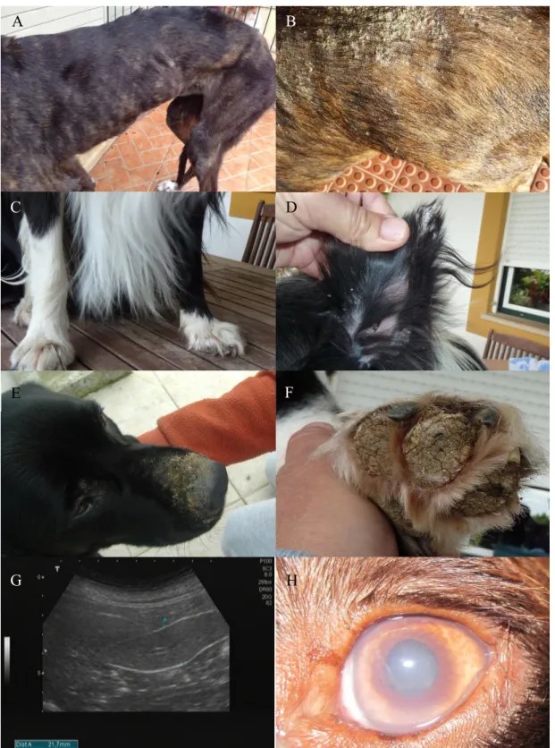
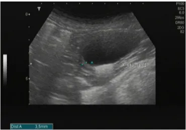
Common clinical signs include weight loss (Fig. 1A) and lymphadenopathy that are observed in 65.1% to 90% of dogs. However, lymph nodes may show normal size or even hypoplasia in advanced disease, particularly in nephritic dogs. Although splenomegaly is not a common clinical finding, since spleen usually does not present a high enlargement to be palpated on physical exam (Slappendel, 1988; Denerolle, 1996; Ciaramella et al, 1997; Koutinas et al, 1999; Saridomichelakis, 2009) it can be detected by imaging techniques (Paltrinieri et al, 2010) (Fig. 1G). The high parasite load frequently observed in the spleen seems to be related to the ineffective immune response mounted by the organ (Engwerda and Kaye, 2000; Lima et al, 2007). The pathological features observed in the bone marrow may explain the anemia, thrombocytopenia and others hematological abnormalities related to CanL. This organ usually shows high parasite load (Foglia et al, 2006; Koutinas and Koutinas, 2014; Momo et al, 2014).
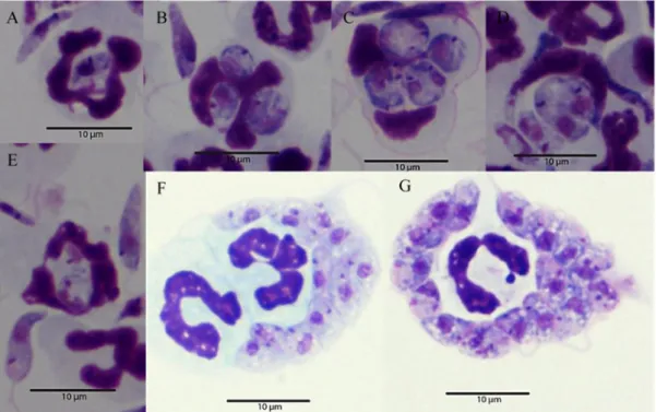
Skin lesions are perhaps the most common clinical signs, observed in 81% to 90% of the cases. Common dermatological entities are non-pruritic exfoliative dermatitis (Fig. 1B, D and E), ulcerative dermatitis, focal or multifocal nodular dermatitis, onychogryphosis (Fig. 1C), hyperkeratosis (Fig. 1F) (Denerolle, 1996; Ciaramella et al, 1997; Koutinas et al, 1999), mucocutaneous proliferative dermatitis and papular dermatitis (Ordeix et al, 2005; Lombardo et al, 2014). Interestingly, nodular and papular dermatitis are considered markers of high and low susceptibility to CanL, respectively. Nodular dermatitis seems to be more prevalent in boxers and has been associated with severely compromised parasite specific cellular immunity (Saridomichelakis and Koutinas, 2014). Conversely, papular dermatitis has been associated with low antibody levels and positive DTH, suggesting a specific immunocompetence and a favorable prognosis (Ordeix et al, 2005; Lombardo et al, 2014).
Leishmania amastigotes are present in both diseased and normal-looking skin of CanL dogs (Solano-Gallego et al, 2004). However, in clinically healthy infected dogs, parasite density in the skin seems to be related to its serologic state. Seronegative dogs do not have histological alterations and the parasite is only detected by PCR, suggesting that
such animals may not be able to transmit the parasite. On the contrary, histological alterations and Leishmania amastigotes are present in seropositive animals, although to a lesser degree compared with CanL dogs (Saridomichelakis and Koutinas, 2014).
The kidney is affected in virtually all dogs with CanL (Costa et al, 2003). However, azotemia is a less frequent laboratory finding (Baneth et al, 2008). Renal disease can progress from asymptomatic proteinuria to nephrotic syndrome and/or end-stage kidney disease (uremic syndrome) (Poli et al, 1991; Plevraki et al, 2006; Solano-Gallego et al, 2009). Renal lesions have been associated with high levels of circulating immune complex (Lopez et al, 1996) and high levels of anti-histone antibodies (Ginel et al, 2008). Occasionally, renal disease is the only CanL manifestation (Ciaramella et al, 1997).
Ocular disease is quite common in dogs (Solano-Gallego et al, 2009; Koutinas and Koutinas, 2014) and can be the only clinical sign. Anterior uveitis (Fig 1H), blepharitis and keratoconjunctivitis are the most frequent manifestations of ocular disease (Peña et al, 2000).
Bleeding disorders are frequently reported, but its pathogenesis has not been completely elucidated. Epistaxis can be the only clinical sign and the cause of death if profuse and uncontrolled (Ciaranmella et al, 1997; Jüttner et al, 2001; Petanides et al, 2008; Koutinas and Koutinas, 2014).
Arthritis is present in some animals (Blavier et al, 2001; Agut et al, 2003; Koutinas and Koutinas, 2014; Sbrana et al, 2014). Osteomyelitis characterized by osteolytic (de Souza et al, 2005) and/or osteoproliferative lesions can also occur. An increased intramedullary opacity on radiography can be seen and corresponds to bone sclerosis (Agut et al, 2003). Muscular involvement can be observed as a masticatory muscle myositis or skeletal muscle polymyositis usually with subclinical evolution (Vamvakidis et al, 2000; Paciello et al, 2009).
Digestive and neurologic signs are less frequent. Lesions of the digestive system can involve the oral cavity (Parpaglia et al, 2007; Viegas et al, 2012), liver (Rallis et al, 2005) or large intestine (Adamama-Moraitou et al, 2007). Various neurological signs have been