ESCOLA UNIVERSITÁRIA VASCO DA GAMA
MESTRADO INTEGRADO EM MEDICINA VETERINÁRIA
Mesenchymal Stem Cells Therapy in Veterinary Medicine:
Useful for Immune Mediated Disorders?
Inês Esteves Dias
Coimbra, julho 2018
i
ESCOLA UNIVERSITÁRIA VASCO DA GAMA
MESTRADO INTEGRADO EM MEDICINA VETERINÁRIA
Mesenchymal Stem Cells Therapy in Veterinary Medicine:
Useful for Immune Mediated Disorders?
Coimbra, julho 2018
Inês Esteves Dias
Aluna do Mestrado integrado em Medicina Veterinária
Constituição do Júri
Presidente do Júri: Professora Doutora Anabela Almeida
Arguente: Professora Doutora Isabel Dias
Orientador: Professor Doutor Pedro Carvalho
Orientador Interno
Professor Doutor Pedro CarvalhoCoorientadores Internos
Dr. Pedro Olivério; Dr. Luís BarrosOrientador Externo
Dr. Pedro OlivérioHospital Veterinário Universitário de Coimbra Dr. João Borges
ii
Dissertação do Estágio Curricular do Ciclo de Estudos Conducente aoiii
“Saímos pelo mundo em busca dos nossos sonhos e ideais. Muitas vezes colocamos em lugares inacessíveis o que está ao alcance das mãos.”iv
AgradecimentosAos meus orientadores, Professor Doutor Pedro Carvalho, Doutor Pedro Olivério e Doutor Luís Barros, por todo o apoio prestado durante a realização deste trabalho, por todos os conhecimentos transmitidos e pela confiança que depositaram em mim.
Aos meus pais que sempre me apoiaram, fizeram o possível e o impossível para que eu pudesse chegar até aqui e por me ensinarem a nunca desistir.
Aos meus avós pelo apoio incondicional em todas as etapas universitárias e pessoais e por estarem sempre presentes.
À minha irmã Carolina pelo apoio nos momentos difíceis e por todas as críticas construtivas que me permitiram evoluir.
A todos os meus amigos pelos magníficos momentos passados juntos, em especial à minha companheira Ana Queirós, Leonor Guimarães, Joana Veiga, Ana Sofia Alexandre, Beatriz Acabado e Carina Sacoor por terem sido a minha segunda família e por estarem sempre a meu lado em todos os momentos bons e especialmente nos menos bons.
A toda a equipa do Hospital Veterinário Universitário de Coimbra pela forma carinhosa com que me acolheu durante os meses de estágio, por todos os momentos de convívio e por todos os conhecimentos transmitidos. Um especial agradecimento às minhas colegas de estágio Isabel e Marcela por toda a amizade e companheirismo.
À Sandra por toda a paciência no esclarecimento de dúvidas e por ter sempre uma palavra amiga a dar.
À Ana Água pela amizade e apoio fundamental na realização deste trabalho.
E finalmente à Escola Universitária Vasco da Gama e a todos os professores e funcionários com quem tive o prazer de contactar e que me ajudaram a percorrer este caminho durante os seis anos de curso.
v
TABLE OF CONTENTSLIST OF FIGURES ... vi
LIST OF TABLES ... vi
LIST OF ABBREVIATIONS ... vii
RESUMO ... 2
ABSTRACT ... 3
1. INTRODUCTION ... 4
2. WHAT IS A STEM CELL? ... 5
3. STEM CELL CLASSIFICATION ... 5
4. MESENCHYMAL STEM CELLS ... 6
4.1. Mesenchymal Stem Cells Characterization ... 6
4.2. Minimal Criteria for Defining Mesenchymal Stem Cells ... 7
4.3. Origin of Mesenchymal Stem Cells ... 8
5. INTERACTION BETWEEN MESENCHYMAL STEM CELLS AND IMMUNE CELLS ... 9
5.1. Mechanisms of Mesenchymal Stem Cells Suppression of Innate Immune Cells ... 10
5.2. Mechanisms of Mesenchymal Stem Cells Suppression of Adaptive Immune Cells ... 10
6. THERAPEUTIC STRATEGIES WITH MESENCHYMAL STEM CELLS IN IMMUNE-MEDIATED DISORDERS ... 11
6.1. Canine Atopic Dermatitis ... 11
6.2. Feline Chronic Gingivostomatitis ... 13
6.3. Inflammatory Bowel Disease ... 16
6.4. Feline Asthma ... 18
7. DISCUSSION ... 222
8. CURRENT STATUS AND FUTURE PROSPECTS ... 24
vi
LIST OF FIGURESFigure 1: Classification of mesenchymal stem cells according to their origin and potentiality. Figure 2: Characterization of mesenchymal stem cells.
Figure 3: Possible origin of mesenchymal stem cells and their differentiation into mesodermal, endodermal and ectodermal cells.
Figure 4: Mechanism of action of mesenchymal stem cells and their interaction with immune cells. Figure 5: Factors that can influence mesenchymal stem cells therapy.
LIST OF TABLES
Table 1: 2015 updated guidelines of acute and chronic atopic dermatitis treatment. Table 2: Clinical trial carried out with mesenchymal stem cells in canine atopic dermatitis. Table 3: Conventional and immunomodulatory therapeutic approach to feline chronic gingivostomatitis.
Table 4: Clinical trials carried out with mesenchymal stem cells in feline chronic gingivostomatitis. Table 5: Conventional and immunomodulatory therapeutic approach to inflammatory bowel disease. Table 6: Clinical trials carried out with mesenchymal stem cells in canine and feline inflammatory bowel disease.
Table 7: Conventional and immunomodulatory therapeutic approach to feline asthma. Table 8: Clinical trials carried out with mesenchymal stem cells in feline asthma.
vii
LIST OF ABBREVIATIONSAD – Atopic Dermatitis
APMVEAC – Associação Portuguesa de Médicos Veterinários Especialistas em Animais de Companhia
ASC – Adipose-derived Stem Cell ATP – Adenosine Triphosphate
BID – Bis In Die (In Latin, two times a day) BMSC – Bone Marrow-derived Stem Cell CCECAI – Canine Chronic Enteropathy Clinical Activity Index
CD – Cluster Differentiation
CIBDAI – Clinical Inflammatory Bowel Disease Activity Index
DCs – Dendritic Cells
FCGS – Feline Chronic Gingivostomatitis FCV – Feline Calicivirus
FeLV – Feline Leukemia Virus FIV – Feline Immunodeficiency Virus HGF – Hepatocyte Growth Factor HO – Hemoxygenase
IBD – Inflammatory Bowel Disease
ICADA - International Committee on Allergic Diseases of Animals
IDO – Indolamine 2,3-dioxygenase IFN-γ – Interferon gamma
Ig – Immunoglobulin IL – Interleukin IM – Intramuscular IV – Intravenous kg - kilogram
LAD – Lower Airway Disease mg - miligram
mgc – microgram
MHC-II – Major Histocompability Complex II MSCs – Mesenchymal Stem Cells
NG2 – Neural/ Glial antigen 2 NK – Natural Killer
NO – Nitric Oxide
NSAID – Nonsteroidal Anti-Inflammatory Drug PDGF-Rβ – Beta-type Platelet-Derived Growth Factor Receptor
PGE2 – Prostaglandin E2
PO– Per os (In Latin, orally) SC – Subcutaneous
SID – Sem’el In Die (In Latin, once a day) TGF-β – Transforming Growth Factor β Th1 – T helper 1
Th2 – T helper 2
TID – Ter In Die (In Latin, three times a day) TNF – Tumor Necrosis Factor
1
Mesenchymal Stem Cells Therapy in Veterinary Medicine:Useful for Immune Mediated Disorders?
Inês Esteves Diasa, Pedro Olivério Pintoa,b, Luís Meireles Barrosa, Pedro Pires Carvalhoa,c
a
Departamento de Medicina Veterinária, Escola Universitária Vasco da Gama, Av. José R. Sousa Fernandes 197, Campus Universitário - Bloco B, Lordemão, 3020-210, Coimbra, Portugal ([email protected]) ([email protected]) ([email protected]) ([email protected])
b
Hospital Veterinário Universitário de Coimbra, Av. José R. Sousa Fernandes 197, 3020-210, Coimbra, Portugal ([email protected])
2
RESUMOAs células estaminais mesenquimatosas (MSCs) são células multipotentes, com capacidade de auto-renovação e diferenciação em diferentes tecidos de origem mesodérmica. Estas células constituem possíveis agentes terapêuticos para doenças auto-imunes, uma vez que apresentam uma notável capacidade imunomoduladora.
As doenças imunomediadas têm alcançado especial interesse em medicina veterinária, devido ao aumento da sua ocorrência na prática clínica. A maioria destas doenças é tratada de forma convencional com terapias imunomodeladoras (como glucocorticóides ou ciclosporina), estando associadas a uma série de efeitos adversos que limitam o seu uso a longo prazo, levando à necessidade de desenvolver novas estratégias terapêuticas mais eficazes e seguras.
Este estudo pretende realizar uma revisão crítica acerca do potencial terapêutico destas células no tratamento de algumas doenças auto-imunes (dermatite atópica canina, gengivostomatite crónica felina, doença inflamatória intestinal e asma felina) em comparação com o tratamento convencional estabelecido para as mesmas.
De acordo com ensaios clínicos realizados para estas doenças, a terapia baseada em MSCs permitiu a melhoria dos sinais clínicos ou mesmo a sua remissão total na maioria dos animais, com excepção da dermatite atópica canina na qual não se observaram melhorias.
Apesar das MSCs apresentarem um futuro promissor no tratamento da maioria destas doenças, a variabilidade nos resultados em alguns ensaios clínicos levaram à actual controvérsia entre autores relativamente à sua eficácia. Atualmente a terapia baseada em MSCs carece de uma análise mais profunda e detalhada, tendo em vista a sua padronização e melhor adequação aos resultados terapêuticos pretendidos, de forma a superar as suas limitações em estudos futuros.
Palavras-chave: Células estaminais mesenquimatosas; Imunomodulação; Dermatite atópica canina; Gengivostomatite crónica felina; Doença inflamatória intestinal; Asma felina
3
ABSTRACTMesenchymal Stem Cells (MSCs) are multipotent cells, with capacity for self-renewal and differentiation into tissues of mesodermal origin. These cells are possible therapeutic agents for autoimmune disorders, since they present remarkable immunomodulatory ability.
The increase of immune-mediated diseases in veterinary medicine has led to a growing interest in the research of these disorders and their medical treatment. Conventional immunomodulatory drug therapy (such as glucocorticoids or cyclosporine) is associated with numerous side effects that limit its long-term use, leading to the need for developing new therapeutic strategies, which are more effective and safe.
The aim of this study is to perform a critical review about the therapeutic potential of these cells in the treatment of some auto-immune disorders (canine atopic dermatitis, feline chronic gingivostomatitis, inflammatory bowel disease and feline asthma) compared with their conventional treatment.
MSC-based therapy in autoimmune diseases has been showing that this approach can improve clinical signs or even cause remission in most animals, with the exception of canine atopic dermatitis in which no improvement was observed.
Although MSCs present a promising future in the treatment of most of these disorders, the outcome variability in some clinical trials has led to the current controversy among authors regarding their efficacy. MSCs-based therapy is currently requiring a deeper and detailed analysis that allows its standardization and better adaptation to the intended therapeutic results, in order to overcome current limitations in future trials.
Key-words: Mesenchymal stem cells; Immunomodulation; Canine atopic dermatitis; Feline chronic Gingivostomatitis; Inflamatory bowel disease; Feline asthma
4
1. INTRODUCTIONRegenerative medicine results from the need of treating diseases for which modern medicine has not come up with an accessible or effective treatment (Ogliari et al., 2014). This kind of therapy relies on the use of ce+lls as therapeutic agents capable of regenerating damaged organs and tissues (Bogers, 2018). Despite the wide range of potential candidates that can be applied in these therapies, stem cells are the ones that have been continuously addressing greatest expectations within the scientific community (Jiménez & Guerrero, 2017).
Mesenchymal Stem Cells (MSCs) are multipotent cells, of non-hematopoietic origin, with self-renewal capacity (Zhao et al., 2016; Klinker & Wei, 2015). They derive from the embryonic layer of the mesoderm and under certain conditions they can differentiate into osteoblasts, chondrocytes, myocytes, β-pancreatic islets cells, neural cells, among others (Gao et al., 2016). These cells were first reported in the literature in 1968 (Friedenstein et al., 1968) and since then the interest in their therapeutic potential has grown, leading to the emergence of new researches.
Currently, MSCs are mainly used in veterinary medicine to treat musculoskeletal system disorders, considering their ability to differentiate into various tissues of mesodermal origin. In human medicine, therapies with MSCs are mostly used in the treatment of immune-mediated inflammatory and ischemic diseases (Schauwer et al., 2013; Figueroa et al., 2012).
The increase of immune-mediated diseases in veterinary medicine (such as canine atopic dermatitis, feline chronic gingivostomatitis, inflammatory bowel disease, feline asthma, among others) (Quimby & Borjesson, 2018; Arzi et al., 2017; Pérez-Merino et al., 2015) has led to a growing interest in the research of these disorders and their medical treatment. Conventional immunomodulatory drug therapy (such as glucocorticoids or cyclosporine) is associated with numerous side effects that limit its long-term use (Carrade & Borjesson, 2013), leading to the need for developing new therapeutic strategies which are more effective and safe.
MSCs have two important effects on the immune system, including an anti-inflammatory and immune-enhancing response. These cells regulate immune responses such as altering antibody production by B lymphocytes, shifting T-lymphocyte subtypes, and inducing immune tolerance to allogeneic transplants (Peroni & Borjesson, 2011). However, their immunomodulatory capacity is not yet fully understood and mixed results concerning immunomodulatory therapies with MSCs still exist, which requires further research and scientific clarification (Gao et al., 2016).
The aim of this study is to perform a critical review about the therapeutic potential, current status and future prospects of these cells in the treatment of immune mediated diseases in veterinary medicine. Another goal is to point out the advantages and disadvantages of a treatment drawn with stem cells in comparison to other conventional treatments of these particular diseases.
5
2. WHAT IS A STEM CELL?In traditional textbooks, stem cells were believed to be at the origin of all main types of tissue and it was also stated that differentiation occurred unidirectionally and irreversibly, once they were totally or partially differentiated in a certain type of cell, they could not differentiate again. Currently there is already knowledge that these cells are characterized by their plasticity and can transform more than what was originally thought (Almeida, 2013; Harman, 2013).
Stem cells are unspecialized cells that share two important characteristics, they can self-renew indefinitely or differentiate into more mature cells with specialized functions (Ogliari et al., 2014; Vogelstein et al., 2002).Their self-renewal capacity allows a limited number of mother cells of a tissue or an organ to be kept undifferentiated for a large period of time, functioning as a reservoir in face of certain physiological and pathological needs (Jiménez & Guerrero, 2017). These cells have been identified in the inner cells mass of the early embryo, in some tissues of the fetus, the umbilical cord and placenta, and in several adult organs and tissues (Vogelstein et al., 2002).
3. STEM CELL CLASSIFICATION
Stem cells can be classified considering their potentiality and origin (Figure 1). According to their potentiality they can be divided into totipotent, pluripotent, multipotent and unipotent cells (Jiménez & Guerrero, 2017; Simara et al., 2013; Spencer et al., 2011).
Totipotent cells are able to originate any type of cell from the three germinal layers (endoderm, mesoderm and ectoderm) as well as extraembryonic annexes(e.g. placenta and umbilical cord). Pluripotent cells are able to originate any type of cell from the three germinal layers, but not from extraembryonic annexes. Pluripotent stem cells do not exist in the adult organism. They can be obtained from embryos (embryonic stem cells) or from the transformation of cells from adult organisms which are modified in the lab – pluripotent induced cells (Ramalho-Santos, 2013).
Multipotent cells are partially specialized cells capable of generating a certain number of cell types, presumably from its own original germinal layer.
Unipotent or somatic cells are able to differentiate in only one type of cell (e.g. spermatogonia).
According to its origin they can be categorized as embryonic, extraembryonic and adult cells. Embryonic cells are present in the blastocyst (inner cell mass of an embryo five days after fertilization of the egg by the sperm) (Ogliari et al., 2014). Despite their enormous potential, their application in cell therapy carries a series of inconveniences. Obtaining these embryonic cells is a much more complex and expensive procedure than other types of stem cell, there is a larger risk of tumorigenesis and immune rejection and there are also moral and legal restrictions to its use (Jiménez & Guerrero, 2017; de Bakker et al., 2013; Harman, 2013).
6
Extraembryonic cells are located in the extraembryonic annexes (e.g. placenta, amnion or umbilical cord) which surround the fetus during pregnancy. These cells are multipotent and have null immunogenicity since they do not reveal major histocompability complex class II (MHC-II), for this reason they can be applied in individuals different from the donor (allogeneic therapy). Unlike embryonic stem cells, these cells are easier to obtain, they do not produce teratomas or suffer moral or legal restrictions (Jiménez & Guerrero, 2017).Adult cells are originated from tissues of endodermal, mesodermal or ectodermal lineages (de Bakker et al., 2013). Adult MSC have been shown to be nontumourigenic and nonimmunogenic. The activation and proliferation of adult stem cells is the most important mechanism of tissue renewal on both human beings and animals (Jiménez & Guerrero, 2017). Of all adult stem cells available, bone marrow (BMSC) and adipose derived (ASC) mesenchymal stem cells are the most commonly used (Gonçalves et al., 2014).
Figure 1 - Classification of mesenchymal stem cells according to their origin and potentiality (adapted from Jiménez & Guerrero, 2017).
4. MESENCHYMAL STEM CELLS
4.1. Mesenchymal Stem Cells Characterization
MSCs, also known as mesenchymal stromal cells, are multipotent cells, of non-hematopoietic origin, with self-renewal capacity present in connective tissue throughout the body (De Witte et al., 2016; Zhao et al., 2016) . They have been isolated from different adult tissues (such as bone marrow, adipose tissue, peripheral blood, muscle, periodontal ligament, articular cartilage, periosteum) and from extraembryonic tissues (for instance umbilical cord blood, membrane and amniotic fluid) (Jiménez & Guerrero, 2017).
7
In veterinary medicine, MSCs are obtained mainly from adipose tissue and bone marrow, taking into consideration their location and harvesting (Gonçalves et al., 2014).MSCs play an important role in the regulation of the immune system. Moreover, they are relatively easy to isolate and can be expanded in culture. Although MSC are not part of the immunologic system according to the prearranged definitions (Hoogduijn, 2015), they interact with all kinds of immunologic cells. They produce a great variety of anti-inflammatory and pro-inflammatory factors, among which are cytokines, chemokines and prostaglandins, which target immune cells and affect their function (De Witte et al., 2016). It is noteworthy that MSCs are safe, non-teratogenic and can be used for tissue regeneration and repair (Zhao et al., 2016).
These cells present remarkable pleiotropic properties, including antiapoptosis, angiogenesis, growth factor production, antifibrosis and chemiotaxis (Glenn & Whartenby, 2014). The MSC ability to differentiate into parenchymal cells of the mesoderm has become one of the main criteria to define their identity. Although recent data suggest that under appropriate culture conditions MSCs may also be induced to transdifferentiate into ectoderm (epithelia and neurons) and endoderm cells (muscle cells, lung cells and gut epithelial cells) (Zhao et al., 2016; Glenn & Whartenby, 2014) (Figure 2).
4.2. Minimal Criteria for Defining Mesenchymal Stem Cells
The pleiotropic nature of MSCs has presented a challenge in their identification (Glenn & Whartenby, 2014). In order to create a broader consensus on the universal characterization of MSCs, and facilitate the exchange of data among investigators, the International Society for Cellular Therapy formulated minimal criteria for defining MSCs. These criteria include plastic adherence, capability for differentiation towards osteoblasts, adipocytes and chondroblasts under standard in vitro conditions, cell surface expression of CD73, CD90, CD105 and absence of CD14, CD19, CD34 and MHC-II (Bateman et al., 2017; Jiménez & Guerrero, 2017; Malhotra et al., 2016; Dominici et al., 2006) (Figure 2).
8
Although stem cell scientists continue to develop more stringent criteria, these basic criteria are generally accepted as the baseline for declaring a cell type as a MSC (Harman, 2013).4.3. Origin of Mesenchymal Stem Cells
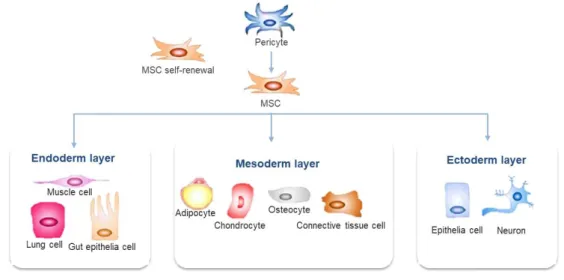
The origin of MSCs and their development is not yet fully understood (Gazdic et al., 2015). Although MSC had formerly been isolated from bone marrow, they have already been withdrawn from the stroma of multiple organs and tissues including the adipose tissue, the tonsils, the umbilical cord, the skin and the dental pulp. Crisan and co-workers suggest as hypothesis that MSC generate from pericytes (Figure 3). Pericytes are perivascular cells which inhabit in multiple organ systems. This group has identified pericytes in multiple human organs (skeletal muscle, pancreas, adipose tissue and placenta) based on the expression of cell markers: CD146, NG2 e PDGF-Rβ. They found out that these cells expressed typical MSC markers and in a specific culture medium they could differentiate into myocytes, osteocytes, chondrocytes and adipocytes (Esteves & Donadeu, 2017; Glenn & Whartenby, 2014; Crisan et al., 2008).
Although the study has not directly monitored the probable in vivo transaction of the pericytes to MSC, they recognized pericytes as potential progenitor cells to non BMSCs (Glenn & Whartenby, 2014; Crisan et al., 2008). Nevertheless, on the regenerative medicine field this hypothesis does not gather everyone’s consensus and acceptance.
Figure 3 – Possible origin of mesenchymal stem cells and their differentiation into mesodermal, endodermal and ectodermal cells (adapted from Glenn & Whartenby, 2014).
9
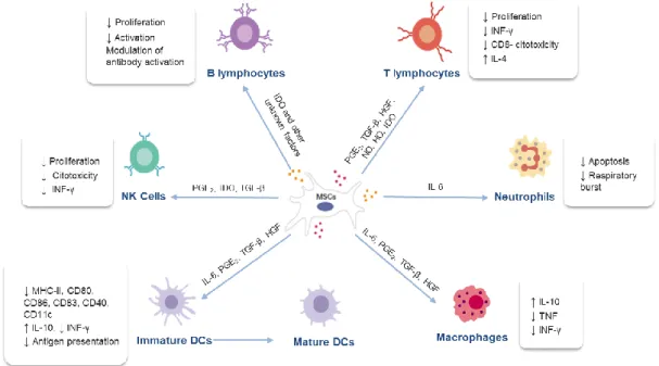
5. INTERACTION BETWEEN MESENCHYMAL STEM CELLS AND IMMUNE CELLSMSCs have two important effects on the immune system, including an anti-inflammatory and immune-enhancing response (Peroni & Borjesson, 2011). These cells release more than 200 bioregulatory products that have antimicrobial, immunomodulatory, antifibrotic, antiapoptotic, hematopoietic stem cells support, chemoattraction, angiogenesis, neuroprotective and mitogenic functions (Jiménez & Guerrero, 2017).
MSCs are capable of interacting with various types of immune cells, including T cells, B cells, natural killer (NK) cells, dendritic cells (DCs), macrophages / monocytes and neutrophils (Li & Hua, 2017) affecting both the innate and humoral immune responses (Peroni & Borjesson, 2011). It is ambiguous whether MSCs should be classified as “immunosuppressive,” suggesting a nonspecific downregulation of the immune system, or rather if they induce an “immune tolerance,” suggesting a more specific suppression of aberrant immune responses. What seems to be clear is the fact that the MSCs’ immunomodulatory ability depends on several factors such as MSC activation, MSC tissue of origin, MSC doses, time of administration of MSCs, and MSC contact with cells of the immune system (Peroni & Borjesson, 2011).
Although the underlying mechanisms of MSC immunomodulation have yet to be elucidated, it is believed that immunomodulation first takes place through paracrine effects by producing immunomodulatory mediators, including nitric oxide (NO), indoleamine 2,3-dioxygenase (IDO) (Yang et al., 2018), transforming growth factor-β (TGF-β), hepatocyte growth factor (HGF), hemoxygenase (HO), interleukin (IL)-6 and prostaglandin E2 (PGE2), and they may also occur through cell-cell direct
contact (Samsonraj et al., 2017; Gao et al., 2016; Kim & Cho, 2015) (figure 4).
Figure 4 – Mechanism of action of mesenchymal stem cells and their interaction with immune cells (adapted from Jiménez & Guerrero, 2017).
10
5.1. Mechanisms of Mesenchymal Stem Cells Suppression of Innate Immune CellsThe innate immune cells play an important role in the organism homeostasis and are the first defence line against invading pathogens such as viruses and bacteria. The cells belonging to this system respond promptly in a non-specific form to pathogens (Glenn & Whartenby, 2014). This defence response is based on inflammation. During inflammation there are certain changes in the tissues caused by microbial invasion or tissue damage, resulting in the increase of blood flow and the local accumulation of cells which may attack or destroy invaders. The innate immunity has a cellular defence line (neutrophils, monocytes / macrophages, DCs and NK cells) and an enzymatic defence line (these enzymes form what is known as the complement system) (Tizard, 2004).
MSCs act on the immunity system through three main mechanisms. One of those mechanisms consists in activating proinflammatory monocytes and macrophages. In the presence of MSCs and their soluble factors (IL-6, PGE2, TGF-β, HGF), the M1 classic macrophages possessing
proinflammatory functions, become active in M2 anti-inflammatory macrophages, which are
characterized by high expression of IL-10, low production of tumour necrosis factor (TNF) and low production of interferon gamma (IFN-y) (Kim & Cho, 2015; Maggini et al., 2010; Kim & Hematti, 2009). MSCs can also inhibit the differentiation of monocytes into mature DCs by releasing soluble factors such as IL-6, PGE2, TGF-β, HGF. These tolerogenic DCs produce high levels of IL-10 and have low
capacity to stimulate proliferation of allogeneic T cells in a mixed lymphocyte reaction (Kim & Cho, 2015; Li et al., 2008)
Finally, MSCs likewise inhibit NK cells proliferation and cytotoxicity, which can require cell-to-cell contact or can be mediated by soluble factors, including mainly PGE2 and IDO, but also TGF-β (Zhao
et al., 2016; Kim & Cho, 2015; Spaggiari et al., 2008).
5.2. Mechanisms of Mesenchymal Stem Cells Suppression of Adaptive Immune Cells
The acquired immune system is responsible for recognizing foreign invaders, destroy them and retain the memory after first antigen contact. If the animal finds the same antigen a second time, the immunologic system will respond in a more effective and prompt way. The acquired immune system is based on two main branches; one is called "humoral immune response" and is mediated by B lymphocytes that produce antibodies against exogenous invaders. The other major branch is called “cell-mediated immune response” and is mediated by T lymphocytes which act directly against endogenous invaders that invade cells (Tizard, 2004).
MSCs are able to suppress the proliferation of T cells and modulate their response through the secretion of several soluble factors (PGE2, TGF-β, HGF, NO, HO, IDO) or through cell-to-cell contact.
In an environment composed by strong inflammatory components, MSCs have the ability to change the T helper 1 (Th1) proinflammatory profile into a T helper 2 (Th2) anti-inflammatory profile (Kim & Cho, 2015; Stagg & Galipeau, 2013).
11
The effects of MSCs on B cells remains contradictory, although there is evidence that MSCs have close interactions with B cells. MSCs are able to inhibit B cell proliferation through cell-to-cell contact and through an arrest in the G0/G1 phase of the cell cycle (Corcione et al., 2006). Moreover, MSCs suppress plasma cell differentiation induced by allostimulation and immunoglobulin (Ig) production. Studies have also suggested that although MSCs are able to suppress B cells which are activated by several stimuli, they are incapable of modulating naive or memory B cells (Zhao et al., 2016; Kim & Cho, 2015; Carrade & Borjesson, 2013; Franquesa et al., 2012).Concluding, MSCs regulate immune responses such as altering antibody production by B lymphocytes, shifting T-lymphocyte subtypes, and inducing immune tolerance to allogeneic transplants due to the lack of MHC-II expression and costimulatory molecules such as CD40, CD80 and CD86, since they escape the recognition and action of T cells and NK receptors (Peroni & Borjesson, 2011). However, their immunomodulatory capacity is not yet fully understood and there are still mixed results concerning immunomodulatory therapies with MSCs (Gao et al., 2016). A deeper understanding of the mechanism through which the MSCs deriving from veterinary species modulate inflammation and contribute to the healing will beneficiate human beings as well as animals (Carrade & Borjesson, 2013).
6. THERAPEUTIC STRATEGIES WITH MESENCHYMAL STEM CELLS IN IMMUNE-MEDIATED DISORDERS
6.1. Canine Atopic Dermatitis
Atopic dermatitis (AD) is the most frequent dermatopathy in dogs and shares many characteristics with the human disease (Jiménez & Guerrero, 2017). This chronic multifactorial disorder affects ≈ 8.7% dogs and is associated with breed predilections, polymorphisms at specific gene loci, altered gene expression and specific allergens (Nuttall, 2010). It is characterized by a dysfunction of the skin barrier due to changes in lipid (ceramides) and protein (filaggrin) composition, modifications in the stratum corneum and loss of water from the transepidermis which predisposes to a higher allergen penetration (Jiménez & Guerrero, 2017; Bizikova et al., 2015a; Bizikova et al., 2015b; Roque et al., 2012; Nuttall, 2010). The skin of atopic dogs tends to produce less antimicrobial agents (defensins, cathelicidins, etc.) usually leading to secondary infection. Infectious agents such as Staphylococcus pseudointermedius, Staphylococcus intermedius and Malassezia pachydermatis worsen the clinical presentation of AD (pyoderma and otitis) and induce a phenomena of allergic sensitisation with large amounts of lgE antibodies (Jiménez & Guerrero, 2017; Pucheu-Haston et al., 2015). When allergenic load overcomes a certain threshold, mast cells will activate leading to consequent allergic response responsible for its clinical presentation (Nuttall, 2010).
12
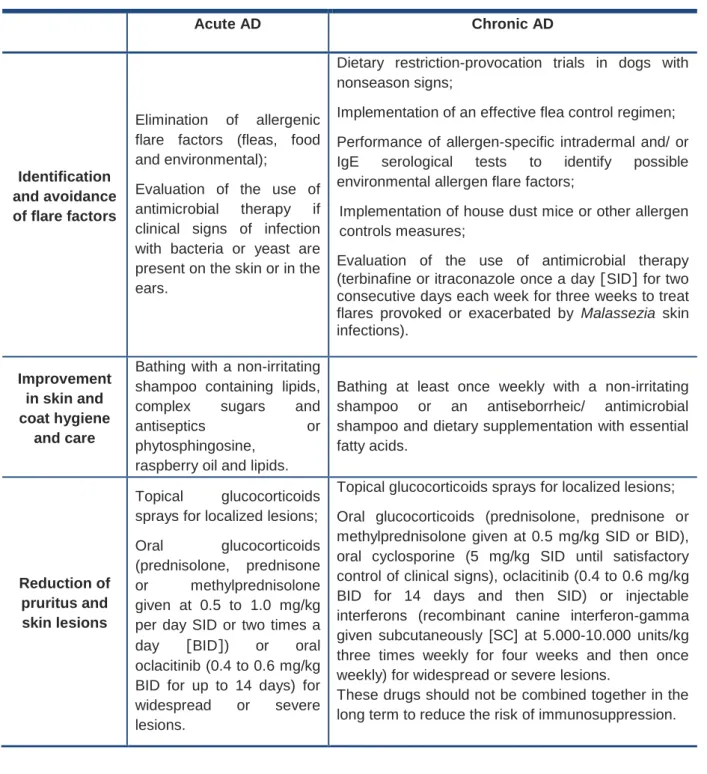
According to the 2015 updated guidelines from the International Committee on Allergic Diseases of Animals (ICADA), the treatment of acute and chronic AD is based on three key points, as described in table 1 (Olivry et al., 2015).Table 1 – 2015 updated guidelines of acute and chronic atopic dermatitis treatment (Olivry et al.,
2015). Acute AD Chronic AD Identification and avoidance of flare factors Elimination of allergenic flare factors (fleas, food and environmental);
Evaluation of the use of antimicrobial therapy if clinical signs of infection with bacteria or yeast are present on the skin or in the ears.
Dietary restriction-provocation trials in dogs with nonseason signs;
Implementation of an effective flea control regimen; Performance of allergen-specific intradermal and/ or IgE serological tests to identify possible environmental allergen flare factors;
Implementation of house dust mice or other allergen controls measures;
Evaluation of the use of antimicrobial therapy (terbinafine or itraconazole once a day [SID] for two consecutive days each week for three weeks to treat flares provoked or exacerbated by Malassezia skin infections).
Improvement in skin and coat hygiene
and care
Bathing with a non-irritating shampoo containing lipids,
complex sugars and
antiseptics or
phytosphingosine, raspberry oil and lipids.
Bathing at least once weekly with a non-irritating shampoo or an antiseborrheic/ antimicrobial shampoo and dietary supplementation with essential fatty acids.
Reduction of pruritus and skin lesions
Topical glucocorticoids sprays for localized lesions;
Oral glucocorticoids
(prednisolone, prednisone
or methylprednisolone
given at 0.5 to 1.0 mg/kg per day SID or two times a
day [BID]) or oral
oclacitinib (0.4 to 0.6 mg/kg BID for up to 14 days) for
widespread or severe
lesions.
Topical glucocorticoids sprays for localized lesions; Oral glucocorticoids (prednisolone, prednisone or methylprednisolone given at 0.5 mg/kg SID or BID), oral cyclosporine (5 mg/kg SID until satisfactory control of clinical signs), oclacitinib (0.4 to 0.6 mg/kg BID for 14 days and then SID) or injectable interferons (recombinant canine interferon-gamma given subcutaneously [SC] at 5.000-10.000 units/kg three times weekly for four weeks and then once weekly) for widespread or severe lesions.
These drugs should not be combined together in the long term to reduce the risk of immunosuppression.
With the aim of preventing the reappearance of clinical signs, some strategies can be developed, such as avoidance of known flare factors, consideration of proactive intermittent topical glucocorticoid therapy and implementation of allergen-specific immunotherapy, if feasible (Olivry et al., 2015).
13
Some adverse effects are seen with these treatments, especially with its long-term use. Unfortunately, due to AD pathophysiology, glucocorticoids are frequently needed. Systemic administration of these drugs might result in polyuria, polydipsia, polyphagia, changes of behaviour (including aggressiveness) and, depending on the initial dose, iatrogenic hyperadrenocorticism (Olivry et al., 2015; Nuttall, 2010).Over the last few years, the immunomodulatory effect of MSCs therapy has been described in animal models and in human beings, showing a significant improvement in the clinical presentation by inhibiting the activation of T and B cells and consequent release of anti-inflammatory cytokines (IL-10, TGF-β), by decreasing the proliferation of IL-4 and INF, and by decreasing the production of lgE (Jiménez & Guerrero, 2017).
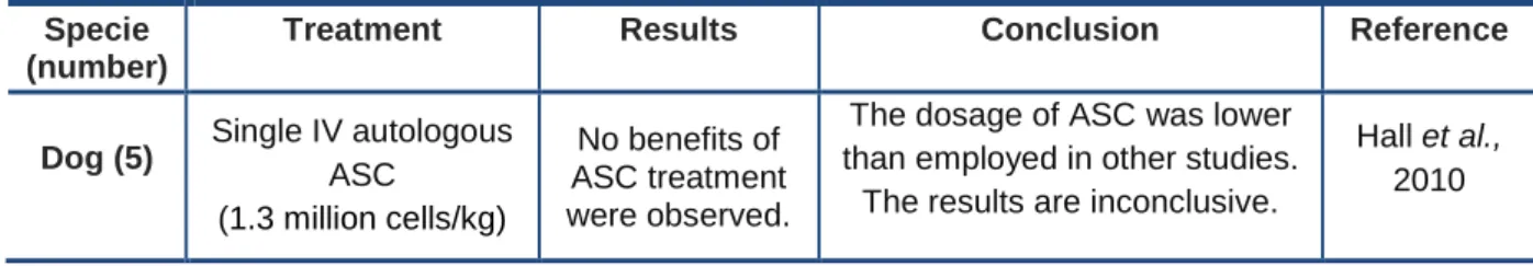
Hall et al. carried out a clinical trial with five AD canine patients (Table 2). All the patients were treated with a single dose of autologous ASC. The dosage of intravenous (IV) 1 x 10⁶ cells (1.3 million cells/kg) applied in this trial was substantially lower than the dosage applied in other trials and lower than the dosages usually applied in human trials (≥2 x 10⁶ / kg of body weight). Although the injections had been considered safe, no signs of progress were observed during this trial with the ASC treatment (Hall et al., 2010).
Jiménez and Guerrero have also published in the Clindervet journal a study on the use of MSC in
veterinary dermatology, where they report a successful case of CAD with MSC-based treatment. They
obtained significant improvement in clinical signs with no complications mentioned (Jiménez & Guerrero, 2017).
Table 2 – Clinical trial carried out with mesenchymal stem cells in canine atopic dermatitis.
Specie (number)
Treatment Results Conclusion Reference
Dog (5) Single IV autologous ASC
(1.3 million cells/kg)
No benefits of ASC treatment were observed.
The dosage of ASC was lower than employed in other studies.
The results are inconclusive.
Hall et al., 2010
6.2. Feline Chronic Gingivostomatitis
Feline chronic gingivostomatitis (FCGS) is a severe, idiopathic, inflammatory oral disease characterized by severe inflammation of the gingiva, buccal mucosa and caudal oral mucosa, that affects approximately 0.7-10% of the general cat population (Greenfield, 2017; Arzi et al., 2016; Winer et al., 2016). The aetiology of FCGS is poorly understood, nonetheless it has been suggested that microbial factors and alterations in the innate immune response may play an important role in the pathogenesis of this disorder (Kouki et al., 2017). Researchers believe that the factor contributing the most to the development of this disorder is the presence of bacterial plaque and that the development of FCGS is due to an immune abnormality, specifically related to the inflammatory mediators produced
14
by lymphocytes and plasma cells in response to infectious agents (e.g. feline calicivirus [FCV], feline leucemia [FeLV], feline herpesvirus, feline immunodeficiency virus [FIV] and Bartonella henselae). However, these infectious agents may not be responsible for the disease, and rather be mere contributors to the patient’s morbidity in the healing phase of the treatment (Greenfield, 2017; Lommer, 2013). Histologically, the injuries in cats are characterized by an inflammation with lymphocytes, mostly effector T cells and B cells (Quimby & Borjesson, 2018).This disorder causes painful mucosal lesions that markedly reduce the quality of life. Clinical signs vary from pain and moderate to severe oral discomfort, inappetence, loss of weight, reduced grooming and ptyalism (Arzi et al., 2017).
According to the Portuguese Association of Veterinarians Specialized in Companion Animals (APMVEAC)1 and collected data from Wild West Veterinary Conference in 2015 (Lappin, 2015), so far there is no treatment against FCGS which is 100% effective, and this may result in euthanasia of several affected cats. Approximately 70% of cats respond to standard treatment consisting of total or partial teeth extraction. The remaining 30% do not react to teeth extraction and require therapy with antibiotics, corticosteroids and other pain relief medication throughout their lives (Arzi et al., 2017; Arzi et al., 2016).
A successful treatment requires oral bacteria minimization (Table 3), therefore the therapeutic plan should be started with the improvement of the animal’s oral hygiene (e.g. chlorhexidine in paste or gel BID) (Greenfield, 2017; Johnston, 2015). Nevertheless this treatment carried out by animal owners is generally not rewarding since cats do not tolerate teeth cleaning and regular mouth washes very well. If the mucosa of canines or incisors is inflamed, these teeth should be removed and total extraction of teeth might be necessary (Table 3). The transition to canned pet food (with an appetite stimulant if necessary) before the surgery is an important step to minimize mouth pain (Greenfield, 2017). For the surgical approach, sedation with buprenorphine (0.02 mg/kg sublingually three times a day [TID] or BID) and gabapentin (5-10 mg/kg BID or SID) is recommended, and a nonsteroidal anti-inflammatory (NSAID) might also be applied (e.g. robenacoxib 1-2 mg/kg orally [PO] SID), during post-surgery. The use of systemic antibiotics instead of partial or total teeth extractions is unwise and only contributes to the patient’s likely resistance to antibiotics (Greenfield, 2017).
The authors define gingivostomatitis refractory to dental extraction when there is no improvement of clinical signs up to 60 days after teeth removal, which comes as a considerable therapeutic challenge for the physician. In refractory situations treatment with anti-inflammatory or immunomodulatory therapies might be considered (Table 3) (Greenfield, 2017; Lommer, 2013; Lewis et al., 2007).
1
15
Table 3 – Conventional and immunomodulatory therapeutic approach to feline chronic gingivostomatitis.Feline Chronic Gingivostomatitis
Teeth extraction
If the mucosa of canines or incisors is inflamed, these teeth should be removed and total extraction of teeth might be necessary (Arzi et al., 2017; Arzi et al., 2016).
Oral hygiene
Chlorhexidine in paste or gel BID (Greenfield, 2017; Johnston, 2015).
Corticosteroids Prednisolone (3-4 mg/kg SID during three to four weeks) (Greenfield, 2017;
Lommer, 2013).
Cyclosporine
Cyclosporine is a potent immunossupressive that minimizes IL-2 expression and subsequently minimizes T cell numbers. Usually microemulsified cyclosporine suspension (2-5 mg/kg PO BID) is used. However a modified cyclosporine has recently been introduced (7.5-10 mg/kg PO SID) and needs to be administrated in higher dosages to attain proper blood levels (Greenfield, 2017; Lommer, 2013).
Feline recombinant interferon omega
This drug has not displayed adverse effects and is licensed to treat retroviral infections. Studies have shown that interferon delivered transmucosally was as effective as prednisolone in decreasing clinical signs (Greenfield, 2017; Lommer, 2013).
CO2 Laser therapy
The purpose of this therapy is to carbonize inflamed tissue, resulting in the formation of scar tissue. This scar tissue is considerably less likely to become inflamed over time. This therapy may be repeated in four to six weeks, if needed (Greenfield, 2017; Lommer, 2013; Lewis et al., 2007).
ASC therapy is another option increasingly used. The ability of MSCs to inhibit T-cell proliferation and induce T-cell anergy suggests that therapy with MSC can be quite promising for the treatment of FCGS. Arzi et al. carried out a clinical trial with seven FCGS patients, non-responsive to total teeth extraction and immunosuppressive therapies. Treatment was based on two IV administrations of 2 x 10⁷ autologous ASC (~5 million cells/kg) with three to four weeks apart whose results are displayed on Table 4. The authors applied flow cytometry to compare CD8 expression with treatment reaction. It was found that cats with <15% CD8 T cytotoxic cells (with low expression of those cells) were 100% responsive to therapy, while cats with >15% did not react to treatment. Relative absence of CD8 cells may be a biomarker to predict the response to therapy using adipose stem cells (Arzi et al., 2016). In 2017 Arzi et al. carried out a similar trial using the same dosage and time spans in seven cats with FCGS, yet the therapy applied used allogeneic ASC (results are displayed on Table 4). These results suggest that autologous therapy may be somewhat more effective, particularly in severely affected cats, and may cause improvement or complete remission of signs more rapidly than with allogeneic therapy (Arzi et al., 2017).
16
Table 4– Clinical trials carried out with mesenchymal stem cells in feline chronic gingivostomatitis.Specie (number)
Treatment Results Conclusion Reference
Cat (7) Two IV autologous ASC
(~5 million cells/kg)
Complete remission (3 cats), substantial improvement (2 cats),
no response (2 cats) Autologous therapy may be slightly more effective Arzi et al., 2016
Cat (7) Two IV allogeneic ASC
(~5 million cells/kg)
Complete remission (2 cats), substantial improvement (2 cats),
no response (3 cats)
Arzi et al., 2017
6.3. Inflammatory Bowel Disease
Inflammatory bowel disease (IBD) is a common term used to describe a group of diseases characterized by intestinal inflammation and persistent or frequent gastrointestinal signs (Malewska et al., 2011; Ettinger et al., 2000). The pathogenesis of IBD involves multiple complex relationships including environmental factors, enteric bacteria, genetic predisposition and immunological abnormalities which leads to an inappropriate autoimmune reaction in the digestive system (Pérez-Merino et al., 2015). Idiopathic IBD is the most common aetiology in dogs and cats, possibly resulting from changes in the immunity of the gastrointestinal mucosa and loss of tolerance to intestinal antigens (Quimby & Borjesson, 2018; Pérez-Merino et al., 2015).
The classification of IBD is determined by the dominant type of inflammatory cells in the lamina propria of the intestinal mucosa (Malewska et al., 2011). The most usual form of this enteropathy is the spontaneous lymphocytic plasmatic enteritis characterised by infiltration of the intestine with lymphocytes and plasmocytes which holds several histopathological and molecular aspects strongly similar to human IBD (Ferrer et al., 2016).
The treatment purpose is to reduce gastrointestinal signs (e.g. vomit and diarrhoea), increase appetite and weight, and reduce intestinal inflammation. When the cause is identified (e.g, dietary, parasitic, bacterial overgrowth, drug reaction, etc), it should be eliminated (Defarges et al., 2010).
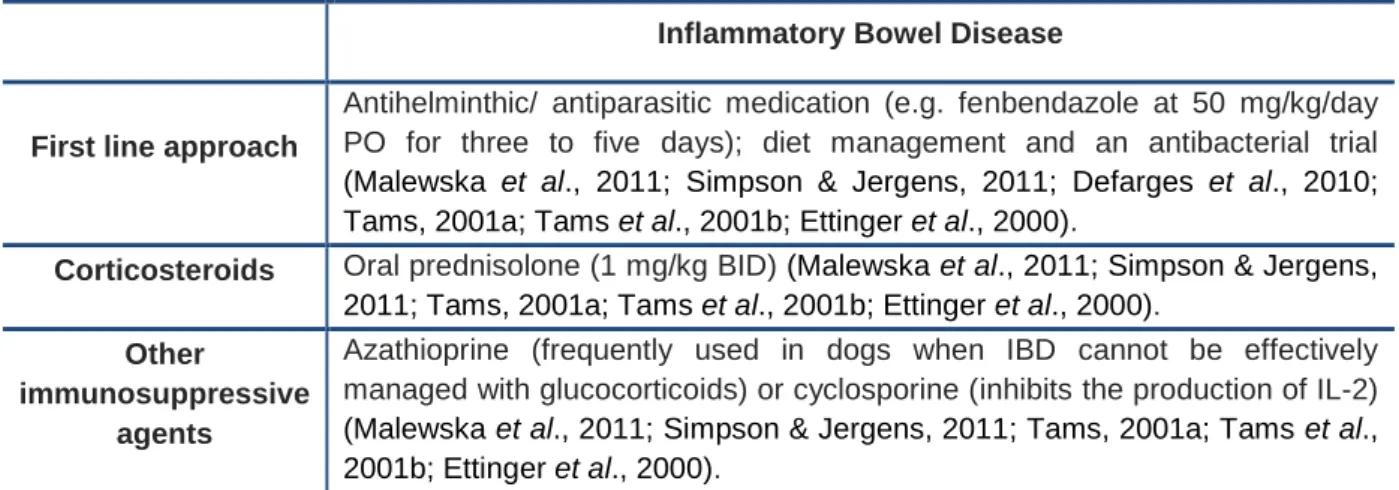
Currently, there are no definitive guidelines for the treatment of IBD so it can undergo individual variability according to the patient’s record and the type of IBD (Table 5). The most frequent conventional treatment (spontaneous lymphocytic plasmatic enteritis) must be started with a first line approach with antihelminthic / antiparasitic medication (e.g. fenbendazole at 50 mg/kg/day PO for three to five days) and with cobalamin and folate supplementation if these are subnormal. This is followed by dietary modification (preferably with an antigen-limited or hydrolyzed protein diet) for three to four weeks, followed by a three to four weeks antibacterial trial (usually tylosin 10 mg/kg PO TID, or metronidazole 10 mg/kg PO BID), and finally a trial with immunosuppressive therapy (initially prednisolone 1 mg/kg PO BID) (Malewska et al., 2011; Simpson & Jergens, 2011; Defarges et al., 2010; Tams, 2001a; Tams et al., 2001b; Ettinger et al., 2000).
17
Corticosteroids are the treatment of choice in most cases, although cushingoid side effects are common but transient as the dosage is reduced. Azathioprine is an alternative drug that has good steroid-sparing properties and is frequently used in dogs when IBD cannot be effectively managed with glucocorticoids or when the glucocorticoid dose has to be reduced. If there is still a poor response, cyclosporine is another alternative agent, because it inhibits the production of IL-2. However its efficacy has not yet been fully validated (Malewska et al., 2011; Simpson & Jergens, 2011; Tams, 2001a; Tams et al., 2001b; Ettinger et al., 2000).Table 5 – Conventional and immunomodulatory therapeutic approach to inflammatory bowel disease.
Inflammatory Bowel Disease
First line approach
Antihelminthic/ antiparasitic medication (e.g. fenbendazole at 50 mg/kg/day PO for three to five days); diet management and an antibacterial trial (Malewska et al., 2011; Simpson & Jergens, 2011; Defarges et al., 2010; Tams, 2001a; Tams et al., 2001b; Ettinger et al., 2000).
Corticosteroids Oral prednisolone (1 mg/kg BID) (Malewska et al., 2011; Simpson & Jergens,
2011; Tams, 2001a; Tams et al., 2001b; Ettinger et al., 2000).
Other
immunosuppressive agents
Azathioprine (frequently used in dogs when IBD cannot be effectively managed with glucocorticoids) or cyclosporine (inhibits the production of IL-2) (Malewska et al., 2011; Simpson & Jergens, 2011; Tams, 2001a; Tams et al., 2001b; Ettinger et al., 2000).
The animal owners’ decision to carry out a treatment for this chronic disease might be a concern because not every animal responds to treatment. For this reason, alternative approaches become necessary (Quimby & Borjesson, 2018). Applying MSCs as an alternative treatment for IBD is still a very recent conception. However, the use of this therapy in clinical trials on human beings with inflammatory gastrointestinal and immune disorder has proven to be effective and safe (Pérez-Merino et al., 2015).
Pérez-Merino et al carried out a clinical trial with eleven dogs with IBD that had received standard treatment (elimination diet, corticosteroids, antibiotics, antidiarrhoeal and antiparasitic drugs) but did not achieved a satisfactory response (Table 6) (Pérez-Merino et al., 2015). These animals went through a period of washout of at least three weeks before the trial was undertaken. All dogs received a clinical score using the Clinical Inflammatory Bowel Disease Activity Index (CIBDAI) and the Canine Chronic Enteropathy Clinical Activity Index (CCECAI) scoring system. Every dog was treated with a single IV ASC infusion (2 × 10⁶ cells/kg bodyweight). After two weeks of ASC therapy, a clinical response occurred in all dogs, but clinical remission (defined by a reduction of initial CIBDAI and CCECAI >75%) occurred in 9/11 dogs at day 42. The remaining two dogs showed a partial response with an initial reduction of 69,2% and 71,4% in CIBDAI and CCECAI respectively. In conclusion, the administration of a single IV infusion of allogenic MSCs was well tolerated by the patients and seemed to produce clinical benefits in dogs with severe IBD.
18
Another study using IV MSC therapy in spontaneous feline enteropathy showed safety and a positive clinical response (Webb & Webb, 2014) as described in Table 6. Seven cats with diarrhoea of no less than three month duration received two IV injections of 2 x 10⁶ cells/kg from cryopreserved feline ASC, while four cats with a similar clinical condition received saline placebo. Improvement of clinical signs was observed in 5/7 cats which were treated with stem cells after one to two months, unlike the placebo group which did not display any progress. With this trial it is possible to conclude that MSC therapy was well tolerated and potentially effective in the treatment of feline chronic enteropathy, although these preliminary results require significant follow-up study.Table 6 – Clinical trials carried out with mesenchymal stem cells in canine and feline inflammatory bowel disease.
Specie (number)
Treatment Results Conclusion Reference
Dog (11) Single IV
allogeneic ASC (2 × 10⁶ cells/kg)
After two weeks of MSC therapy, a clinical response occurred in all dogs
ASC was well tolerated and appeared to produce clinical
benefits in dogs and cats with confirmed IBD
Pérez-Merino
et al., 2015
Cat (7) Two IV allogeneic ASC
(2 x 10⁶ cells/kg)
Improved clinical signs in 5/7 MSC-treated cats and 0/4
placebo cats
Webb & Webb, 2014
6.4. Feline Asthma
Asthma is a common lower airway inflammatory disease (LAD) in cats that is associated with substantial morbidity and occasional mortality (Reinero, 2016; Trzil et al., 2016; Trzil & Reinero, 2014). The word asthma suggests reversible bronchoconstriction and a prevailingly eosinophilic inflammation of the airways and primary symptoms include cough, wheeze and respiratory distress (Sharp, 2014; Reinero, 2011).
The main factors responsible for triggering asthma are extensive and complex and they include infectious, environmental, allergic and genetic elements (Rosenberg & Druey, 2018). In cats there is evidence that asthma is mediated by an allergic response after exposure to inhaled aeroallergens. These aeroallergens induce stimulation of a Th2 response and lead to production of a variety of cytokines that trigger molecular switches leading to pathologic changes in airways (Reinero, 2016; Trzil & Reinero, 2014; Reinero, 2011).
Currently there is no curative treatment for feline asthma. Treatment goals consist of reducing airway inflammation, reducing airway hyperreactivity and bronchoconstriction (which relieves airflow limitation), ameliorating airway remodeling and removing the underlying cause, if known (Reinero, 2016; Sharp, 2014). Treatment of acute dyspnea associated with LAD in cats starts with oxygen supplementation and minimal handling / stress reduction. The conventional therapeutic approaches for acute and chronic asthma, described in Table 7, are based on glucocorticoids (which are the gold
19
standard of therapy for reducing airway inflammation) and bronchodilators (Sharp, 2014; Trzil & Reinero, 2014).Table 7 – Conventional and immunomodulatory therapeutic approach to feline asthma.
Acute Asthma Chronic Asthma
Glucocorticoids
Dexamethasone (0.15-1
mg/kg intramuscular [IM] or IV) is indicated in cats that show signs of acute dyspnea (Sharp, 2014; Trzil & Reinero, 2014).
Oral prednisolone (0.5-1 mg/kg BID) is
recommended for the first seven to fourteen days. Once clinical signs are well controlled, the dose can be gradually reduced over two to three months to once a day.
Inhaled fluticasone (110 mcg BID for two to three weeks) is an alternative although it is not useful in a crisis because it takes about 10 to 14 days to become effective and, since pets cannot be trained to inhale correctly, administration of aerosolized drugs requires the use of mask.
Injectable methylprednisolone acetate (10-20 mg/cat IM or SC every four to twelve weeks) may also be used (Sharp, 2014; Trzil & Reinero, 2014).
Bronchodilators
β2-receptor agonists, such as terbutaline (0.01 mg/kg IM or SC), or albuterol (90 mcg inhaled) to reduce bronchoconstriction and relieve airflow limitation (Sharp, 2014; Trzil & Reinero, 2014).
The most generally used are β2-receptor agonists, namely terbutaline (0.1-0.2 mg/kg PO TID or BID), and less commonly methilxanthine derivates such as theophylline (the recommended dose of sustained-release theophylline in cats is 20 to 25 mg/kg PO SID, and for non-sustained-release theophylline 4 mg/kg TID or BID) (Sharp, 2014; Trzil & Reinero, 2014).
Allergenic-specific immunotherapy
Intravenous or subcutaneous allergenic-specific immunotherapy was proved to decrease eosinophilia airway inflammation and is generally associated with minimal side effects (Trzil & Reinero, 2014; Lee-Fowler et al., 2009).
Inhibitors of tyrosine kinase
Inhibitors of tyrosine kinase are small molecules that block ATP-binding site of kinases. During a trial model of feline asthma, this therapy has proven to be efficient in reducing airway inflammation (Trzil & Reinero, 2014; Lee-Fowler et al., 2012).
Cyclosporine
Cyclosporine inhibits T-cell activation and blocks the development of a Th2 phenotype and the associated Th2-eosinophil interactions. In a feline asthma experimental study, Mitchell et al. demonstrated that cyclosporine did not inhibit the early phase response to allergen challenge, but it was effective at reducing airway hyperresponsiveness to acetylcholine and airway remodeling (Reinero, 2016; Byers & Dhupa, 2005; Mitchell et al., 1998).
20
Although conventional therapy reduces inflammation and dyspnea in a considerable number of cats, there is still no therapy capable of preventing or reversing all pathological aspects of asthma. Some cats continue resisting to this therapy with persistent clinical signs and eosinophilia in the airways. Other cats may be affected by concurrent diseases (e.g. cardiac disease or diabetes mellitus) against which the use of glucocorticoids is not advised. This is the reason why new therapies for the treatment of asthma become necessary (Trzil et al., 2016; Trzil & Reinero, 2014) namely allergenic-specific immunotherapy, inhibitors of tyrosine kinase, cyclosporine and MSCs therapy (Table 7).Therapy with MSCs would be ideal given their immunomodulatory abilities (they modulate Th2 lymphocyte activity) and their capability to pass through the lung when administered intravenously (Quimby & Borjesson, 2018). Murine asthma models have demonstrated that stem cells can reduce airway eosinophilia, airway hyper-responsiveness and airway remodeling (Trzil & Reinero, 2014). Two pilot trials carried out in cats have been published to test the efficiency of MSCs therapy. Both trials involved sensitization to Bermuda grass allergen, which resulted in the development of an asthmatic phenotype with airway eosinophilia and airway hyper-responsiveness.
The first study involved six cats with acute asthma, 4/6 received five intravenous infusions of allogeneic MSCs (infusions varied between 2 x 10⁶ and 1 x 10⁷ cryopreserved MSCs per cat) and 2/6 received a saline placebo (Table 8). Cats treated with MSCs achieved a decrease in airway eosinophilia and diminished airway hyper-responsiveness at day 133 when compared to the group that was exclusively administrated with placebo. In this trial lung attenuation and bronchial wall thickness were also assessed by computerized tomography and it was verified that the score of these parameters was substantially reduced in cats treated with MSCs nine months later (Trzil et al., 2016). The second study involved nine cats with chronic asthma, 5/9 received six intravenous infusions of allogeneic MSCs (with range amplitude of 0.36–2.5 x 10⁷
MSCs/infusion) and 4/9 received a saline placebo (Table 8). Unlike the previous trial, cats suffering from chronic asthma which were treated with MSCs did not experience a decrease in airway eosinophilia and diminished airway hyper-responsiveness compared with placebo group.However, there was a significant reduction in the lung attenuation and in the bronchial wall thickness observed through computerized tomography eight months after treatment, once again indicating a positive effect on airway remodeling (Trzil et al., 2014).
Therapy with ASC proved to have a positive effect on remodeling airways in the two pilot trials controlled by placebo and using feline asthma models. Moreover, the results attained from this cell therapy were more favourable when treatment was carried out in an acute stage of the asthma.
21
Table 8 – Clinical trials carried out with mesenchymal stem cells in feline asthma.Specie (number)
Treatment Results Conclusion Reference
Cat (6) Acute asthma Five IV allogeneic ASC (varied between 2 x 10⁶ and 1 x 10⁷per cat) Decreased in: airway eosinophilia, hyper-responsiveness and airway remodeling ASC therapy had a positive effect on airway remodeling Trzil et al., 2016 Cat (9) Chronic asthma Six IV allogeneic ASC (with range amplitude of 0.36– 2.5 x 10⁷ MSCs/infusion) Decreased in airway remodeling Trzil et al., 2014
22
7. DISCUSSIONMSCs immunomodulatory properties make them a unique cell type capable of repairing tissue and organ injuries caused by chronic inflammation or autoimmune disorders.
According to the results of trials aforementioned, it is possible to verify a positive balance in the use of MSCs for the treatment of FCGS, IBD and feline asthma. In what concerns FCGS and IBD, one or two infusions of stem cells were sufficient to attain considerably favorable results, as most of the animals involved experienced improvements of the clinical signs and none of them revealed any adverse effects.
In feline asthma, the clinical trials required a higher number of stem cells infusions (five to six) to provide for a proper passage of these cells towards the lung, and the results were equally satisfactory. Furthermore, the results achieved from this cell therapy were more favourable when treatment was carried out with autologous MSCs and in acute asthma.
Canine atopic dermatitis has not displayed results as satisfactory as the other observed diseases. However, only one clinical trial has been carried out in a sample of five dogs, which included a sole administration of IV MSCs at a lower dosage than the dose used in other similar trials. This might be one of the reasons for the failure of the therapy. Meanwhile, there are several unpublished studies that claim success in treating AD by injecting MSCs both IV and IM on the main areas affected by the disease. Despite this claim, these results still lack validation and standardization of the total number of cells injected, as different studies report using different numbers of cells per Kg of body weight.
Although MSCs can bring a promising future to the treatment of the majority of these disorders, the considerable variability of their quality derived from different donors (autologous vs allogeneic therapy), different tissues, different administration routes, different dosages, individual variabilities of each patient (e.g. age of the patient) and state of the disease, might limit its therapeutic benefit (Figure 5).
23
Canine atopic dermatitis, feline chronic gingivostomatitis, inflammatory bowel disease and feline asthma are relatively common diseases among veterinary medicine, which is why they were widely considered throughout this revision. Nevertheless, the same rational used in these disorders may be applied to several other with a similar immunomediated nature, namely systemic lupus erythematosus, pemphigus foliaceus, perianal fistula and immune mediated cutaneous vasculitis (Jiménez & Guerrero, 2017; Jang et al., 2016; Jayne & Tyndall, 2004). All these pathologies are characterized by a dysfunction of the immune system with a pathologic response mediated by Th1 cells and inflammatory mediators. Since MSCs are able to suppress the proliferation of T cells and modulate their response (change the Th1 proinflammatory profile into a Th2 anti-inflammatory profile) through the secretion of several soluble factors or through cell-to-cell contact (Kim & Cho, 2015), they will certainly become an important therapeutic approach to treat any of these pathologies. Nevertheless, sufficient clinical trials have not yet been performed to confirm the success and validity of the therapy. The main obstacle to MSCs therapy is individual and species diversity, and also the inconsistency of protocols applied to date, since there are still very few clinical trials performed in veterinary medicine and most of them have used small samples. Both physicians with expertise in this field and regulatory agencies need to work together towards the standardization and quality assurance of cellular therapies being applied clinically. The way we practice medicine is changing and evolving rapidly, and although it is difficult to predict where we will be in the near future, cellular therapies certainly seem to have come to stay.24
8. CURRENT STATUS AND FUTURE PROSPECTSThe multipotent and non-teratogenic properties of MSCs led to initial findings stating that these cells could be clinically used to regenerate injured tissues and to treat immune-mediated disorders due to their immunomodulatory potential. Over the last few years some researchers have questioned the efficiency of the treatment with stem cells, as well as the purpose for which they were created (Klinker, 2015). There is sparse evidence suggesting that the primary function of MSCs is its differentiation in in vivo new tissues, questioning the importance of differentiation to the therapeutic properties of such cells when injected in a naive state (Berglund et al., 2017).
There are still several questions concerning the use of MSCs: their immunomodulation mechanism has not yet been completely understood and the combined results of this therapy require a better scientific clarification. The use of allogeneic vs. autologous MSC therapy might contribute to the differences in efficacy observed in some clinical trials. On the other hand, the in vitro expansion of MSCs before clinical usage might take weeks before it provides enough cells to administrate, resulting in loss of stemness (Berglund et al., 2017).
In order to overcome these obstacles to treatment, careful evaluation of appropriate cell sources, good quality control systems, standardized protocols for cell culture and their differentiation, expansion and cryopreservation are necessary. Therefore, the regulation mechanism of MSCs to produce soluble factors and the way these factors are capable of modulating cells of the immune system are key questions that underlie the successful immunomodulation effects of MSCs. All these factors combined with genetically modified MSCs might open a way for the development of an effective cell therapy for multiple animal and human immune disorders.
Despite current contradiction found among researchers concerning the efficacy of MSCs, this revision has demonstrated a generally positive result of the use of these cells in the treatment of immune-mediated disorders in veterinary medicine. Even so, MSCs-based therapy is currently requiring, more than ever before, a complete analysis and reconsideration in the hope to overcome its limitations in future trials. In the future this approach could be more cost-effective than actual conventional treatments due to the recent development and continuous success regarding MSCs application in immunomodulated diseases.