Mestre em Biotecnologia
Structural and Functional studies on the
Reactivity of CORMs with plasma
proteins
Dissertação para obtenção do Grau de Doutor em Bioquímica, Especialidade Bioquímica Estrutural
Orientador: Doutora Teresa Sacadura Santos-Silva,
Investigadora Auxiliar, FCT/UNL
Co-orientador: Doutora Maria João Lobo de Reis Madeira
Crispim Romão, Professora Catedrática, FCT/UNL
Júri
Presidente: Prof. Doutora Maria Paula Pires dos Santos Diogo Arguentes: Doutor João Domingos Galamba Correia
Doutora Isabel Maria Travassos de Almeida de Jesus Bento
Vogais: Prof. Doutor Carlos José Rodrigues Crispim Romão Prof. Doutor João Emídio da Silva da Costa Pessoa
Prof. Doutor Eurico José da Silva Cabrita
Doutor Tiago Miguel Guerra Miranda Bandeiras Doutora Teresa Sacadura Santos Silva
.
“Structural and Functional studies on the Reactivity of CORMs with plasma proteins”
“Copyright” em nome de Marino Filipe Alves dos Santos, da FCT/UNL e da UNL
.
O trabalho apresentado nesta Dissertação foi realizado com o apoio financeiro da Fundação para a Ciência e Tecnologia (FCT–MEC), através da atribuição da Bolsa de Doutoramento individual SFRH/BD/77894/2011 e do projecto PTDC/QUI-BIQ/117799/2010.
O trabalho desenvolvido ao longo da presente Dissertação resultou na publicação dos seguintes artigos em revistas internacionais:
1) – Marino F. A. Santos, João D. Seixas, Abhik Mukhopadhyay, Patrícia M. Reis, Maria J. Romão, Carlos C. Romão, Teresa Santos-Silva, New insights into the chemistry of fac-[Ru(CO)3]2+ fragments
in biologically relevant conditions: The CO releasing activity of [Ru(CO)3Cl2(1,3-thiazole)], and the
X-ray crystal structure of its adduct with lysozyme, Journal of Inorganic Biochemistry2012, 117, 285-291.
2) – Sameena Mehtab, Gisela Gonçalves, Somnath Roy, Ana Isabel Tomaz, Teresa Santos-Silva,
Marino F.A. Santos, Maria J. Romão, Tamás Jakusch, Tamás Kiss, João Costa Pessoa, Interaction of vanadium (IV) with human serum apo-transferrin, Journal of Inorganic Biochemistry2013, 121, 187-195.
3) – João Costa Pessoa, Gisela Gonçalves, Somnath Roy, Isabel Correia, Sameena Mehtab, Marino F.A. Santos, Teresa Santos-Silva, New insights on vanadium binding to human serum transferrin, Inorganica Chimica Acta2014, 420, 60-68.
4) – Marino F. A. Santos, Isabel Correia, Ana R. Oliveira, Eugenio Garribba, João Costa Pessoa, Teresa Santos-Silva, Vanadium Complexes as Prospective Therapeutics: Structural Characterization of a VIV Lysozyme Adduct, European Journal of Inorganic Chemistry2014, 2014(21), 3293-3297.
5) – João D. Seixas, Marino F. A. Santos, Abhik Mukhopadhyay, Ana C. Coelho, Patrícia M. Reis, Luís F. Veiros, Ana R. Marques, Nuno Penacho, Ana M. L. Gonçalves, Maria J. Romão, Gonçalo J. L. Bernardes, Teresa Santos-Silva, Carlos C. Romão, A contribution to the rational design of Ru(CO)3Cl2L complexes for in vivo delivery of CO, Dalton Transactions2015, 44, 5058-5075.
6) – João Costa Pessoa, Eugenio Garribba, Marino F.A. Santos, Teresa Santos-Silva, Vanadium and proteins: uptake, transport, structure, activity and function, Coordination Chemistry Reviews 2015, 301-302, 49-86.
I
Agradecimentos
Acknowledgements
“O passado é a chave do fu
turo.
”
Foi assim que comecei os Agradecimentos da minha Tese de Mestrado. Quatro anos volvidos, e porque há tradições que se devem manter e preservar, assim começo os Agradecimentos da minha Tese de Doutoramento.Obrigado a todos. E, usando esta tão simples e sucinta expressão, podia ficar por aqui. Todavia, parece-me que se gerou uma infundada expectativa sobre esta secção pelos mais variados quadrantes da sociedade civil que, pese embora o seu manifesto exagero, me faz sentir na obrigação de não a defraudar por simples desistência. Em alternativa, será defraudada lenta e gradualmente à medida que a leitura da secção avance. Quer isto dizer que se não pretendem ficar desiludidos com o conteúdo destas parcas linhas, aconselho vivamente que parem a leitura por aqui embora, claro, sejam livres para continuar por vossa conta e risco.
Se, após todo este palavreado, ainda estão a ler, merecem os meus parabéns pela persistência e o meu reparo pela teimosia. Perguntei-me várias vezes qual seria a melhor maneira de começar esta narrativa. Como não a encontrei, opto por uma saída diplomática sublinhando o meu apreço pelo Benfica e por História. O que tem isso a ver com os Agradecimentos? Pouco, mas não se devem
perder oportunidades de mostrar o quanto “me envaidece” a fé inabalável no clube que “nunca encontrou rival neste nosso Portugal”. Já a História, tem que aparecer em qualquer apontamento
biográfico da minha parte até porque, qual gloriosa epopeia camoniana, esta secção não pretende mais do que registar para a posterioridade todos aqueles que contribuíram de forma decisiva, quiçá, estrutural – um excelente jogo de palavras – para o desenrolar deste trabalho. Posto isto, comecemos (finalmente) os Agradecimentos devidos.
À Teresa, como não podia deixar de ser, ficam as primeiras palavras pela entusiástica e abrangente orientação ao longo destes seis anos. Enquanto seu primeiro aluno de Mestrado e de Doutoramento, disse-lhe, algures no espaço e no tempo, algo como que eu seria “o seu primeiro ciclo de refinamento” enquanto orientadora. Muitos alunos depois, acho que tal previsão se confirmou. Espero que, para primeiro refinamento, tenha obtido uns valores de R e Rfree aceitáveis.
À professora Maria João Romão, por ter possibilitado a realização desta Tese no grupo de Cristalografia de Proteínas assim como pela sua co-orientação. No meio de tantos assuntos burocráticos, conseguiu nunca deixar o laboratório e respectiva investigação para trás.
Ao professor Carlos Romão, agradeço não apenas a sua acção vital no desenvolvimento do projecto dos CORMs, mas também a sua boa disposição e optimismo. À Catarina por todo o (muito) auxílio durante a minha breve estadia no mundo da síntese organometálica.
Ao professor João da Costa Pessoa, pela oportunidade de ter contactado com o vasto “mundo do vanádio” e pela prontidão com que sempre respondeu às nossas (muitas) dúvidas. À Isabel, pela medição dos espectros de EPR e disponibilidade para as minhas dúvidas.
II
Às “minhas” alunas de Projecto – Joana, Rita e Marta – pela valiosa oportunidade de exploração desumana que me proporcionaram (deve ler-se “pela sua contribuição para os resultados”).
To Abhik for his availability to help me, first in the lab and then directly from the PDBe headquarters. To Shabir for the tips and patience in my first trips to the Synchrotron and for the dinners in one of the fanciest French restaurants: Pizza Hut at Lyon Airport.
Ao Jorge, pelos seus mordazes e pertinentes comentários da vida em geral bem como por todo o “apoio moral” que nunca me negou nos (raros a tender para raríssimos) resultados menos conseguidos do Benfica. Que a Estrutura esteja contigo!
À Cecília, por todas as encomendas, orçamentos, cristais congelados, soluções, dicas de cristalização, manutenção e operação do robot, envios de dewars e organização logístico-moral de tantos e tantos eventos. Tomara o Quartzy conseguir tal eficiência.
A um excelente Trio d’Ataque pós-doutoral sempre com “a genica que as engrandece”: à Benedita, à Catarina e à Márcia. À Benedita, pelas eloquentes conversas de fim de tarde e salutares tentativas (vãs é certo) de me instruir em diferentes variantes sócio-artístico-culturais. À Catarina, pela organização de vários pequenos/grandes aspectos do dia-a-dia do laboratório e pelo passeio ao SLS. À Márcia, pelas peripécias no Diamond (já incluindo o Miguel), pelas (muitas) dicas de SAXS e pela já longínqua paciência enquanto “orientadora” aquando da minha chegada ao laboratório.
À Diana Vieira, pela ajuda de longa data que se prolongou pelo doutoramento – em Portugal (desde as aulas até ao passeio a Coimbra) ou em Inglaterra (com o jantar de boas-vindas). Note-se a total ausência de recursos de stand-up sublinhando, tristemente, a minha total inaptidão para tal arte.
Aos meus dois prezados “companheiros de armas” que durante algum tempo completaram
com a minha pessoa um outro Trio d’Ataque: o dos alunos de Doutoramento. À Rita, pelo baptismo (de fogo) nas idas ao Sincrotrão e por tentar melhorar o desolador desterro que é a Margem Sul com a sua presença – cá estarei quando chegar a tua vez. Ao Hugo, pela épica viagem à BM30, pelas muitas boleias e por todas as dicas cristalográficas e afins – deixaste uma importante marca neste caminho e lamento que não assistas à sua conclusão; sem grandes palavras, deixo apenas um obrigado por tudo.
Às duas futuras experts em açúcares e afins, Diana e Viviana, seja pela aula de microarrays ou pelo passeio a Grenoble (que, com mais ou menos sono, foi, obviamente, magnifique!).
Ao Francisco que, chegando a meio desta Tese, rapidamente fez parecer que já estava no grupo desde há largos anos. Uma aquisição de enorme potencial, tendo sempre na “alma a chama imensa” e um excelente formando (e formador) que adoptou os valores da FCT e da Margem Sul com distinção e classe. Um orgulho!
Ao Filipe, por todos e mais alguns motivos que, se fossem exaustivamente mencionados, iriam ocupar tanto ou mais espaço que a restante Tese. Desde finais de 2008, nunca nenhuma dúvida, observação, reparo ou nota de âmbito académico, profissional, lúdico ou de avaliação de situações e comportamentos (estes então…) deixou de ser analisada e respondida. Sem a eloquência do ROC, resta-me tentar demonstrar o meu profundo reconhecimento invocando um simples lema que, ainda assim, poderá ser tão grande como os maiores da Europa: “Esforço, Dedicação, Devoção e Glória”!
III
À Filipa, seja em Portugal, em Espanha, na Holanda, em Inglaterra ou na Suíça, pelas várias visitas à casa-mãe e pelo frequente despoletar dos e-mails comuns dos supracitados heróis de BCM. Estou certo que as noções laboratoriais adquiridas no XTAL te ajudam a brilhar por esta Europa fora.
À Tânia Perestrelo, já que nunca é demais lembrar, pela sua providencial escolha da BII (com mais ou com menos juízo) além dos muitos trabalhos (e outras tantas “sessões de estudo”) na
licenciatura, pelas visitas à casa-mãe e pela constante “presença não-presencial” com as modernas
modernices comunicacionais dos nossos dias. Seja “no” Machico (ou outras zonas de Marrocos como o aeroporto Ronaldo), em Lisboa, em Coimbra ou no outro lado do Atlântico – seja com caneta, lápis ou porta-minas – mais um exemplo da formação XTAL a singrar pelo mundo.
À Tânia Leandro, pela sua intervenção decisiva na conversão de almas menos crentes para a causa de História da Ciência, pelas (muito) frequentes visitas à casa-mãe e, sobretudo, pelas várias sessões terapêuticas nas já referidas modernices comunicacionais sobre muitos e variados aspectos da vida seja a recordar o passado, comentar o presente ou projectar o futuro. Mantendo a convicção que, mais tarde ou mais cedo, a tão ansiada colaboração se vai concretizar, resta-me terminar (já que
“aprecias” de sobremaneira o idioma) com um singelo (mas sentido) gracias!
Ao Rui, pelo facto de, resumidamente, me ter feito uma licenciatura, um mestrado e ainda ter tempo para me apresentar ao mundo da investigação. E assim se resumem os últimos dez anos. Todavia, muito mais há a dizer. O profissionalismo irrepreensível na elaboração de numerosos resumos, apresentações, discussões, relatórios, portfólios e outros trabalhos afins em 30 disciplinas
partilhadas entre dois ciclos de estudos. A mítica frase “amanhã às 8h” e respectivas sessões de estudo
intensivas na sala de BCM. O esmiuçamento de tantos programas e/ou segmentos cómicos e a criação dos nossos próprios, vamos lá, peculiares sketches. As profícuas visitas ao 3º e/ou ao 6º piso para a actualização detalhada dos pormenores e curiosidades da vida. E tantos outros episódios de relevo.
Temos, em breve, de avançar com um livro de memórias, mas, para já, fica um simples “Danke schön e que continuemos a ’legislelar bué’ por mais umas boas (e longas) décadas”.
Não podia terminar tão nobre secção sem uma referência à minha família que, indubitavelmente, possibilitou a realização desta Tese. Em especial aos meus pais (José e Ilda) e aos meus avós (Daniel e Céu) pelos 28 anos de trabalho constante (seja à semana, Sábados, Domingos ou feriados) para eu poder andar a brincar por estes lados. Uma igualmente importante palavra para a minha avó Álida e para a Maria sempre presentes nas visitas à terrinha nas mui nobres e académicas proximidades de Coimbra. Por último, fica uma simbólica palavra de saudade para o meu avô Eduardo que, não tendo a oportunidade de ver o culminar deste longo trajecto, foi estando presente em grande parte do mesmo.
E, muitos caracteres depois, parece-me que estou a chegar ao fim dos Agradecimentos. Para alguma dúvida ou esclarecimento adicionais, façam o obséquio de entrar em contacto com a minha pessoa. Contudo, não posso terminar sem uma breve, mas sentida, palavra para um grupo de bons rapazes que resolveram subir ao lugar mais alto do futebol europeu de selecções na véspera da minha defesa – o que um golo (até do Éder) pode fazer pela confiança e alegria de um país.
E, se comecei esta secção com a auto-citação inicial de 2011, parece-me apropriado finalizá-la com a auto-citação terminal da mesma publicação:
“muito
obrigado a todos os que ajudaram
neste caminho”
.E pluribus unum
“Não somos 11, Somos 11 milhões”
IV
V
Abstract
Drug design is a multidisciplinary field involving several methodologies namely X-ray crystallography. This Thesis reports the results obtained in two drug design projects related to the interactions of putative metal-based drugs (Carbon Monoxide Releasing Molecules, CORMs, and vanadium complexes) with different proteins.
Carbon monoxide, beyond its toxic potential, is a signaling molecule with biological and potential therapeutic roles. CORMs are able to transport CO in the blood stream delivering it at the damaged tissues and their pharmacokinetics strongly depends on the interactions with blood proteins (hemoglobin, albumin and transferrin).
Soaking and co-crystallization trials have been tried and crystal structures were obtained, namely two 1.9 Å resolution hemoglobin structures and a 2.7 Å resolution transferrin structure soaked with ruthenium- and iridium-based CORMs. Several [Ru(CO)3Cl2L] CORMs have been soaked with
lysozyme (HEWL) to understand their mechanism of action. The 1.5 Å resolution HEWL•ALF850
structure shows the adduct [His15•Ru(CO)(H2O)4]2+ revealing the importance of the stepwise decay of
[Ru(CO)3]2+ for the delivery of CO in vivo. The subsequent 1.7 Å resolution structures corroborate such proposal: an adduct [His15•RuII(COOH)(CO)(H
2O)3], found in the HEWL•ALF486 structure,
corresponds to a metallacarboxylate intermediate of the decarbonylation pathway. This suggests that the aqueous chemistry of these complexes is governed by the water-gas shift reaction initiated with the nucleophilic attack of HO− on coordinated CO.
Vanadium is an element with biological and therapeutic roles namely as insulin-enhancer agent and transferrin has been proposed as a natural carrier. The conformation adopted by transferrin upon vanadium binding was addressed by X-ray crystallography, urea gel electrophoresis and SAXS but no definitive answers were obtained.
HEWL was also used in different soaking experiments and a 1.3 Å resolution structure with picolinate was solved showing that VIVO(pic)
2 covalently binds to the COO– group of the side chain of
Asp52 in the active site. The long VIV=O bond is explained by the reduction of VIV to VIII during
exposure to the X-ray beam as further confirmed by EPR and DFT. Picolinate was used with trypsin and similar results have been obtained: the 1.1 Å resolution structure exhibits a VIVO(pic)
2 adduct
bound to Ser195 in the active site. The ability of picolinate complex to bind to the active center of proteins – glycoside hydrolases and serine proteases – has been demonstrated suggesting that similar vanadium compounds can act as protein inhibitors, accounting for eventual side effects derived from the use of these metal complexes as therapeutic agents.
VII
Resumo
O processo de drug design é uma área interdisciplinar envolvendo várias técnicas nomeadamente a cristalografia de raios-X. A presente Tese aborda dois projectos de drug design relacionados com as interacções de potenciais fármacos – Moléculas Libertadoras de Monóxido de Carbono (CORMs) e complexos de vanádio – com diferentes proteínas.
O monóxido de carbono, além do seu potencial tóxico, é uma molécula sinalizadora com efeitos biológicos e terapêuticos. Os CORMs transportam o CO na corrente sanguínea libertando-o nos tecidos afectados e a sua farmacocinética depende das interacções estabelecidas com proteínas sanguíneas (hemoglobina, albumina e transferrina).
Realizaram-se ensaios de soaking e de co-cristalização com estas proteínas obtendo-se estruturas de hemoglobina (resolução de 1.9 Å) e transferrina (resolução de 2.7 Å) com diferentes CORMs de ruténio e de irídio. Usaram-se diferentes compostos [Ru(CO)3Cl2L] em ensaios de soaking
com lisozima (HEWL) para compreender o seu mecanismo de acção. A estrutura de HEWL•ALF850
(resolução de 1.5 Å), mostra o aducto [His15•Ru(CO)(H2O)4]2+ sugerindo que a descarbonilação
progressiva de [Ru(CO)3]2+ é fundamental para a libertação do CO in vivo. Outras três estruturas
(resolução de 1.7 Å) reforçam tal ideia: o aducto [His15•RuII(COOH)(CO)(H
2O)3], identificado na
estrutura HEWL•ALF486, corresponde a uma espécie intermediária da descarbonilação indicando que tal processo é iniciado pelo ataque nucleofílico do grupo HO- ao grupo CO.
O vanádio possui diversas acções biológicas e terapêuticas nomeadamente como mimetizador da insulina sendo que a transferrina foi proposta como o transportador natural. A sua conformação após a ligação ao vanádio foi investigada por diferentes metodologias – cristalografia de raios-X, electroforese em gel de ureia e SAXS – mas sem resultados conclusivos.
Realizaram-se diversos ensaios de soaking com a lisozima obtendo-se uma estrutura com picolinato (resolução de 1.3 Å) mostrando o aducto VIVO(pic)
2 covalentemente ligado ao grupo COO
-da cadeia lateral do Asp52 no centro activo. A longa ligação VIV=O deve-se à redução do VIV a VIII
durante a exposição ao intenso feixe de raios-X como posteriormente mostrado por EPR e DFT. O mesmo composto foi também usado com a tripsina obtendo-se resultados semelhantes com o aducto VIVO(pic)
2 ligado à Ser195 no centro activo (resolução de 1.1 Å). A capacidade de ligação do
complexo de picolinato ao centro activo de proteínas – glicosil hidrolases e proteases de serina – foi demonstrada sugerindo que compostos de vanádio semelhantes possam actuar como inibidores de proteínas o que, por sua vez, pode interferir no seu uso como agentes terapêuticos.
Termos chave: Monóxido de carbono e Moléculas Libertadoras de Monóxido de Carbono (CORMs) •
Vanádio e complexos de vanádio • Desenvolvimento de fármacos • Cristalografia de raios-X •
IX
Table of Contents
Agradecimentos……….I
Abstract………....V
Resumo………..VII
Table of Contents………IX
Figures Index………...XIII
Tables Index……….XIX
Abbreviations and symbols………..XXI
Chapter 1 – Introduction………1
1.1 – Drug Design and Development………3
1.1.1 – General concepts and historical perspective ………..3
1.1.2 – Principles of Drug Design………...8
1.1.2.1 – Drug Discovery steps……….10
1.1.2.2 – Drug-like molecules properties……….14
1.1.2.3 – Pharmacokinetics and pharmacodynamics………16
1.1.3 – Metal-based pharmacological compounds……….19
1.2 – Carbon monoxide –Biological and therapeutic applications………22
1.2.1 –General concepts and toxicity………...22
1.2.2 –Endogenous production of carbon monoxide………...24
1.2.3 –Biological effects of carbon monoxide……….27
1.2.3.1 – Carbon monoxide as a signaling molecule………28
1.2.4 –Therapeutic application of carbon monoxide………...33
1.2.4.1 – Carbon Monoxide Releasing Molecules – CORMs………..34
1.2.4.1.1 – CORMs and transition metals………..35
1.2.4.1.2 – Mechanisms of CO release from CORMs………...36
1.2.4.1.3 – Early CORMs………..38
1.2.4.1.4 – Drug-like CORMs………40
1.3 – Vanadium – Biological and therapeutic effects……….42
1.3.1 – General concepts and historical perspective……….42
1.3.2 – Vanadium and vanadium complexes……….43
1.3.3 – Vanadium in Biology and Medicine……….45
1.3.3.1 –Vanadium and proteins………...46
1.3.3.1.1 – Vanadium uptake and transport………....48
1.3.3.1.2 – Vanadium in protein activity and function………...49
X
1.3.3.2 –Therapeutic applications of vanadium………...52
1.3.3.2.1 – Anti-diabetic activity………54
1.3.3.2.2 – Other therapeutic actions ………57
1.4 – X-ray crystallography………58
1.4.1 – General concepts and historical perspective ……….58
1.4.2 –Crystals and protein crystallization………61
1.4.3 – X-ray diffraction and data collection……….66
1.4.4 –The “phase problem” and structure solution………..69
1.4.5 – Refinement and structure validation………..70
Chapter 2 – Interaction of proteins with Carbon Monoxide Releasing Molecules……….73
2.1 – Introduction and objectives………75
2.2 – Structural and functional studies with proteins………..76
2.2.1 – Ruthenium-based CORMs……….76
2.2.1.1 – Synthesis of Cs2[Ru(CO)2Cl4] complex (ALF_MS1)……….76
2.2.1.1.1 – Materials and Methods……….76
2.2.1.1.2 – Results and Discussion………..77
2.2.1.2 – Interactions with Hen Egg White Lysozyme………..78
2.2.1.2.1 –Materials and Methods……….79
2.2.1.2.2 –Results and Discussion……….80
2.2.1.2.2.1 – ALF850………...80
2.2.1.2.2.2 – ALF475, ALF486 and ALF487………..86
2.2.1.2.2.3 – ALF_MS1………...93
2.2.1.3 – Interactions with Bovine Hemoglobin……….95
2.2.1.3.1 –Materials and Methods………..95
2.2.1.3.2 –Results and Discussion………..96
2.2.2 – Iridium- and platinum-based CORMs………101
2.2.2.1 – Synthesis of Cs2[Ir(CO)Cl5] complex (ALF_MS2) and Cs[Pt(CO)Br3] complex (ALF_MS3)………..102
2.2.2.1.1 – Materials and Methods………102
2.2.2.1.2 –Results and Discussion………103
2.2.2.2 – Interactions with Hen Egg White Lysozyme………....105
2.2.2.2.1 –Materials and Methods………105
2.2.2.2.2 –Results and Discussion………106
XI
2.2.2.3 – Interactions with Human and Bovine Serum Albumin……….112
2.2.2.3.1 –Materials and Methods………113
2.2.2.3.2 –Results and Discussion………...114
2.2.2.3.2.1 – Human serum Albumin………115
2.2.2.3.2.2 – Bovine serum Albumin……….119
2.2.2.4 – Interactions with Human Serum Transferrin……….120
2.2.2.4.1 –Materials and Methods………120
2.2.2.4.2 –Results and Discussion………121
2.2.3 – Other metal-based CORMs………..126
2.2.3.1 –Materials and Methods……….126
2.2.3.2 –Results and Discussion………127
Chapter 3 – Interaction of proteins with vanadium compounds………131
3.1 – Introduction and objectives………..133
3.2 – Structural and Functional studies of vanadium complexes with proteins………...133
3.2.1 –Human Serum Transferrin………...134
3.2.1.1 –Materials and Methods……….134
3.2.1.2 –Results and Discussion………....136
3.2.1.2.1 – Crystallization and preliminary X-ray diffraction analysis……....136
3.2.1.2.2 – Conformation determination by urea gel electrophoresis………...137
3.2.1.2.3 –Conformation determination by SAXS………...139
3.2.2 –Hen Egg White Lysozyme………..143
3.2.2.1 –Materials and Methods……….143
3.2.2.2 –Results and Discussion………....144
3.2.2.2.1 –HEWL and vanadium picolinate complex………..145
3.2.2.2.2 – HEWL and other vanadium compounds……….149
3.2.3 –Bovine Trypsin……….153
3.2.3.1 –Materials and Methods………...154
3.2.3.2 –Results and Discussion………..155
Chapter 4 – General conclusions and future perspectives………..163
XII
Appendix………..193
Appendix 1 – Structure 1 screen (Molecular Dimensions)………...195
Appendix 2 – Structure 2 screen (Molecular Dimensions)………...197
Appendix 3 –In-house sparse matrix screen (80!)………...199
XIII
Figures Index
Figure 1.1– Compounds in clinical development by therapeutic area between 1996 and 2011……….3
Figure 1.2–Piece of the Ebers Papyrus………..5
Figure 1.3– Cover of De Materia Medica(edition dated from 1554, in Latin)………..5
Figure 1.4– Summary of some important findings on drug discovery over the last 200 years………..7
Figure 1.5–Schematic representation of the major periods of drug development……….8
Figure 1.6 – Graphical representation of the approval of new drugs by the FDA between 1950 and 2008………..8
Figure 1.7– Schematic representation of the different stages involved in the drug design process from the target identification until the market release………...9
Figure 1.8– Workflow of the stages involved in the discovery of putative drug candidates…………10
Figure 1.9– Evolution of the type of microtiter plates used in HTS assays through the years……….12
Figure 1.10–Scheme of the steps of FBDD methodology………...12
Figure 1.11– Schematic representation of the three methods used in the hit-to-lead phase………….13
Figure 1.12 – Schematic representation of the concepts related to pharmacokinetics and pharmacodynamics……….16
Figure 1.13– Schematic representation of the steps of pharmacokinetics………17
Figure 1.14–Schematic representation of the “Medical Periodic Table”……….20
Figure 1.15–Schematic representation of the mode of action of cisplatin………...22
Figure 1.16– Graphical correlation of the harmful effects of CO with its concentration and percentage of COHb in human health………...24
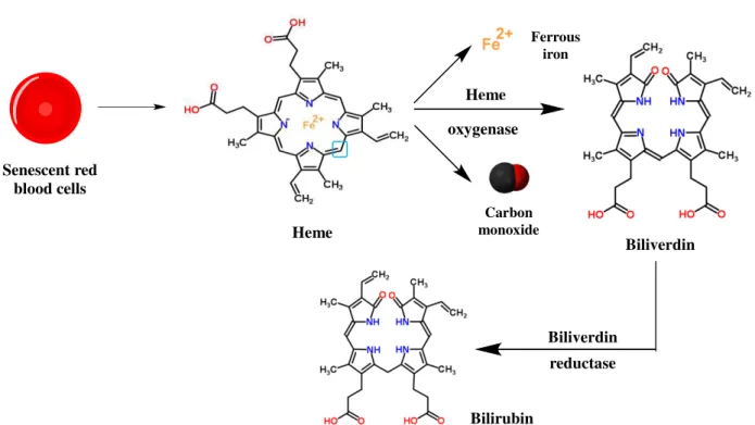
Figure 1.17– Schematic representation of the products generated from the heme degradation by heme oxygenase………25
Figure 1.18– Structural representation of the human HO-2 in the apo-form (PDB: 2Q32, green) and heme-bound form (PDB: 2QPP, orange)………26
Figure 1.19 – Representation of the mechanisms in which the gasotransmitters are involved: nitric oxide (left), carbon monoxide (middle) and hydrogen sulfide (right)………28
Figure 1.20–Schematic representation of the cellular mechanisms in which CO is involved……….29
Figure 1.21– Schematic representation of the reaction catalyzed by soluble guanylyl cyclase……...30
XIV
Figure 1.23 – Carbon monoxide effects on cell proliferation verified after a vascular balloon
angioplasty………..33
Figure 1.24– Schematic representation of a M–CO bond……….36
Figure 1.25 – Schematic representation of possible trigger mechanisms responsible for CO release from a general MCC represented by LnM-CO………37
Figure 1.26–Structural representation of the paradigmatic early CORMs………..39
Figure 1.27– Schematic representation of a drug-like CORM……….40
Figure 1.28–Structural representation of ALF794………...41
Figure 1.29 – Minerals containing vanadium: vanadinite (left), carnonite (center) and descloizite (right)………..42
Figure 1.30 – Structural representation of some VIII aqueous monomeric species: [V(H 20)6]3+ (left), [V(OH)(H20)5]2+ (center) and [V(OH)2(H20)4]+(right)………..44
Figure 1.31– Structural representation of a VIV aqueous species: [VO(H 20)5]2+………..44
Figure 1.32 – Structural representation of two VV vanadates: monovanadate [(VO 4) 3-] (left) and decavanadate [(V10O28)6-] (right)………45
Figure 1.33–Schematic representation of the analogy between phosphate and vanadate(V)………..46
Figure 1.34– Structural representation, in cartoon, of vanabin2 (PDB: 1VFI)……….48
Figure 1.35 – Structural representation of the adduct formed between the CiVCPO and the vanadate(V) moiety (PDB: 1VNC)……….50
Figure 1.36 – Structural transition state analogues models of the reaction mechanism of PTP1B……….51
Figure 1.37– Structural representation of the vanadate(V)-RNase A adduct (PDB: 1RUV)…………52
Figure 1.38 – Schematic representation of the uptake, distribution and excretion of vanadium and vanadium compounds in the organism………...53
Figure 1.39 – Schematic representation of the molecular mechanisms responsible for the normal glucose uptake (left) or the absence of the glucose uptake (right)……….55
Figure 1.40– Schematic representation of the action of vanadium as insulin-enhancing agent……...56
Figure 1.41 – Schematic overview of the major steps involved in the determination of a protein structure by X-ray crystallography……….61
Figure 1.42 – Schematic representation of the concepts of asymmetric unit, unit cell and crystalline lattice………...62
Figure 1.43–Schematic representation of the unit cell constants……….62
XV
Figure 1.45–Crystallization phase diagram……….64
Figure 1.46 – Schematic representation of the vapor diffusion technique: hanging-drop (left) and sitting-drop (right)………...65
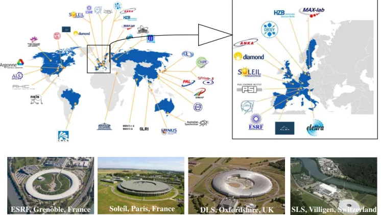
Figure 1.47–Synchrotron facilities in the world………..66
Figure 1.48–Geometric representation of the “Bragg’s Law”……….68
Figure 1.49 – Schematic representation of the constructive and destructive interference (top and
bottom, respectively)………..68
Figure 2.1 – Reaction scheme of the synthesis of the compound ALF_MS1 according the followed experimental procedure………...77
Figure 2.2–FTIR spectrum of solid ALF_MS1 in KBr pellet………..78
Figure 2.3– Structure of ALF850 complex, [Ru(CO)3Cl2(1,3-thiazole)]………..80
Figure 2.4 – FTIR spectrum of lyophilized dialysate of the adduct of ALF850 with HEWL, in KBr
pellet………81
Figure 2.5–HEWL crystals with ALF850 after 24 hours of soaking………...82
Figure 2.6–Diffraction pattern of the HEWL•ALF850 crystal………82
Figure 2.7– Overall structure of HEWL bound to Ru fragments derived from ALF850………..84
Figure 2.8–Structural representation of Ru•His15 adduct………...85
Figure 2.9– Schematic representation of adducts formed between the histidine residue of HEWL and CORM-3 (A) and ALF850 (B)………...86
Figure 2.10–Structures of ALF475 (left), ALF486 (center) and ALF487 (right) complexes………..87
Figure 2.11 – Structural representation of HEWL•CORM complexes at the site with the highest Ru
occupation, obtained by soaking HEWL crystals with ALF 475 (left), ALF486 (center) and ALF487
(right)………..89
Figure 2.12– Reactivity of [RuII(CO)
3L3]2+CORMs in aqueous, aerobic solutions……….91
Figure 2.13 – Proposed mechanism for the interaction of [RuII-(CO)
3Cl2L] with cells or biomolecules………...92
Figure 2.14–HEWL crystals with ALF_MS1 after 24 hours of soaking……….93
Figure 2.15 – Structural representation of the Ru•His15 adduct obtained by soaking HEWL crystals
with ALF_MS1………...95
XVI
Figure 2.17 – Diffraction pattern of hemoglobin•CORM-3 (left) and hemoglobin•ALF475 (right) crystals………97
Figure 2.18–Overall structure of hemoglobin•CORM-3 model………..99
Figure 2.19 – Structural representation of the Ru adduct obtained in the hemoglobin•CORM-3
model………..99
Figure 2.20–Overall structure of hemoglobin•ALF475 model………..100
Figure 2.21 – Structural representation of the Ru adduct obtained at the interface of chains B and D in the hemoglobin•ALF475 model………...101
Figure 2.22 – Reaction scheme of the synthesis of the compounds ALF_MS2 (top) and ALF_MS3 (bottom) according the followed experimental procedure………103
Figure 2.23– FTIR spectra of solid ALF_MS2 (top) and ALF_MS3 (bottom) in KBr pellets……...104
Figure 2.24 – HEWL crystals with ALF_MS2 (left), ALF_MS3 (center) and ALF_MS4 (right) after
24 hours of soaking………...106
Figure 2.25–Structural representation of the HEWL•ALF_MS4 adduct………...108
Figure 2.26–Structural representation of the HEWL•ALF_MS2 adduct………...109
Figure 2.27–Structural representation of the HEWL•ALF_MS3 adduct………...110
Figure 2.28 – FTIR spectra of solid native HEWL (blue), HEWL•ALF_MS3 (red) and
HEWL•ALF_MS4 (green) in KBr pellets………111
Figure 2.29 – Comparison of the amino acid sequence of human serum albumin (UniProt entry:
P02768) and bovine serum albumin (UniProt entry: P02769)………..112
Figure 2.30 – Purification of human serum albumin by gel filtration chromatography using a
Superdex S75 column………...115
Figure 2.31 – Purification of human serum albumin by gel filtration chromatography using a
Superdex S200 column……….117
Figure 2.32 – Native HSA crystals obtained before and after the optimization of the found
crystallization hit (left and right, respectively)……….118
Figure 2.33– Native BSA crystals obtained with 0.2 M calcium acetate, 20% PEG4K, 0.1 M Tris-HCl
pH 6.5 (condition 2)………..119
Figure 2.34– Native hs-apoTF crystals obtained with 15% glycerol, 20% PEG 4K, 0.2 M ammonium
citrate pH 7………122
Figure 2.35 – Native hs-apoTF crystals obtained with 13% glycerol, 17.5% PEG 4K, 0.2 M
ammonium citrate pH 6.73………...123
Figure 2.36– Soaked hs-apoTF crystals with ALF_MS2………123
Figure 2.37 – Structural representation of the iridium moiety found at the surface of the N-terminal
XVII
Figure 2.38– HEWL crystals with the tested manganese-, iron- and molybdenum-based CORMs after
24 hours of soaking………...127
Figure 2.39– Bovine hemoglobin crystals with the tested manganese-, iron- and molybdenum-based
CORMs after 24 hours of soaking………127
Figure 3.1 – Molecular formulas of maltol (A), Hdhp (B), picolinic acid (C) and dipicolinic acid
(D)……….137
Figure 3.2 – Urea-polyacrylamide gel electrophoresis of: 1) hs-apoTF, 2) hs-apoTF+VIVO, 3)
hs-apoTF+VIVO+Maltol, 4) hs-apoTF+VIVO+Hdhp, 5) hs-apoTF+VIVO+Hpic, 6)
hs-apoTF+VIVO+H
2dipic and 7) hs-holoTF………..138
Figure 3.3 – Experimental SAXS data of native hs-apoTF (blue) and hs-apoTF+VIVO+Hpic (red)………...140
Figure 3.4– Scattering profiles generated by CRYSOL from transferrin crystal structures 2HAV and 3V83 and respective comparison with the scattering profiles of the native apoTF (a) and hs-apoTF+VIVO+Hpic (b) samples………141
Figure 3.5 – Superposition of the calculated most likely molecular envelope of native hs-apoTF (a) and hs-apoTF+VIVO+Hpic (b) samples with the X-ray structure 2HAV………..141
Figure 3.6 – Experimental SAXS scattering profiles of hs-apoTF+VIVO+Hpic obtained by long
incubation (red) and short incubation (green)………...142
Figure 3.7–HEWL crystals with different vanadium complexes after 24 hours of soaking………..144
Figure 3.8– Overall representation of HEWL with the VIVO(pic)
2 complex close to the Asp52 residue
at the enzyme active site (A) and the detailed structural representation of the respective VIVO(pic) 2– Asp52 fragment (B)………..147
Figure 3.9–Molecular formulas of salicylic acid (left) and proline (right)………150
Figure 3.10 – HEWL crystals soaked (24 hours) with VOSO4 and salicylic acid (left) and proline (right)………150
Figure 3.11– Molecular formulas of VO(acac)2 (left), 1,10-phen (center) and bipy (right)………...150
Figure 3.12– HEWL crystals soaked (24 hours) with different vanadium complexes: VO(acac)2 (left),
VOSO4+1,10-phen (center) and VOSO4+bipy (right)………..151
Figure 3.13–Preliminary structural representation of the adduct observed in the HEWL•VOSO4•bipy structure………153
Figure 3.14 – Bovine trypsin crystals with different vanadium complexes after 24 hours of
soaking………..156
Figure 3.15 – Overall representation of bovine trypsin with the VIVO(pic)
XVIII
Figure 3.16– Structural representation of VIVO(pic)
2-Ser195 adduct……….159
Figure 3.17 – Bovine trypsin crystals soaked with different vanadium complexes: VOSO4+salicylic
acid (left, top), VOSO4+proline (right, top), VO(acac)2 (bottom, left), VOSO4+1,10-phen (bottom,
XIX
Table Index
Table 1.1– List of some metals and the respective medical applications………..21
Table 1.2– Physical properties of carbon monoxide……….23
Table 1.3– List of some agents responsible for the activation of HO-1………26
Table 1.4–Correlation between the CO concentration and the respective effect……….27
Table 1.5 – List of organ successfully transplanted organs (in rats) and the respective CO
concentration used in the procedure (in parenthesis, the administration method is indicated)………..33
Table 1.6–List of some of the most common minerals containing vanadium………..42
Table 1.7–Summary of function and/or reaction of proteins related to vanadium………...47
Table 1.8– List of Nobel Prizes winners associated with X-ray crystallography……….60
Table 1.9– List of crystal systems, Bravais lattices and space groups allowed for proteins………….63
Table 2.1– Data collection and refinement statistics for HEWL•ALF850 adduct crystal………83
Table 2.2– Occupancy and B factors of ruthenium atoms in the HEWL•ALF850 model………84
Table 2.3 – Data collection and refinement statistics for HEWL•ALF475, HEWL•ALF486 and
HEWL•ALF487 adduct crystals……….88
Table 2.4 – Metal binding sites found in the crystal structures of HEWL soaked with ALF475, ALF486 and ALF487, describing the ligands at each Ru binding site………...90
Table 2.5 – Data collection and current refinement statistics for HEWL•ALF_MS1 adduct
crystal………..94
Table 2.6 – Data collection and refinement statistics for hemoglobin•CORM-3 and hemoglobin•ALF475 adduct crystals……….98
Table 2.7 – Data collection and refinement statistics for HEWL•ALF_MS2, HEWL• ALF_MS3 and
HEWL•ALF_MS2 adduct crystals………..107
Table 2.8–Optimization of the crystallization condition of HSA………...118
Table 2.9– Optimization of the crystallization condition of hs-apoTF………...122
Table 2.10 – Data collection and refinement statistics for hs-apo-TF•ALF_MS2 adduct
crystal………124
Table 2.11 – X-ray diffraction results obtained with the soaked HEWL and bovine hemoglobin crystals with ALF21, ALF58, ALF73, ALF153 and ALF157………...128
XX
Table 3.1 – SAXS data collection and scattering-derived parameters of native apoTF and
hs-apoTF+VIVO+Hpic samples……….139
Table 3.2 – X-ray diffraction results obtained with the best HEWL crystals soaked with different
vanadium complexes……….145
Table 3.3 – Data collection and refinement statistics for HEWL•VOSO4•Hpic adduct crystal………146
Table 3.4 – Distance between V and the coordinating ligands for the complete data set, the first 350 images, the first 500 images and the last 500 images collected………...148
Table 3.5 – Data collection statistics for HEWL•VOSO4•Hpic sub-sets: images 1-350, images 1-500
and images 501-1000………148
Table 3.6 – Data collection and refinement statistics for HEWL•VOSO4•bipy adduct crystal………152
Table 3.7 – X-ray diffraction results obtained with the best bovine trypsin crystals soaked and
co-crystallized with different vanadium complexes………..157
Table 3.8 – Data collection and refinement statistics for trypsin•VOSO4•Hpic adduct crystal………158
Table 3.9 – X-ray diffraction results obtained with the best bovine trypsin crystals soaked with
XXI
Abbreviations and symbols
|Fhkl| Amplitude
1,10-phen Phenanthroline
ADME Absorption, Distribution, Metabolism and Excretion
ADMET Absorption, Distribution, Metabolism, Excretion and Toxicity
AOX Aldehyde oxidase
Bipy 2,2'-bipyridine
BK Large-conductance Ca2+-and voltage-gated K+ channels
BPG 2,3-biphosphoglycerate
BSA Bovine Serum Albumin
CaV Voltage-gated Calcium Channels
cGMP Cyclic Guanosine Monophosphate
CO Carbon monoxide
COHb Carboxyhemoglobin
CORM Carbon Monoxide Releasing Molecule
CORM-1 Dimanganese decacarbonyl, [Mn2(CO)10]
CORM-2 Tricarbonyldichloro ruthenium (II) dimer, [Ru(CO)3Cl2]2
CORM-3 Tricarbonyldichloro(glycinato)ruthenium (II), [Ru(CO)3Cl(glicinate)]
CORM-A1 Sodium boranocarbonate, [Na2H3BCO2]
Cryo-EM Cryo Electron Microscopy
CSD Cambridge Structural Database
Da Dalton
DCM Dichloromethane/Methylene chloride
DFT Density Functional Theory
dhkl Distance between the planes in the crystal lattice
DLS Diamond Light Source
EMA European Medicines Agency
ERK Extracellular signal Regulated Kinases
ESRF European Synchrotron Radiation Facility
ET-CORM Enzyme-triggered CORM
FBDD Fragment-based Drug Discovery
Fcalc Calculated Structure Factor
FDA Food and Drug Administration
FDT Free Drug Theory
XXII
Fobs Observed Structure Factor
FTIR Fourier Transform Infrared Spectroscopy
GI tract Gastrointestinal tract
GPCRs G-protein-coupled receptors
GTP Guanosine Triphosphate
H2dipic Dipicolinic acid
H2S Hydrogen Sulfide
Hdhp 1,2-dimethyl-3-hydroxy-4-pyridinone
HEPES 4-(2-hydroxyethyl)-1-piperazineethanesulfonic acid
HEWL Hen White Egg Lysozyme
HO Heme oxygenase
HO-1 Heme oxygenase-1
HO-2 Heme oxygenase-2
Hpic Picolinic acid
HSA Human Serum Albumin
Hs-apoTF Human Serum Transferrin
HSF1 Heat Shock Factor 1
HSP70 Heat Shock Protein 70
HTS High Throughput Screening
I/R Ischemia/Reperfusion
IL-10 Interleukin-10
IL-1β Interleukin-1β
IRS Insulin Receptor Substrate
ITC Isothermal Titration Calorimetry
JNK c-Jun NH2-terminal kinases
KCa Calcium-activated Potassium Channels
logP Logarithm of the octanol-water partition coefficient
MAD Multiple-wavelength Anomalous Dispersion
Maltol 3-Hydroxy-2-methyl-4H-pyran-4-one
MAPK Mitogen-activated protein kinase pathway
MCC Metal Carbonyl Complex
MIP-1α Macrophage Inflammatory Protein-1α
MIR Multiple Isomorphous Replacement
MR Molecular Replacement
NaVO3 Sodium metavanadate
XXIII
NO Nitric Oxide
p38 p38 MAPK
PD Pharmacodynamics
PDB Protein Data Bank
PEG Polyethylene glycol
PK Pharmacokinetic
PKG cGMP-dependent Protein Kinase or Protein Kinase G
PTPase Phosphatases with affinity to tyrosine-phosphate proteins
RO5 Rule-of-five or Lipinski’s rule
ROS Reactive Oxygen Species
SAD Single-wavelength Anomalous Dispersion
SAR Structure-Activity Relationship
SAXS Small Angle X-ray Scattering
sGC Soluble Guanylyl Cyclase
SLS Swiss Light Source
SPR Surface Plasmon Resonance
TNFα Tumor Necrosis Factor
Tris Tris-(hydroxymethyl)-aminomethane
TS Fluorescence-based Thermal Shift
V Vanadium
VBPO Bromoperoxidases
VCPO Chloroperoxidases
VHPO Vanadium-containing Haloperoxidases
VO(acac)2 Vanadyl acetylacetonate
VOSO4 Vanadyl sulfate tetrahydrate
θ Angle between the incident wave and the crystal plane
λ Wavelength
1
HAPTER
1
I
NTRODUCTION
3
1.1
–
Drug Design and Development
1.1.1
–
General concepts and historical perspective
The 20th century, as well as the earlier years of the 21st century, led to deep and significant
geographic, political, economic, technologic and social changes in the world. Greatly connected with such modifications, it is commonly recognized that the current life conditions are much better than those 100 years ago, particularly in the developed countries in Europe and North America.
The improvement of the health conditions was an essential step for such progress as proven by the raising of the life expectancy. This improvement is partially explained by a better nutrition and basic hygiene procedures but the development of new, better and safer drugs was absolutely vital for such achievement. Remarkably, former deathly diseases as smallpox, poliomyelitis and diphtheria were largely eradicated, a large range of antibiotics were made available and many other chronic or minor conditions – as simple as a cold or a headache – could be effectively treated or, at least, controlled (Figure 1.1).1,2
Figure 1.1 – Compounds in clinical development by therapeutic area between 1996 and 2011.2
Oncology is the more representative area with 26% of the total of the products in development in 2011.
At this point, one very important question emerges: what is a drug? In general, a drug can be
defined as “an agent that has a desired biological effect on the human body or some other living system” or, even in a more broad definition, agents that “interact with a biological system and produce
a biological response”.3 These definitions can be ambiguous covering many compounds. Can
4
associated to addiction and habituation, do not have medicinal goals (heroin and cocaine can be cited as belonging to this group).
However, such classification is not entirely satisfactory. The line between both sides is very flexible meaning that the same compound could be classified as a “good drug” or as a “bad drug” depending on the situation. A classic example is morphine: known as a potent analgesic, but also addictive and responsible for respiratory problems.3 Other illustrative example is heroin: one of the
most well-known “bad drugs”, heroin is a very effective painkiller used in terminal cancer cases as
diamorphine.3,4
These two straightforward examples illustrate that, by one hand, is very hard, or virtually
impossible, to distinguish between “good drugs” and “bad drugs” and, by the other hand, that no drug
is entirely safe. Helping to explain these facts, it should be taken into account the famous citation of Paracelsus – the “father” of Toxicology – in the 16th century: “All things are poison and nothing is
without poison; only the dose makes a thing not a poison” or, more simpler, “The dose makes the poison” (Sola dosis facit venenum in Latin).5
Apart from all the possible discussions related with this nomenclature, the terms drug development and drug design are usually associated to the research conducted by large pharmaceutical companies with very significant financial costs (around US $800 million according some studies).1,6
Nevertheless, long before the foundation of these companies, scientists were already involved in this subject. The following paragraphs aim to give a brief historical perspective on the evolution of drug discovery.
The search for medicinal substances is nearly as old as man himself and there are evidences of the use of drugs in the Neolithic period.7 Natural products were the source of the drugs used –
particularly herbs and other vegetal products, but also arising from animal and mineral origin – and nowadays continue to be important in the drug discovery processes.5,8
Moving to some ancient civilizations (Mesopotamia, Egypt, India and China, among others), it is possible to asseverate the use of drugs thanks to the available documents and artifacts that survive until modern ages.5,7That’s the case of several pieces of clay from Mesopotamia, dated around 1700
B.C., found about one century ago and interpreted by R. Campbell Thompson.7 Also importantly, an
Egyptian document was also found in 1872; written in c. 1550 B.C., the so-called Ebers Papyrus (named in honor to George Ebers who found it) contains information on prescriptions, drugs and medicinal plants (Figure 1.2). Regarding the Oriental cultures, some compositions should be also mentioned: the Indian Ayurveda(“Science and Knowledge of Life’’) and the Chinese Shen Nong Ben Cao Jing(“Shen Nong’s Canon on Materia Medica”).5,7
5
mentioned as Hippocrates (the “Father of Medicine” and leader of the Cos school, he is associated to the Hippocratic Oath and the Hippocratic Corpus where medical and ethical questions are considered), Aristotle and Teophrastus (the authors of Historia Plantarum and De Causis Plantarum, respectively, which present a classification for over 500 plants and describes the vegetal physiology).5,7
In Rome, in the first and second centuries after Christ, Pedanius Dioscorides and Claudius Galen or Ganelus should be also highlighted. Dioscorides was the author of De Materia Medica (Figure 1.3) which presents a detailed study not only on over 900 drugs (from animal, vegetal and mineral sources) but also on some elementary chemical techniques (as distillation). Galenus was the author of Opera Omnia which describes several drugs and respective applications; he also developed the use of the so-called “galenicals” – complexes resulting from the junction of several ingredients.5,7
After the end of the West Roman Empire (5th century) and the beginning of the Medieval
period, the focus on science was moved to the Arab world, merging the occidental and the oriental knowledge in different areas. It should be emphasized that Iberian Peninsula was particularly influenced by the Arabian culture due to several centuries of occupation (from 711 to 1492) as proven, for instance, by the vocabulary, architecture and technology adopted by the Portuguese and Spanish people. Concerning pharmacology, the Arabian thinking is similar to the Greek one; Rhazes (Abu Bakr Razi), Avicenna (Abu Ali Hussain ibn Abdallah ibn Sina) and Abulcasis (Abu Qasim al-Zahrawi) are some of the greatest names in the 10th and 11th centuries. For instance, in first years of the
11th century, Avicenna published the book al-Qanun fi at-tibb–Canon Medicinae– containing, among
other details, the description of the uses and efficacy of 760 drugs alphabetically ordered.5,7
During the Middle Ages, Europe is not recognized as a fruitful and fertile land for the development of the scientific questions with exceptions of some medicinal works namely the book
Figure 1.3 – Cover of De Materia Medica (edition dated from 1554, in Latin).5
The original edition was written in Greek and, due to the details presented on several drugs and some of the side effects, it was one of the main pharmacology books until the 18th and 19th centuries.
Figure 1.2– Piece of the Ebers Papyrus.5
6
published in the last half of the 13th century Thesaurus Pauperum attributed to the Portuguese Pope
John XXI (born Pedro Julião and also known as Pedro Hispano Portucalense).9,10
The Renaissance was the stage for a noteworthy change in this paradigm and science, among architecture, painting and sculpture, gained new life. The already referred Paracelsus (born Philippus Teophastrus Bombastus von Hohenheim) was one of the exponents of this period; in addition to the introduction of the concept of dose, Paracelsus used metal salts for the treatment of some disorders in what can be considered as an ancestor of medicinal chemistry.5
One of the facts closely related to such development was the Portuguese Discoveries (the famous Descobrimentos brilliantly described in the equally famous Portuguese epic poem Os Lusíadas by Luís Vaz de Camões). Following the example of Portugal, some other European countries also started its discoveries namely Spain (the Treaty of Tordesillas – by which the two Iberian countries divided the world between them, shortly after the discovery of America – became well-known). Starting in 1415 and prosecuting into the 15th and 16th centuries, the Portuguese people were able to
reach very widespread regions such as Africa, Orient (India, China and Japan) and Brazil. As result, in addition to the expansion of the Portuguese Empire, it was possible to contact with other civilizations as well as to study new herbs and other pharmacological agents introducing them in Europe. New compendia were written as “The Colloquies on the Simples and Drugs of India” (Coloquios dos simples, e drogas he cousas mediçinais da India) published in 1563 by Garcia de Orta.11
The progressive advances in biology and chemistry that occurred in the following centuries paved the way into the modern drug discovery processes from the 19th century until nowadays (Figure
1.4).5,12,13 In fact, by the 19th century, more than the simple use of herbs or other agents with medicinal
properties, the goal was to identify, isolate and characterize the respective active substances. This approach was determinant for the development of the pharmacology as an experimental and systematic scientific field thanks to the work conducted by some European scientists as François Magendie, Claude Bernard, Rudolph Buchheim and Oswald Schmiedeberg. The foundation of specialized journals has also contributed for the mentioned development; as example, Schmiedeberg and Bernhard Naunyn founded, in 1873, the journal Archiv für experimentelle Pathologie und
Pharmakologie which is still active nowadays under the name “Naunyn-Schmiedeberg’s Archives of Pharmacology”.5
The first example of this new methodology took place in the first years of the 19th century
when Friedrich Sertürner was able to isolate morphine from opium testing it in dogs. From this point, several other alkaloids were identified and isolated like emetine (by Pierre Pelletier), quinine and caffeine (both by Pierre Pelletier and Joseph Caventou), atropine (by Philipp Geiger), papaverine (by Georg Merck) and cocaine (by Albert Niemann).5,7 As important as these findings, was the raising of
7
Shortly after, some other factories were created or reconverted for such purpose as Merck – still a world giant of the pharmaceutical industry nowadays.5
Figure 1.4– Summary of some important findings on drug discovery over the last 200 years. Adapted from 12 Starting with the isolation of the active principles from plants, the significant advances in Chemistry,
Molecular Biology, Genetics, Bio-informatics and Biochemistry were essential for the development of new drugs.
In the subsequent years, different approaches led to the emergence of new drugs as represented in Figure 1.5. During the 19th century, the introduction of different vaccines, as anthrax, rabies and
cholera by Louis Pasteur, was crucial avoiding thousands of deaths by year. In the first years of the 20th century, new concepts were introduced, as the term chemoreceptor by Paul Ehrlich, which is on
the basis of the modern studies of ligand-receptor interactions.5,12
Moving to the second period (from the middle of 1930s to nowadays), the discovery of antibiotics – penicillin was the first to be identified by Alexander Fleming –was also a remarkable progress in the evolution of drug discovery. Penicillin played an important role during the Second
1815
Isolation of morphine from opium extract
1870
Publication of Periodic Table
1872
Establishment of chemotherapy
1910
Rational design of arsphenamine
1929
Discover of penicillin
1953
Discover of the structure of DNA (double helix)
1973
First applications of DNA Recombinant
techniques
1998
Isolation of human embryonic stem cells
2003
Complete map of the human genome
2007
8
World War and, from then to now, several other natural and semisynthetic antibiotics were produced helping to reduce the number of deaths caused by bacterial infections, around the world. A large range of new drugs has been developed in the last 40 years covering an increasing number of conditions. The expansion of the drug discovery process is closely related to the improvement of the available methodologies and tools: molecular biology, computational chemistry, genetic engineering and structural biology (namely X-ray crystallography) are some examples of the techniques that contributed to the boost of this already well established field.5,12
Figure 1.5– Schematic representation of the major periods of drug development. Adapted from 5The
modern drug discovery process is divided into two periods which are subdivided in different generations of drugs spanning from the beginning of the 19th century until the 21st century. Vaccines and antibiotics are some of
the most representative compounds in the drug development area.
Nevertheless, this amazing journey is not finished or not even close to its end. Research is conducted daily in order to try to improve our knowledge on different diseases – cancer is, perhaps, the most paradigmatic case – leading to the identification of new potential usable drugs.
1.1.2
–
Principles of Drug Design
As previously mentioned, the development of new drugs is a very expensive and time-consuming process.6 In fact, from the thousands potential compounds studied, only a minor percentage
will reach the market after approval by some legal institutions as the US Food and Drug Administration (FDA) or the European Medicines Agency (EMA): from 1950 to 2008, only 1222 new drugs were approved by the FDA (Figure 1.6).14,15
Figure 1.6 – Graphical representation of the approval of new drugs by the FDA between 1950 and 2008.14 Out of a total of 1222 approved drugs, 1103 correspond to small molecules and 119 are biological
9
The low number of successfully approved drugs is a clear indicator of the complexity of the process. Several steps are required in order to obtain a suitable therapeutic agent (Figure 1.7) and all of them must be entirely satisfactory. That means that, for instance, a drug candidate could be very effective against a given disorder but is useless if it is also toxic or not bioavailable. In fact, bioavailability and toxicity are the common factors for rejecting the use of different molecules as drugs, covering 39% and 21% of the causes of the unsuccessful cases, respectively.4,16,17
The creation of a new drug could be divided into two different stages: discovery and development. The first stage leads to the identification of a drug candidate (after identifying the target and optimizing the lead) while the second stage encompasses the preclinical, clinical (phases I, II and III) and post-clinical trials in animals and humans
Only after all these steps, which usually take 10-15 years to be completed, a molecule can be considered as a safe drug and reach the market. Regardless of the importance of the clinical trials, they will be not addressed herein. Instead, a brief outline on the discovery process of a new drug candidate will be covered.
Figure 1.7– Schematic representation of the different stages involved in the drug design process from the target identification until the market release. Adapted from 17 Following the identification of a
10
1.1.2.1
–
Drug Discovery steps
As previously referred, before starting the clinical trials phases, it is necessary to determine suitable drug candidates. Figure 1.8 summarizes the principal steps involved in the discovery of drug candidates: target identification and validation, hit identification, hit-to-lead and lead optimization.18
Figure 1.8 – Workflow of the stages involved in the discovery of putative drug candidates. Adapted from 17 For each stage, different methodologies are required in order to achieve the desired goal: obtaining a
new and safe drug. The great number of required steps and respective associated techniques explains partially why drug design is an expensive and time-consuming process.
o Target identification and validation
Keeping in mind the current paradigm, a given disease should be characterized as accurately and detailed as possible. Therefore, it makes sense thinking in the target identification as the first stage of the drug design process.
A target could be defined as a biological molecule – usually proteins, DNA or RNA – able to interact with a potential drug (small molecules or other biomolecules like antibodies) producing a beneficial effect. An important consequence from this definition is the existence of a binding site – a particular region of the target capable of accommodating the drug through covalent interactions or, more commonly, intermolecular interactions (for example, ionic bonds, hydrogen bonds, van der Waals interactions and hydrophobic interactions). A “druggable” target must follow some specific
characteristics namely to be directly involved in a given disorder, to be locally expressed in some tissues or organs and to be functionally and structurally characterized.3,18,19,20,21
Currently, proteins are the most well studied targets being grouped into different classes such as enzymes, receptors, ion channels and transport proteins. Particular attention has been given to proteases and kinases and also to G-protein-coupled receptors (GPCRs).22 The progressive studies on
the area allow to identify an increasing number of targets.19,20,21 Iterative medicinal chemistry


![Figure 1.30 – Structural representation of some V III aqueous monomeric species: [V(H 2 0) 6 ] 3+ (left), [V(OH)(H 2 0) 5 ] 2+ (center) and [V(OH) 2 (H 2 0) 4 ] + (right)](https://thumb-eu.123doks.com/thumbv2/123dok_br/16472760.731777/75.892.128.773.425.569/figure-structural-representation-aqueous-monomeric-species-center-right.webp)