Licenciada em Física
Monte Carlo simulations for dosimetric
verification in photon and electron beam
radiotherapy
Dissertação para obtenção do Grau de Doutor em Engenharia Biomédica
Orientador: Doutora Grisel Mora Paula, CFNUL/FC
Co-orientador: Professora Doutora Adelaide de Jesus, FCT/UNL
Professor Doutor Nuno Teixeira , FCM/UNL
Júri:
Presidente: Doutor Fernando José Pires Santana Arguente(s): Doutor José Pedro Miragaia Trancoso Vaz Doutor Carlos Manuel Azevedo Sousa Oliveira
Vogais: Doutor Francisco José Cerqueira Alves Doutor Nuno José Coelho Gomes Teixeira
Doutora Maria Adelaide de Almeida Pedro de Jesus Doutora Grisel Margarita Mora Paula
of making real their ”always” dream...
Forte n ˜ao ´e aquele que nunca cai mas sim
... October 2005, the beginning of a new adventure ...
Back stay the cold Germany, the country where I began my interest in the physics research. New
year, new life and why not Lisbon? All the stars were showing me the way to Portugal; an unknown
country for me at that time, but nowadays my home country.
I arrived in Lisbon with an adventure in mind: to do my Ph.D. work, an ”always” dream. I knew
that it would not be an easy task, but I was sure that it would be an excellent opportunity for my
professional and personal life. Now, after 6 years of changes and efforts, I finish this adventure; an
adventure which would have been not possible without the support, help and love of a long list of
people:
First of all, I would like to express my gratitude to my supervisor Doctor Grisel Mora for the
inestimable support in pedagogical aspects and also in theoretical and practical supports that she
has always manifested along this work. A special thanks for supplying the necessary criticism to
evaluate the papers and this thesis.
Professor Dr. Adelaide Pedro de Jesus for having orientated this work and, particularly, for
believing in me from the first time I met her at Bochum (Germany) in 2004 until today. She gave
me the opportunity to join the Lisboa group in 2005, introducing me to the exciting field of nuclear
astrophysics in Portugal for one year. I want also to thank her for allowing me to reach a dream in
the medical physics area, the present Ph.D. work. Her constant support and advices during all the
phases of my work is gratefully acknowledged.
I would like to thank Professor Dr. Nuno Teixeira for having oriented the experimental part of
this thesis and for opening the door of the radiotherapy departments where this research has been
developed.
Chaves, SAMS of Lisbon and Hospital de Santa Maria. A special thanks to Enga. Iola Cardoso,
Engo. Luis Madureira, Enga. Ana Catarina Souto and Enga. Dalila Mateus for their great patience
in listening the mountains of doubts that I had during this work. Thanks for the fruitful discussions
we had.
To Fundac¸ ˜ao para a Ci ˆencias e Tecnologia for its financial support through the grant.
To Centro de F´ısica Nuclear da Universidade de Lisboa for its financial and technological
sup-ports to do my Ph.D.
To my recent colleagues of the ENAPG group, a great thanks for their support in the last phase
of the work. A special thanks to Enga. Ana Henriques, Engo. Jorge Machado and Dr. Pamela
Teubig for believing in me during the last and difficult phase of the work. I’ll never forget our coffee
breaks.
To my ”scientific father” and friend, Daniel Galaviz Redondo, for introducing me in the world of
physics research in 2003 at Darmstadt (Germany). ”Dani, aqu´ı la tienes. Gracias por haberme dado
a conocer el mundo de la investigaci ´on en f´ısica y especialmente por haber estado cerca en los
momentos m ´as dif´ıciles del camino”.
To my best portuguese friends Micaela Fonseca and Cecilia Borges for all the positive energy
and strength that they always transmitted me along this work.
To D. Luisa, Sr. Vitor, Vitor Luis, Susie and my lovely Leonardo and Miriam, for the special
moments I spent with all of them during this time. They are a wonderful family and I’ll always take
them in my heart. ”Obrigado pela amizade e o carinho que me ofereceram durante estes anos”.
To you, Valter, for being always close even a thousand miles of distance away. We can shine!
”Obrigada por teres acreditado sempre no meu valor e me teres feito perceber que posso brilhar”.
And, finally, I cannot finish my acknowledgments without thanking to my family who constantly
supported me with their love and allowed me to complete this work. This thesis is dedicated to them.
”A mi familia por la fuerza y el cari ˜no que me regalaron durante esta fase de mi vida. Pap ´a,
mam ´a, Carlos y abuelos, siempre estuvisteis presentes en mi mente y en mi coraz ´on a pesar de la
distancia. Luchasteis por hacer realidad mis sue ˜nos y nunca tendr ´e amor suficiente para compensar
todo aquello que hicisteis por m´ı. Esta tesis es vuestra.”
One of the primary requirements for successful radiotherapy treatments is the accurate
calcu-lation of dose distributions in the treatment planning process. Monte Carlo (MC) dose calcucalcu-lation
algorithms are currently recognized as the most accurate method to meet this requirement and to
increase even further dose accuracy.
The improvements in computer processor technology and the development of variance reduction
techniques for calculations have led to the recent implementation and use of MC algorithms for
radiotherapy treatment planning at many clinical departments.
The work conducting to the present thesis consists of several dosimetric studies which
demon-strate the potential use of MC dose calculations as a robust tool of dose verification in two different
fields of external radiotherapy: electron and photon beam radiotherapy.
The first purpose of these studies is to evaluate dose distributions in challenging situations
where conventional dose calculation algorithms have shown some limitations and it is very
diffi-cult to measure using typical clinical dosimetric procedures, namely in regions containing tissue
inhomogeneities, such as air cavities and bones, and in superficial regions.
A second goal of the present work is to use MC simulations to provide a detailed
characteriza-tion of photon beams collimated by a multileaf collimator (MLC) in order to assess the dosimetric
influences of these devices for the MC modeling of Intensity Modulated Radiotherapy (IMRT) plans.
Detailed MC model of a Varian 2100 C/D linear accelerator and the Millenium MLC incorporated in
the treatment head is accurately verified against measurements performed with ionization chambers
and radiographic films.
Finally, it is also an aim of this thesis to make a contribution for solving one of the current
prob-lems associated with the implementation and use of the MC method for radiotherapy treatment
planning, namely the clinical impact of converting dose-to-medium to dose-to-water in treatment
planning and dosimetric evaluation. For this purpose, prostate IMRT plans previously generated by
a conventional dose algorithm are validated with the MC method using an alternative method, which
involves the use of non-standard CT conversion ramps to create CT-based simulation phantoms.
THESIS AIM AND OUTLINE xix
I
INTRODUCTION
1 Introduction to external beam radiation therapy 3
1.1 Brief introduction . . . 3
1.2 Techniques in external radiotherapy . . . 5
1.3 The importance of accuracy in radiation delivery . . . 8
2 Fundamentals of radiotherapy physics and dosimetry 11 2.1 Interaction of ionizing radiation with matter . . . 11
2.1.1 Photon interactions in matter . . . 12
2.1.1.1 Types of interaction mechanisms . . . 12
2.1.1.2 Attenuation coefficients . . . 18
2.1.1.3 Relative predominance of individual effects . . . 20
2.1.2 Electron and positron interactions in matter . . . 21
2.1.2.1 Types of interaction mechanisms . . . 21
2.1.2.2 Stopping power and range . . . 23
2.1.2.3 Restricted stopping power . . . 26
2.2 General concepts of clinical radiation dosimetry . . . 27
2.2.1 Basic dosimetric quantities . . . 27
2.2.2 Basic theorems and principles . . . 31
2.2.2.1 Inverse-square law . . . 31
2.2.2.2 Charged particle equilibrium . . . 32
II
MATERIALS AND METHODS
3 Radiation treatment and dosimetry equipment 41
3.1 Medical linear accelerator . . . 41
3.1.1 General description of an electron linear accelerator . . . 42
3.1.2 Treatment head components . . . 43
3.2 Radiation dosimetry equipment . . . 47
3.2.1 Ionization chamber dosimetry . . . 48
3.2.2 Film dosimetry . . . 53
3.2.2.1 Radiographic film . . . 54
3.2.2.2 Radiochromic film . . . 57
4 Monte Carlo simulation techniques in radiation therapy 61 4.1 General fundamentals of the Monte Carlo method . . . 61
4.2 Why use Monte Carlo in radiotherapy? . . . 63
4.3 Simulation of Photon and Electron Transport . . . 65
4.4 The EGSnrc Code System . . . 73
4.4.1 General Description . . . 73
4.4.2 BEAMnrc: A linac modeling tool . . . 77
4.4.3 DOSXYZnrc: Phantom dose calculation tool . . . 78
4.4.4 Variance Reduction Techniques and Efficiency Improvements . . . 81
III
ELECTRON BEAM RADIOTHERAPY APPLICATION
5 Dosimetric effect of shallow air cavities in high energy electron beams 87 5.1 Motivation . . . 875.2 Material and Methods . . . 89
5.2.1 Air cavity phantom . . . 89
5.2.2 Monte Carlo simulations . . . 90
5.2.3 Measurements . . . 100
5.2.3.1 Home-built PMMA phantom . . . 100
5.2.3.2 Radiochromic film dosimetry . . . 101
5.3 Results . . . 104
5.3.1 Air cavity effect for standard 10 x 10 cm2field . . . 104
5.3.1.1 Central-axis PDD variation due to the presence of air cavity . . . . 104
5.3.1.2 Off-axis dose variation due to the presence of the air cavity . . . 108
5.3.1.3 Influence of air cavity on spectral distributions . . . 110
5.3.2 Air cavity effect for fields shaped by Cerrobend blocks . . . 115
5.4 Conclusions . . . 117
IV
PHOTON BEAM RADIOTHERAPY APPLICATION
6 Monte Carlo modeling of a 6 MV Varian 2100C/D medical accelerator 123 6.1 Linear accelerator model . . . 1236.2 Determination of primary electron beam parameters . . . 131
6.3 Beam spectral characterization at phantom surface . . . 143
6.4 Conclusions . . . 152
7 Varian Millennium 120-leaf MLC in Monte Carlo simulations 153 7.1 Monte Carlo MLC model description . . . 153
7.2 Experimental commissioning of MLC model . . . 157
7.2.1 Monte Carlo simulations . . . 157
7.2.2 Experimental measurements . . . 160
7.2.3 Benchmark tests and results . . . 161
7.3 Conclusions . . . 173
8 Assessing Multileaf Collimator effect on the build-up region for 6 MV photon beams 175 8.1 Motivation . . . 175
8.2 Monte Carlo evaluation of the MLC effect . . . 178
on field size . . . 181
8.2.3 MLC effect on spectra at the phantom surface . . . 184
8.3 Experimental evaluation of the MLC effect . . . 188
8.3.1 Ionization chamber measurements . . . 188
8.3.2 MLC effect on depth ionization curves: dependence on field size . . . 189
8.4 Experimental validation of MC calculated dose in the build-up region . . . 191
8.5 Conclusions . . . 194
9 The use of non-standard CT conversion ramps for Monte Carlo verification of 6 MV prostate IMRT plans 197 9.1 Motivation . . . 197
9.2 Material and methods . . . 200
9.2.1 Treatment planning . . . 200
9.2.2 Monte Carlo calculations . . . 202
9.2.3 CT conversion ramps . . . 205
9.2.4 Dose to medium to dose to water conversion . . . 207
9.3 Results and discussion . . . 208
9.3.1 Material composition and density effect on MC dose distributions . . . 208
9.3.2 Eclipse TPS and MC dose comparison . . . 214
9.4 Conclusions . . . 217
V
CONCLUSIONS
1.1 Schematic illustration of three different radiotherapy modalities . . . 6
1.2 Dose dependence of tumor control probability (TCP) and the probability of normal
tissue complication (NTCP) . . . 9
2.1 Schematic diagrams of the main interaction processes of the photons with matter . . 13
2.2 Mass attenuation coefficient for water . . . 19
2.3 Relative importance of the three major types of photon interactions . . . 20
2.4 Parameters in an electron collision with atom . . . 22
2.5 Stopping power for electrons in tissue and bone compact (ICRU): Collision and
radia-tive components . . . 25
2.6 Relationship between absorbed dose and collision kerma for a megavoltage photon
beam as function of depth in a medium . . . 33
2.7 Diagram illustrating the definition of the percentage depth dose and transversal dose
profiles in a rectangular phantom . . . 37
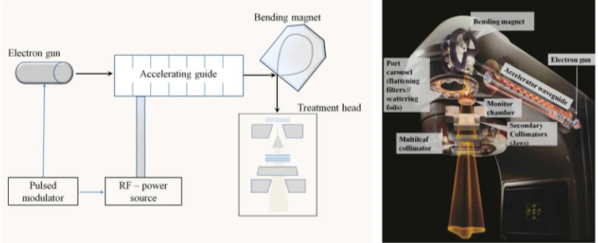
3.1 Schematic drawing of the main components incorporating a medical linear
accelera-tor and the collimaaccelera-tor systems . . . 42
3.2 Diagram showing the components of a typical medical linear accelerator head
work-ing in both photon and electron mode . . . 44
3.3 Scheme of an ionization chamber emerged in water . . . 48
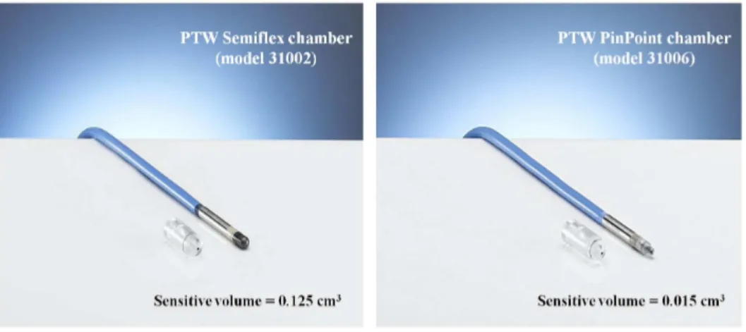
3.4 Pictures of the PTW Semiflex of 125 cm3volume and PTW PinPoint chambers . . . 50
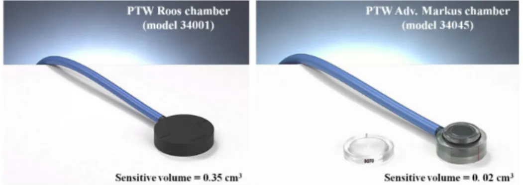
3.5 Pictures of the parallel-plate PTW Roos and Adv. Markus chambers . . . 51
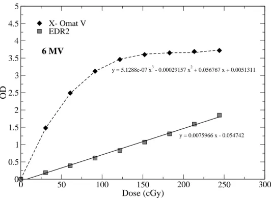
3.6 Typical response curve, i.e. net optical density versus dose curves of radiographic
films for direct x-ray exposure . . . 57
condensed history . . . 68
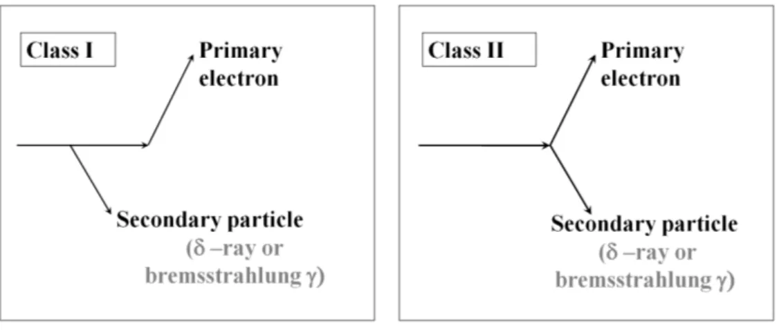
4.2 Representation of the methodologies (class I and class II) adopted by MC algorithms of an electron discrete event . . . 70
4.3 Electron pathlength correction in a MC simulation . . . 71
4.4 Schematic drawing of the boundary problem in the condensed-history method for the simulation of the electron transport . . . 72
4.5 The structure of the EGSnrc code system . . . 76
4.6 DOSXYZnrc default ramp for converting CT-number to material density . . . 80
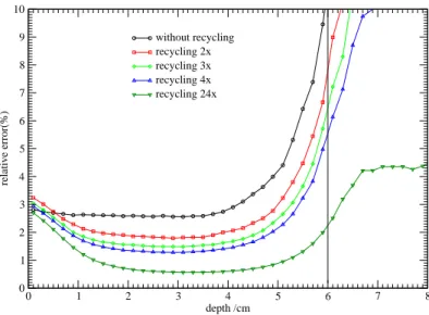
5.1 Recycling effect on DOSXYZnrc dose calculations . . . 92
5.2 Isodose distribution for a 12 MeV electron beam shaped by a cerrobend block . . . . 93
5.3 Comparison of the water-air stopping power ratio given in the IAEA protocol (TRS-398) and those calculated for realistic Siemens Primus electron beams of 12 and 18 MeV using the SPRRZnrc code . . . 95
5.4 On-axis depth dose in a water phantom relative to the dose maximum and lateral pro-files at depths below the water surface for the 12 and 18 MeV beams from Siemens Primus with 10 x 10 cm2field (100 cm SSD) . . . 96
5.5 Energy distributions and mean energy profiles of electrons and photons present in 12 and 18 MeV beams from Siemens Primus with 10 x 10 cm2field (100 cm SSD) . . . 98
5.6 Fluence profiles and angular distributions of electrons and photons present in the 12 and 18 MeV beams from Siemens Primus with 10 x 10 cm2field (100 cm SSD) . . . 99
5.7 Scheme of sagittal and transverse views of the home-built PMMA phantom including an air cavity with area S and thickness L used for measurements and modeled for MC dose calculations . . . 100
5.8 EBT film samples exposed by the 18 MeV electron beam in a heterogeneous phan-tom containing the cavity of 1 x 1 x 2.8 cm3 . . . 102
5.9 Energy dependence of the calibration curve for Gafchromic EBT film . . . 104
5.10 On-axis PDD curves measured and MC calculated in heterogeneous phantom in-cluding an air cavity of varied area and thickness for an electron beam of 12 MeV . . 105
an heterogeneous phantom for a 12 MeV electron beam . . . 109
5.13 MC calculated and measured X dose profiles at 3.3 cm depth in an heterogeneous
phantom for a 18 MeV electron beam . . . 110
5.14 Electron energy spectra calculated at a depth of 3.3 cm in a PMMA phantom with and
without an air cavity of varying dimensions, area S or thickness L, irradiated by a 12
MeV electron beam . . . 112
5.15 Angular spectra of electrons calculated at a depth of 3.3 cm in PMMA homogeneous
and heterogeneous phantoms with an air cavity of varying dimensions, area S or
thickness L, irradiated by a 12 MeV and 18 MeV electron beams . . . 114
5.16 MC calculated PDD curves in heterogeneous phantoms irradiated with an 12 MeV
electron beam collimated using a cerrobend cutout . . . 115
5.17 Calculated lateral profiles (3.3 cm) in heterogeneous phantoms irradiated with an 12
MeV electron beam collimated using a cerrobend cutout . . . 116
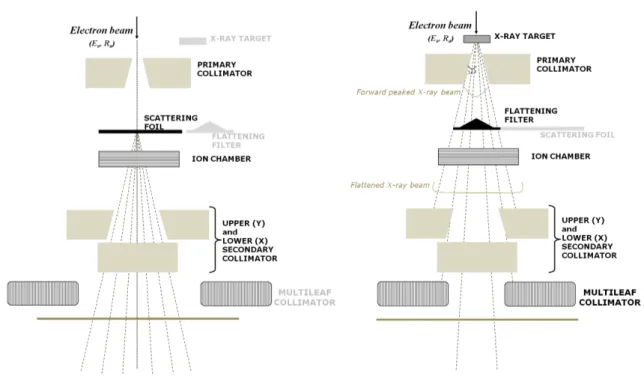
6.1 Schematic drawing of Varian 2100C/D linac components modeled in Monte Carlo
simulations . . . 126
6.2 Schematic draw of (a) the bremsstrahlung target of W/Cu and (b) flattening filter
ge-ometry for 6 MV photon beam . . . 128
6.3 Bremsstrahlung spectra generated by a 6.0 MeV electron beam with 1.0 mm radius . 129
6.4 Flowchart of the procedure to find the accurate description of the incident electron
beam at the target, namely electron energy Eiand radius Ri. . . 133
6.5 Influence of the energy of incident electron beam on PDD curves for a 30 x 30 cm2field138
6.6 Influence of the energy of incident electron beam on lateral profiles for a 30 x 30 cm2
field . . . 140
6.7 Influence of electron beam radius on Y dose profile for a 30 x 30 cm2field . . . 141
6.8 Lateral dose profile for 4 x 4 cm2 and 10 x 10 cm2 at several depths for the final
benchmarked 6 MV photon beam . . . 143
6.9 Planar fluence vs off-axis distance calculated with contributions from photons and
electrons reaching a plane at 100 cm across 10 x 10 cm2field for a 6 MV photon beam144
scored inside the 10 x 10 cm field for a 6 MV photon beam . . . 148
6.12 Comparison of photon energy spectra (on-axis) for 3 different field sizes at 100 cm
SSD and 6 MV photon beam . . . 149
6.13 Mean energies of the photons and electrons as function of the distance to the central
beam axis for a 6 MV photon beam and a 10 x 10 cm2field at SSD of 100 cm . . . . 150
6.14 Angular distribution of photons and electrons at SSD = 100 cm inside a 10 x 10 cm2
field for a 6 MV photon beam . . . 151
7.1 Varian Millennium 120-leaf MLC geometry . . . 154
7.2 Geometry of DYNVMLC CM modeling the Millennium MLC. . . 155
7.3 The effects of the interleaf air gap on the leakage profiles for a 6 MV photon beam . . 163
7.4 The effects of leaf density on the leakage profiles for a 6 MV photon beam . . . 165
7.5 Film measured and MC calculated leakage transmission dose profile through
oppos-ing leaves completely closed at the central axis of a 10 x 10 cm2field defined by jaws
for a 6 MV beam . . . 167
7.6 Depth-dose and off-axis profiles (X and Y axis) measured with the PinPoint ion
cham-ber and calculated with MC in water for MLC defined static fields . . . 169
7.7 Film measured and MC calculated profile in water irradiated for a ”alternate even and
odd” leaf shape with a 6 MV photon beam . . . 170
7.8 Measured and calculated dose profiles for a pyramid intensity pattern delivered using
a dynamic mode for a 6 MV photon beam . . . 172
8.1 Schematic representation of the simulated geometry of the Varian 2100C/D linac
head and water phantom, showing also the location of the two phase space scoring
planes considered for the simulations. . . 178
8.2 Simulated water phantom geometry for surface and build-up dose calculations . . . . 180
8.3 MC calculated depth dose curves in the build up region for the MLC defined field size
of 10 x 10 cm2, 4 x 4 cm2, 2 x 2 cm2in comparison to the MLC open field ones . . . 182
8.4 MC calculated depth dose curves in the build up region for the MLC defined field size
to the beam axis for different MLC defined field sizes . . . 185
8.6 Contribution of particles and electrons scattered from jaws and MLC to relative planar
fluence profile (X axis) for 10 x 10 and 2 x 2 cm2MLC defined fields . . . 186
8.7 On-axis energy spectra of electrons reaching the scoring plane for 2 x 2 cm2and 10
x 10 cm2MLC defined fields and the respective MLC open fields . . . 187
8.8 Comparison of measured PDIs in the dose build-up region of a water phantom for
different MLC defined field and the respective MLC open field . . . 190
8.9 Measured PDIs compared to MC calculated PDDs in the dose build-up region in
water with a cylindrical and a Roos parallel-plate chambers for different MLC defined
fields . . . 191
8.10 Water to air stopping power ratios against depth calculated for different MLC defined
fields and 6 MV photon beam . . . 194
9.1 Transversal CT slices for patient 3 illustrating the delineations of the PTV volumes for
each treatment phase, the femoral heads and the bladder) . . . 201
9.2 Schematic diagram of set-up for the MC simulation of IMRT treatment plans
per-formed using BEAMnrc code . . . 203
9.3 The CT ramp for the conversion of CT values to material type and densities according
to the conventional CTCREATE ramp . . . 206
9.4 Comparison of dose profiles calculated along the X axis for MC phantoms (y = -16.13
cm) . . . 209
9.5 Comparison of dose profiles calculated along the X axis for MC phantoms (y = -20 cm)210
9.6 Comparison of dose profiles calculated along the Y axis for MC phantoms (x= -6.03
cm and x= -0.78 cm) . . . 211
9.7 DVHs of the PTV, rectum and left femoral head calculated by MC in patient phantoms
built using different CT conversion ramps . . . 213
9.8 Comparison of isodose distribution for the Phase I of the IMRT treatment in patient 1
calculated by Eclipse TPS and by Monte Carlo . . . 215
9.9 Comparison of DVH curves calculated by Eclipse TPS and by Monte Carlo for the
1.1 Estimates of uncertainty in absolute dose for a complete radiotherapy procedure . . 10
4.1 CT numbers and density range for the four materials used in the ramp for converting
CT numbers to material parameters . . . 80
5.1 Variations of the thickness L and square area S of the air cavities considered in the
study . . . 89
6.1 Energy and radius combinations of incident electron beam impinging on target . . . . 133
6.2 Summary of the number of particles scored in the phase space file and the CPU time
used for the BEAMnrc simulations for various field sizes using an electron beam of
6.2 MeV and 1.5 mm radius impinging on the bremsstrahlung target . . . 134
6.3 Transport parameters and variance reduction techniques selected in the commission
process of 6 MV photon beam . . . 136
6.4 Photon fluence along the X-axis of photons and electrons contributions in a plane at
100 cm (SSD) for a 10 x 10 cm2field for a 6 MV photon beam . . . 146
6.5 Photon fluence along the X-axis of photons and electrons contributions in a plane at
100 cm (SSD) for a 4 x 4 cm2field for a 6 MV photon beam . . . 146
6.6 Photon fluence along the X-axis of photons and electrons contributions in a plane at
100 cm (SSD) for a 30 x 30 cm2field for a 6 MV photon beam . . . 147
7.1 Film measured and MC calculated MLC transmission with different MLC leaf densities
for a 6 MV photon beam . . . 164
7.2 MLC transmission through the closed leaves measured and MC calculated with
defined field sizes . . . 184
8.2 Geometric characteristics of the ionization chambers used for the surface and
build-up dose measurements . . . 189
8.3 MC calculated and measured doses at depths of 0.25 and 0.52 cm in a water
phan-tom for a 6 MV beam and three different MLC defined fields . . . 192
9.1 Typical number of beams, beam angles and jaws openings of IMRT treatment plans . 202
9.2 CT conversion ramps used to build Monte Carlo phantoms from the CT data set of
Symbols
A Mass number
AE Electron Threshold Energy for Explicit Electron Interaction Modeling
AP Photon Threshold Energy for Explicit Electron Interaction Modeling
CAX Central Axis
C M Component Module
CPE Charged Particle Equilibrium
CS DA Continuous Slowing Down Approximation
CT Computed Tomography
de f f Shift of effective point of measurement
DICOM Digital Imaging and Communications in Medicine
Dmax Maxima Absorbed Dose
dmax Depth of Maximum Dose
Dmed Absorbed dose in a medium
DV H Dose Volume Histogram
ECUT Electron Cutoff Energy
EGS Electron Gamma Shower
Gy Gray
I Intensity of radiation
ICRP International Commission on Radiological Protection
(L/ρ)air Restricted mass collision stopping power ratio of the medium-to-air
m Mass
MeV Mega electron-volt
MC Monte Carlo
MERT Modulated Electron Radiation Therapy
MLC Multileaf Collimator
MU Monitor Units
MV Megavoltage
PBC Pencil Beam Convolution
PCUT Photon Cutoff Energy
PDD Percentage depth dose
PM MA Polymethyl methracrylate, ”Lucite”
PRES T A Parameter reduced electron-step transport algorithm
PT V Planning Target Volume
ST Total unrestricted stopping power in medium
Sc Collision unrestricted stopping power
Sr Radioactive unrestricted stopping power
(S/ρ)medair Mass stopping power ratio of the medium-to-air
S AD Source-Axis Distance
S S D Source-Surface Distance
T PS Treatment Planning System
Z Atomic number
µ Energy absorption coefficient in medium
Motivation
External radiotherapy is currently the most common technique used for the treatment of many
types of cancer. The main objective of radiotherapy lies in the delivering of a very accurate dose to a
well-defined target volume with minimal absorbed dose to the surrounding normal tissue, especially
highly radiosensitive organs. In order to achieve this goal, one of the primary requirements is to
ensure that the treatments are delivered in accordance with the dosimetric intentions. It has been
clearly demonstrated that, an overdosage of radiation can lead to severe side effects, while an
underdosage can reduce significatively the probability for a patient cure.
The calculation of radiation dose distributions plays an important role in the treatment of
pa-tients requiring external radiotherapy. The accuracy of dose calculations is crucial to the quality of
treatment planning and consequently to the effectiveness of the radiotherapy treatment.
Most of the conventional dose calculation algorithms implemented in the majority of the
treat-ment planning systems at radiotherapy departtreat-ments are known to be inaccurate when radiation
disequilibrium conditions exist, such as near tissue inhomogeneities, for small radiation fields or for
dose gradient regions (e.g. superficial regions). Examples of these algorithms are pencil-beam and
superposition-convolution algorithms.
The validation of these dose calculation algorithms is commonly performed by comparisons
with measured data. The reliability of measured data sets is however very dependent on several
aspects, such as the stability of the accelerator (e.g., energy, output, flatness, and symmetry) or on
the choice of detector and experimental set-up. These limitations may thereby restrict the number
of comparison points and introduce dosimetric problems to the verification.
as 3D - conformal and Intensity Modulated Radiotherapy (IMRT) techniques. These techniques
involve higher complexity in the planning and validation of the treatments due to the use of
high-dose gradients regions, small fields and the incorporation of new dynamic collimation devices such
as multileaf collimator (MLC), which can introduce important effects on the final dose output. Under
these circumstances, the requirements of high accuracy for the dose calculations plays a more
important role.
For the last years, dose calculation algorithms based on the Monte Carlo (MC) method have
been shown to be a powerful tool to overcome the existing limitations, not only of conventional
algorithms, but also of experimental procedures. These algorithms are currently recognized as the
most accurate method to calculate dose, since particle histories are simulated explicitly based on
physical interaction probabilities inside an arbitrary media for a wide range of complex radiation
treatment conditions.
Due to the ability of the MC method to accurately compute dose for complex delivery scenarios
along with the improvements in computer technology, this method has now the potential to replace
conventional dose calculation algorithms in radiotherapy treatment planning systems and also to
be used as alternative method to measurements for quality assurance of treatments, specially for
those treatments delivered with advanced techniques. Currently, MC treatment planning systems
are quickly becoming a real possibility in clinical settings.
The present thesis uses the MC method for the verification of calculated dose in two challenging
situations: tissue inhomogeneity and surface regions. It aims, on one side, to circumvent problems
associated with conventional procedures of dosimetric verification of radiotherapy treatments and,
on the other side, enables the study of dosimetric and physical characteristics which are difficult or
impossible to assess from measured data or conventional dose calculation algorithms.
In the context of the thesis, a detailed characterization of electron and photon beams of different
energies and field configurations has also been performed using Monte Carlo simulations. The
ade-quate beam characterization represents an essential component of the accuracy of dose calculation
and it has become even more important with the implementation of more conformal and advanced
radiotherapy techniques, such as Intensity of Modulated Radiotherapy (IMRT). The introduction of
the dynamic conformation devices, such as multileaf collimators, in the accelerator heads to provide
work aims also to introduce a detailed assessment of the characteristics of the Varian Millenium
120-leaf MLC used for the MC modeling of IMRT plans.
It is also an objective of this thesis to contribute to the implementation of Monte Carlo based
plan-ning systems, participating thus to the widespread effort performed by the international radiotherapy
community to overcome long-standing problems found in the implementation of this algorithms at
clinic settings.
Thesis Outline
This dissertation is organized in five parts. The first part provides a general introduction to
radiotherapy with emphasis in external beam therapy (Chapter 1) and, on the other hand, to the
physics of the radiation transport, including the basic particle interaction processes in matter and
the definition of basic quantities describing the effects of these interactions (Chapter 2).
In the second part of this dissertation, the materials and methods used for the present
investiga-tion are introduced. First, it is given a brief overview of the producinvestiga-tion of electron and x-ray beams by
a medical linear accelerator (linac) for use in external beam radiotherapy (Chapter 3). Second, the
basic concepts of ionometric and film dosimetry are described. This second part concludes with a
general description of Monte Carlo simulation of radiation transport, in particular the EGSnrc Monte
Carlo code, which is the code used for the dose calculations in this work (Chapter 4). A discussion
of variance reduction techniques and efficiency enhancing methods integral to MC calculations is
also given.
The third and the fourth parts contain the main body of the dissertation, that is, the description
of the dosimetric studies developed during the course of this research.
The third part is dedicated to electron radiotherapy and it presents a systematic study assessing
air cavities perturbation on electron dose distributions by using both Monte Carlo simulations and
experimental measurements (Chapter 5).
The fourth part is focused on photon radiotherapy. Chapter 6presents the MC modeling of the
Varian 2100C/D linear accelerator as well as the experimental validation of this model through
com-parison against experimental measurements. A detailed description of the Monte Carlo model used
for the multileaf collimator (MLC) embedded in the previous accelerator is discussed in chapter 7.
in chapter 8 using MC simulations and measurements carried out with several types of ionization
chambers. Finally,chapter 9presents a clinical study assessing the dosimetric differences between
dose-to-medium and dose-to water for prostate IMRT plans calculated by Monte Carlo methods.
The fifth and last part of the dissertation summarizes the main conclusions arisen from the
Introduction to external beam radiation
therapy
1.1
Brief introduction
The discovery of x-rays by Wihelm R ¨ontgen in 1895 marked the beginning of a new physics branch:
medical physics, which is concerned with the application of physics to medicine. This field covers
a broad range of technologies and applications, ranging from diagnostic methods (x-ray imaging,
x-ray computed tomography, nuclear medicine, ultrasound imaging, etc.) over techniques for the
treatment of human disease (radiation therapy, image guided therapy, laser treatment techniques)
to supportive fields like medical image processing, quality assurance and radiation dosimetry. From
all these areas of speciality, the field of interest of this thesis is radiotherapy, although radiation
dosimetry will also be briefly referred along the course of the investigations here presented.
Radiotherapy (RT) is an important form of cancer treatment used for more than 100 years. The
goal of radiotherapy is the eradication of tumor cells with the use of ionizing radiation, while
mini-mizing the damage to the surrounding healthy tissue. In fact, the ionizing radiation deposits energy
in the tumor cells as result of the ionization caused by the radiation interaction with the medium.
This deposited energy damages the genetic material (DNA) or other important biological molecules,
leading to the destruction of these cells or inhibiting further cell division.
in the early 1900s mainly due to the important contribution of the Nobel Prize winner scientist Marie
Curie, who discovered the radioactive elements, polonium and radium. This discovery marked the
beginning of a new era. The radium started to be used in various forms to treat cancer disease
until the mid 1900s, when cobalt and cesium units were introduced as the new landmarks for cancer
research and treatment. Since then, many alternative medical procedures were developed, namely
medical linear accelerators (linacs) which are currently the most frequently devices used for
radio-therapy.
With the discovery of the computed tomography (CT) in the 1970s [Houn79], the delivery of
three-dimensional (3D) radiotherapy became a possibility and, at the same time, CT improved the
ability of physicians to directly define the dose delivered to a treatment volume previously defined
on the patients anatomy.
During the last two decades, most of the developments in radiation medicine were related to the
integration of computers in imaging, the development of digital diagnostic imaging techniques and
the incorporation of computers into therapeutic dose delivery with high-energy linear accelerators,
among others. The recent improvements of imaging technologies along with the development of new
radiation delivery equipments has resulted in the emergence of more advanced techniques. These
advanced techniques represent an important change in radiotherapy, since they allow the design
and implementation of relatively complex radiotherapy treatments where the delivery of radiation to
the tumor volume can be achieved with high levels of dose and a high degree of accuracy without
affecting surrounding tissues.
Generally, radiotherapy can be delivered through three different forms depending on the location
of the radiation source: externally (teletherapy or external radiotherapy), internally by a radioactive
source inside the body (brachytherapy) or by administration of radiopharmaceuticals (metabolic
ra-diotherapy). The subject of this thesis is related to external beam radiotherapy and further discussion
will be restricted to this method.
External radiotherapy is the most commonly used form of radiotherapy, where the radiation
di-rected to the tumor comes from outside the body. Electrons and photons (x- and γ-rays) are the
two type of ionizing radiation widely used in external radiotherapy in the present days. The photons
are usually provided by a radioactive source as cobalt-60 emitting two gamma rays of 1.17 and 1.33
the range 4 - 25 MeV are also generated from linacs.
More recently, heavy charged particles (hadron radiotherapy) as protons and carbon ions are
be-ing used to treat cancer patients at some radiotherapy departments [Wil46, Jak03]. Although hadron
therapy has shown many advantages over electron and photon beams (e.g. more precision of dose
location and high effectiveness of treatment), its use is still limited at few radiotherapy departments
due to the complexity and the high cost of the devices required for the beam production.
Neverthe-less, the effort of the research community to investigate the viability of these type of particles for
external radiotherapy has increased noticeably in the last years.
1.2
Techniques in external radiotherapy
Different techniques have been developed for the delivery of external radiotherapy treatments
us-ing electron and x-ray beams produced by linear accelerator machines. This section gives a brief
overview of the development of these techniques, with a special attention on IMRT for photon beams
which is the technique used in the context of this thesis.
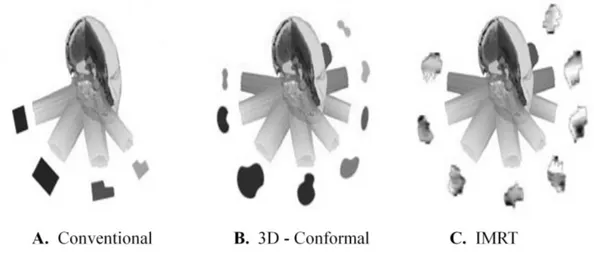
2D conventional radiotherapy
In the early days of radiotherapy, conventional radiotherapy consisted of 2D planning methods that
involved a radiographic film or a image localization procedure. Rectangular fields with shielding
and beam-modifying devices (blocks and wedges) were used to obtain conformity and minimize the
dose in the normal tissue (figure 1.1A). The treatment plans using this technique mainly consists
of a single beam with an uniform intensity delivered from several directions to the treatment volume
(target volume). This technique was well established at most radiotherapy centers, because it is
generally quick and reliable. However, its use has shown to be limited to the cases where the tumor
is symmetrically shaped and centrally located in the body with minimal number of surrounding critical
organs. Since normally tumors have no rectangular shape, this technique was not very satisfying
because, depending on the actual shape of the tumor, a lot of healthy tissue could be irradiated,
leading to a high radiation toxicity of healthy tissues close to the target volume.
Three-dimensional (3D) conformal radiotherapy
By the mid-1990s, the advances in technology and software allowing the calculation and delivery
of non-uniform fluence maps on three-dimensional (3D) patient volumes enabled the clinical
imple-mentation of new delivery techniques, 3D-conformal radiotherapy. This impleimple-mentation was also
motivated by the development achieved in medical imaging with the emergence of the computed
tomography.
The goal of this technique is to conform the profile of each radiation beam to the shape of
the target volume by using a variable number of static beams. In this case, the radiation beams
normally have a uniform intensity across the field and they are shaped with irregular geometries
using the projection of the target volume (figure 1.1B). The final contribution of all beams lead to
a high conformity of dose to the the target volume. In order to shape the beam, different types
of device can be used as MLC for photon beams or Cerrobend blocks inserted in the applicator
for electron beams, among others. The great advantage of 3D - conformal radiotherapy in relation
to conventional radiotherapy is the reduction of the relative toxicity of radiation to the surrounding
normal tissues due to the improvement of dose conformity to the target volume. As consequence,
higher doses of radiation can be used to treat the tumor and thereby to increase the effectiveness
of the treatment.
Intensity Modulated Radiotherapy (IMRT)
IMRT is a more advanced development of 3D conformal radiotherapy where, in addition to the shape
of the radiation beam, the intensity of the beam can be also modulated [Webb03], as it can be
schematically seen in figure 1.1C. Unlike 3-D conformal radiation therapy that is delivered using a
single radiation beam, IMRT is delivered as a sequence of many small beams that enter the body
from many angles. These multiple thin beams allow that the intensity of radiation within each field
can be modulated to achieve a better conformity to the tumor. This leads again to higher doses in
the tumor and lower doses to the surrounding sensitive structures and organs at risk.
The technique of IMRT was initially developed using photon beams; however, recent research
has also investigated the plausibility of this techniques for electron radiation therapy, known as
Mod-ulated Electron Radiotherapy or MERT [Ma00a].
Of the various alternatives proposed for the delivery of IMRT, the most frequently used is that
modality based on the use of multileaf collimators (MLC). This type of collimator is made up of
individual leaves of a high atomic number material, usually tungsten, that can move in and out of the
field to produce a sequence of complex field shapes or beam apertures.
3D conformal radiation therapy also uses a multileaf collimator to customize the shape of the
beam. However, the leaves of the collimator are not allowed to move in and out within that particular
field, therefore, the shape of the beam for each field stays the same during the treatment.
Two types of MLC-based IMRT delivery modes are clinically used, namely ”step-and-shoot or
static (SMLC)” and ”sliding window or dynamic (DMLC)”. The differences between both basically
relates to the different ways used to deliver the radiation, segmented and dynamic, respectively.
In the step-and-shoot mode, the intensity of the modulated fields are delivered with a sequence
of small segments or subfields, each one with an uniform intensity. The multiple segment fields are
set up at selected orientations of the gantry and the beam is only turned on when the leaves of the
MLC are stationary in each of the specific segment positions, i.e. the MLC does not move while the
beam is on.
In the dynamic mode, the fields are delivered in a dynamic way with the leaves of the MLC
moving during the irradiation of the patient. For a fixed gantry position, the position of the MLC
leaves is swept across the target volume with the beam turned on to produce the desired fluence
map.
inverse treatment planning is used to find the optimal position and movement for the leaves of the
MLC during irradiation. In inverse treatment planning a certain target volume must be defined in the
treatment planning system and a desired dose for this target must be prescribed. The treatment
planning system will then try to optimize the positions and movements of the MLC leaves so that the
prescribed dose is homogeneously distributed across the target volume only. Usually not only the
target volume but also the organs at risk are defined and dose volume constraints are given by the
planner in order to further enhance the result of the dose optimization algorithm.
New different approaches to deliver IMRT have been developed during the last years: Intensity
Modulated Arc Therapy (IMAT), volumetric arc therapy (VMAT), Cyberknife, Image-guided radiation
therapy (IGRT), among others. IMAT is a rotational approach in which both the gantry and the leaves
of the MLC move during arc beam delivery. More recently introduced, the Cyberknife integrates a
compact photon beam linear accelerator mounted on a robotic arm with advanced image guidance
technology to deliver concentrated beams of radiation from multiple positions and angles.
1.3
The importance of accuracy in radiation delivery
It has previously been mentioned that the main goal of the radiotherapy is focused on the destruction
of tumor cells with ionizing radiation while limiting the damage to the surrounding healthy tissue. The
destruction of tumor cells occurs through the ionization of the medium by the radiation, which leads
to the deposition of energy. The extent of the damage caused by the radiation is therefore a direct
result of the amount of energy deposited per unit mass, i.e. the absorbed dose. Hence, a central
point for the success of any radiotherapy treatment stays on the exact knowledge of the radiation
dose delivered to the patient.
The importance of accuracy in radiation delivery is most apparent by observing biological
ef-fects. In particular, the dose dependence of two biological parameters are usually evaluated for
this purpose, namely the local tumor control probability (TCP) and the normal tissue complication
probability (NTCP) as a function of the absorbed radiation dose in tissue. The irradiated volume in a
patient contains usually both targeted and normal tissues; thereby the optimal radiotherapy plan will
maximize TCP while minimizing NTCP. Figure 1.2 illustrates the sigmoidal dependence of both TCP
and NTCP values on the radiation dose.
while the NTCP rises sharply at a slightly higher absorbed dose. Thus, any uncertainty on delivered
dose may either result in an underdosage of the tumor or a complication for normal tissue. In fact,
as stated in the AAPM Report No 85 [AAPM85], a dose error of 5 % may lead to a change in TCP
of 10 or 20 % and to an even larger change of 20 to 30 % in NTCP. The need of accurate dose
calculation is thus imperative.
Figure 1.2: Dose dependence of tumor control probability (TCP) and the probability of normal tissue compli-cation (NTCP). The vertical line indicates a certain dose in the steep part of both curves. Uncertainties in delivered dose might worsen the clinical outcome due to either reduction of TCP or increase of NTCP.
In a radiotherapy treatment, a large number of steps are involved between the dose
prescrip-tion and the final delivery of the dose, e.g. machine calibraprescrip-tion, dose calculaprescrip-tion, acquisiprescrip-tion of
patient-specific tumor information, patient positioning, patient motion, etc. During each of this steps,
small uncertainties are involved, accumulating to a large overall uncertainty for the full process of
dose delivery. These uncertainties may be categorized as random and non random (systematic)
uncertainties and they are combined in quadrature to obtain the overall uncertainty of the complete
radiotherapy process. The uncertainties that occur during the treatment planning will impact on the
entire treatment and are therefore systematic. On the other hand, those uncertainties that occur
during the treatment delivery will be however random errors as they will affect the treatment by
dif-ferent amounts at each fraction of the treatment delivery. Their estimates are summarized in table
1.1.
In 1976, the ICRU Report 24 [ICRU24] concluded that an uncertainty less than 5 % at the 2σ
each step involved in the radiotherapy treatment must be performed with an accuracy much better
than 5 %.
Table 1.1: Estimates of uncertainty (in terms of one standard deviation) in absolute dose in the patient for the complete treatment procedure using megavoltage photons [AAPM85].
Source of Uncertainties Uncertainty(%) Dose at the calibrated point in water 2.5 Additional uncertainty for other points 0.6 Beam Monitor stability 1.0
Beam flatness 1.5
Patient data 1.5
Patient setup and organ motion 2.5 Overall (excluding dose calculation) 4.3 Dose calculation algorithm 1.0/2.0/3.0/5.0
TOTAL 4.4/4.7/5.2/6.6
After evaluating the contribution of each step, it was observed that the use of an extremely
accurate dose calculation algorithm will not automatically lead to very low uncertainties in clinical
dose delivery, since several other factors contribute significantly to the overall uncertainty. However,
it was claimed that the dose calculation step should be accurate to within 2-3 % to achieve the
5 % requirement of the overall uncertainty. The accuracy of the dose calculation algorithm plays
therefore an important role in a radiotherapy treatment.
More details about the accuracy of the existing algorithms used for the dose calculation will
be briefly referred in the chapter 4, where it is also stressed the Monte Carlo method as the most
Fundamentals of radiotherapy physics
and dosimetry
This chapter provides a brief introduction to the basic physics of the radiation interaction with
matter and introduces several basic quantities used in characterizing radiation.
2.1
Interaction of ionizing radiation with matter
Ionizing radiation is characterized by the ability to ionize matter through electromagnetic interactions[Att86].
Typically, ionizing radiation can be classified into two main categories depending on the mode of
ion-ization:
• directly ionizing radiation: Charged particles (electrons/positrons, alpha particles, etc.) which
deposit energy in the medium through direct one-step processes involving inelastic
Coulomb-interactions with orbital electrons and other charged particles present in the medium.
• indirectly ionizing radiation: Neutral particles (photons and neutrons) which interact with the
medium following a process of two steps: 1) energy transfer to charged particles in medium
and 2) energy deposition in medium by released charged particles.
Both directly and indirectly ionizing radiations are actually used in the diagnosis (medical
which is the topic of the present thesis, photons and electrons are considered the main choice for the
treatment of disease with radiation and they are used in more than 90 % of radiotherapy treatments.
Next sections introduce briefly the different mechanisms of interaction of photons and electrons
with matter and discuss the quantitative probabilities of each of these mechanisms occurring in
different regions of energy. This is helpful, not only to understand better how the radiation interacts
with the living tissue, but also for later discussion of the results.
2.1.1
Photon interactions in matter
2.1.1.1 Types of interaction mechanisms
As previously referred, photons ionize matter indirectly, i.e. the photon interactions in a medium
re-lease charged particles (electrons or positrons), which in turn deposit energy through direct Coulomb
interactions with the orbital electrons of the atoms.
The five major interaction processes which the photons can undergo when interacting with matter
are:
• Photoelectric absorption effect
• Incoherent (Compton) scattering
• Pair production
• Coherent (Rayleigh) scattering
• Photonuclear reactions
In general, the probability of occurrence of the above interactions depends both on photon
en-ergy and atomic number of the medium. For the enen-ergy range applied in radiotherapy, the four first
interactions show the highest probability to occur. However, only the first three interactions lead to
energy deposition, as they result in the transfer of energy to electrons which will then be imparted
to matter in small Coulumb-force interactions along their tracks. The Rayleigh scattering, which is
sometimes referred to as coherent scattering, is an elastic interaction where the photon looses
not contribute to the transfer of energy to the medium. Figure 2.1 illustrates a scheme of these four
first processes.
The photonuclear reactions are commonly ignored in dosimetry considerations, since this kind
of process occurs with a very low probability and is just dominant for higher energies above 10 MeV.
A review of each of the above processes is presented next, with a special attention to the three
first interactions since they play the major roles at the energies commonly used in the radiotherapy.
In addition, it is briefly discussed the relative dependence of each process on energy and material
as well as the respective contribution to the total probability.
Figure 2.1: Schematic diagrams of the main interaction processes of the photons with matter: Rayleigh scattering, Photoelectric effect, Compton scattering and Pair production.
Photoelectric effect
The photoelectric effect is the predominant mode of interaction for photons of low energy, in the
energy range of several eV to around 0.1 MeV. In this process, the incident photon interacts with a
tightly bound electron (inner shells as K, L, M or N) and it is completely absorbed in the interaction
In order for the photoelectric effect to occur, the incident photon energy has to be higher than
the binding energy of the electron. Thus, part of the photon energy is used to overcome the binding
energy and free the electron from the atom and the residual energy is transferred to the kinetic
energy of the escaping electron. The photoelectron appears thus with an kinetic energy given by:
Ee=E−Ui (2.1)
where E is the energy of the incoming photon andUi is the binding energy of the electrons in the
atomic shell.
As result of the emission of the electron, the atom is left in an excited state with a vacancy in the
ionized shell. This vacancy can be quickly filled through the capture of an outer orbital electron and,
therefore, one or more characteristic x-ray photons (fluorescent photons) may also be generated.
In some fraction of the cases, the emission of Auger electrons may substitute for the characteristic
X-ray in carrying away the atomic excitation energy.
The probability of occurrence of the photoelectric effect varies roughly whit the energy of the
incident photon and the atomic number Z of the medium, as follows:
τ∝ Z
n
(hν)3 (2.2)
where the exponent n varies between 3 and 4 over the photon energy region of interest. The units
of the cross sectionτare cm−2.
According to this equation (2.2), it can be observed how the photoelectric effect will be enhanced
for photons of relatively low energy and for materials of high atomic number Z.
The angular distribution of the emitted electrons depends on the energy of the incident photon.
For low photon energy, the electrons are predominantly ejected at 90◦
relative to the photon direction.
With increasing the photon energy, the electrons are emitted in more forward directions [Att86].
Compton effect
The Compton effect is the dominant mode of interaction in the energy range from several hundred
keV to several MeV and therefore it represents the major mechanims of interaction for most photon
energies used in radiotherapy.
electron and it is deflected through an angle θwith respect to its original direction (see figure 2.1).
In contrast to the photoelectric effect, the electron struck by the incoming photon is a lightly bound
electron, i.e. an outer shell electron, and it is thus assumed that it is initially free and at rest.
In the collision, the photon transfers a portion of its energy to the electron, which will departs at
angleθewith a kinetic energy given by:
Ee= E−E′ (2.3)
whereE=hνandE′ =hνare the energy of the incident and scattered photons, respectively.
Simple energy and momentum conservation constraints can be used to derived the relation
between the energy of incident and scattered photons, given by the next equation:
E′= E
1+( E moc2
)(
1−cosθ)
(2.4)
in which themois the electron’s rest mass andcis the speed of light in vacuum.
This relation shows that the energy of the scattered photon depends not only on the energy of
incident photon but also on its scattering angleθ. From equation 2.4, it is clear observed that as the
energy of the scattered photon increases, the photon is deflected to more and more forward
direc-tions. For a given incident photon energy, there exits a minimum energy for the scattered photons
(corresponding to a maximum energy for the scattered electron), corresponding to the backward
direction atθ=180◦
Emin′ = moc
2/2
1+moc2/2E
(2.5)
Finally, the angleθof the emitted electron is related to the energy and angle of incident photon
through the next equation:
cotθe=
(
1+ E
moc2
)
tan
(θ
2 )
(2.6)
From equation 2.6, it is interesting to notice that the electron angle is thus always confined to the
forward direction (0≤θe≤90◦), whereas the photon can be scattered to any direction. On the other
side, it is also observed that, as the energy of the incident photon increases, the electrons tend to be
Compton scattering is the only type of interaction that is not highly dependent on the Z of the
medium, but it depends on the incident energy and the density of the material. In particular, the
probability of occurrence of this effect decreases with increasing photon energy and it shows to
be proportional to the material density. The total cross-section is derived according to the Klein
-Nishina formalism [Eva55], assuming for that unpolarized and unbound electrons, as follows:
σ∝Zσe ∝ Z
E (2.7)
whereσeis the the total Klein - Nishina cross section per electron [Att86]. The units of the cross
sectionσare cm−2.
Pair production process
Pair production refers to the creation of an electron and a positron pair from a photon in the field of
an atomic nucleus (see figure 2.1). In order to this interaction to occur, the photon energy should be
greater than the rest energy of the electron-positron pair, that is, E≥2mc2= 1.022 MeV.
When this process takes place, the massive nucleus recoils whit negligible energy and, therefore,
the photon energy is converted into mass rest energy (2mc2) plus kinetic energy of the electron (E−)
and positron (E+):
E=2mc2+E−+E+ (2.8)
The kinetic energy received by the electron and positron is not necessarily equal, but it can be
estimated an average kinetic energy (T) of:
T = E−1.022MeV
2 (2.9)
For photon energies close to the threshold energy 2mc2, the created electron and positron travel
almost in opposite directions to each other. For energy above this threshold, the pair can travel in
a more forward direction. In this last case, the average angle of the particle emission relative to the
original photon direction is roughly:
θ mc
2
The probability of occurrence of the pair production process is governed by the theory ofBethe
and Heitler. According to this formalism, the probability increases rapidly as the photon energy
increases and it is also strongly dependent on the atomic number asZ2:
κ∝Z2logE (2.11)
The units of the cross sectionκare cm−2.
Pair production can also occur in the field of an atomic electron, but the probability is considerably
smaller and the energy of the photon has to be higher than 4mc2. This process is usually know as
triplet production, since three particles are resulting from the interaction: electron/positron plus the
orbital electron.
Rayleigh scattering
It has been previously mentioned that the Rayleigh scattering is a elastic scattering where the photon
loose none of its energy and it is just redirected through a small angle θ (see figure 2.1). The
probability of occurrence of this process decreases with the incident photon energy, but it increases
with the atomic number of the medium as follows:
σR∝
Z2
(hν)2 (2.12)
The units of the cross sectionσR are cm−2.
The relative importance of the Rayleigh scattering is in the low energy regime, but it contributes
only a few percent or less to the total attenuation cross section. On the other side, it is important to
point out that this mechanism of photon interaction does not contribute to the kerma or dose, since
no energy is transferred during this interaction. Based on that, this kind of process is more important
in imaging applications than in radiotherapy.
Photonuclear reactions
At energies above 10 MeV photonuclear reactions can occur, in which the high-energy photon is
ab-sorbed by the atomic nucleus and a nucleon is then emitted. The most likely result of this interaction
is the emission of a single neutron through a (γ,n) reaction, even though the emissions of charged
occur with less probability. Generally, the contribution to the total attenuation cross-section of the
photonuclear reactions is very small (about 5 %) and therefore they do not play a role in general
photon attenuation studies. On contrary, they are of considerable importance for shielding
calcula-tions as consequence of the neutron emission. In this particular work, this process does not play an
important role since the maximum energy of photons used for the investigation is around 6 MeV.
2.1.1.2 Attenuation coefficients
When the photons travels thought the matter, they can undergo one or a combination of the above
processes depending on their energy and also they can be transmitted out of the medium without
undergoing any interaction.
This transport through matter is ruled statistically by the probability per unit distance traveled
by the photon, calledlinear attenuation coefficient and denoted byµ. This coefficient is frequently
known as macroscopic cross section (cm−1) and it can be expressed as the product of the atomic
density N =ρNA/Aand the total cross sectionσtotal(cm−2) as:
µ=Nσtotal ⇔µ=
ρNA
A σtotal (2.13)
Based on this coefficient, the number of photons passing a certain thickness x of a medium
decreases following an exponential function as:
N= Noe−µx (2.14)
withNobeing the incident number of photons.
In general, the total linear attenuation coefficient is represented as the sum of attenuation
coef-ficients for all individual interactions that a photon of given energy may have with atoms of a specific
material. As discussed above, the interactions of interest in the therapeutic energy range are
ba-sically three: the photoelectric effect, the Compton scattering and the pair production; thereby the
linear attenuation coefficient relative to these three process will compose the total linear attenuation:
µ=µph+µcomp+µpair=
ρNA
A
(
τ+σ+κ
)
(2.15)
Comp-ton and pair production, respectively. The corresponding cross section for theses interactions are
denoted byτ,σandκ, as previously discussed.
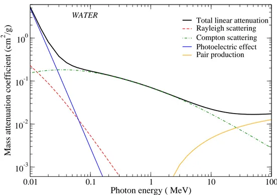
Figure 2.2 shows the total mass attenuation coefficient for water plotted against photon energy.
In addition to the total coefficient, the coefficients for the individual components (photoelectric effect,
Compton scattering, pair production and Rayleigh scattering) are also shown.
0.01 0.1 1 10 100
Photon energy ( MeV)
10-310-2 10-1 100
Mass attenuation coefficient (cm
2
/g)
Total linear attenuation Rayleigh scattering Compton scattering Photoelectric effect Pair production
WATER
Figure 2.2: Linear attenuation coefficient of photons of different energies in water (equivalent to body tissue). The relative contribution of photoelectric, Compton scattering and pair production processes are illustrated. The data were extracted from the NIST/XCOM: Photon Cross Sections Database (www.nist.gov/pml/data/xcom/index.cfm).
As seen from equation 2.13, the linear attenuation coefficient of a given material is directly
proportional to the density of the material. In order to eliminate the density dependence, themass
attenuation coefficient,µ/ρ(where theρis the mass density of the medium ), is used instead.
The mass attenuation coefficient can be divided into two parts, namely the energy transfer
coef-ficient (µtr/ρ) related to the transfer of energy to charged particles and the energy scatter coefficient
(µs/ρ) which applies to the energy converted into scattered photons. It can also happen that a part
of the energy transfer to the electrons is not deposited locally within the medium along the electron
track, being lost by emission of bremsstrahlung photons. This fact is described by the mass energy
µen
ρ =(1−g)
µtr
ρ (2.16)
where g refers to the fractional energy of the electrons that is lost as bremsstrahlung. This fraction
g is negligible for photons of low energy, but it becomes significant at high energies and in materials
of high atomic number Z.
2.1.1.3 Relative predominance of individual effects
The linear attenuation coefficient is characteristic of both the medium and the photon energy. Figure
2.3 shows an overall picture of the dependence of the relative magnitude of the different interaction
processes on energy E and atomic number Z. Curves of Z vs E corresponding to equal probabilities
of the photoelectric and the Compton processes (left) and of the Compton and the pair production
processes (right) are also presented.
As shown, the photoelectric effect is dominant at low energy range. As the energy increases,
the Compton effect becomes the most important process and, at higher energies (> 5 MeV), the
pair production is the interaction more likely to occur. Additionally, it can also be seen in this figure,
that the middle interval, with the Compton scattering predominance, is broader for media with low
atomic number.
In particular, for water and tissue with an effective atomic numberZe f f ective ≈ 7.0, this region
ranges from ∼ 20 KeV up to ∼ 20 MeV, indicating thus that for most of radiotherapy studies and
treatments, the most important interaction of photons with tissues is the Compton scattering. On the
other hand, the interval of influence of the photoelectric effect and the pair production is increased
for high-Z materials.
2.1.2
Electron and positron interactions in matter
Electrons play an important role in medical physics. They are used directly as beams for cancer
ther-apy, but also they are responsible for the energy deposition in matter by photon beams. Therefore,
these charged particles govern the experimental and theoretical aspects of radiation dosimetry.
Contrary to photons which can pass through the matter with no interactions at all, charged
par-ticles2cross the medium loosing almost continuously their energy through ionization and excitation
of atoms and through Coulomb interactions with the external nuclear field until they come to rest.
Additionally, it is important to point out that, compared to heavy charged particles, electrons
and positrons have a different behavior when passing through matter, although they undergo the
same kind of interactions. Because of their small mass, electrons (and positrons) can loose a large
fraction of their energy in a single collision with an atomic electron (which have equal mass as
the incident electron) and, they can also be scattered into relatively large angles. Furthermore, in
contrast to heavy charged particles, the electrons have a high probability of being sharply deflected
and accelerated by the nuclei resulting in the emission of bremsstrahlung photons. Next paragraphs
are focused on the interaction of electrons and positrons in matter and further details about this
subject are presented.
2.1.2.1 Types of interaction mechanisms
In general, when electrons pass through a medium they interact through Coulomb forces with nuclei
and orbital electrons. The collisions they can undergo may be elastic, when only a change of
direc-tion occurs, or inelastic when energy is also transferred. The type of interacdirec-tion will depend on the
energy of the incident electron and the distance of approach of the electron to the atom, that is, the
impact parameter b vs. the atomic radius a (figure 2.4).
2