ABSTRACT:It is well known that phytochromes mediate a wide range of photomorphogenic processes in plants. In addition, many studies have demonstrated the involvement of phytochromes as part of abiotic stress signaling responses. However, little is known about cadmium (Cd) stress regulation by phytochromes. Thus, in this study, we used the phyA (far red-insensitive; fri), phyB1 (temporary red-insensitive; tri) and phyB2 (phyB2)tomato (Solanum lycopersicum L.) mutants to investigate the roles of these three phytochromes on Cd stress responses. The plants were grown over a 21-d period in the presence of Cd. We evaluated plant growth, Cd and chlorophyll
BASIC AREA -
Article
The role of phytochromes in cadmium stress
responses in tomato
Lucas Aparecido Gaion1*, Paulo Guilherme Lorevice1, Carolina Cristina Monteiro1, Marina Alves Gavassi1,
Victor D’Amico-Damião2, Priscila Lupino Gratão1, Eduardo Custódio Gasparino1, Rogério Falleiros Carvalho1
1.Universidade Estadual Paulista “Júlio de Mesquita Filho” - Faculdade de Ciências Agrárias e Veterinárias - Departamento de Biologia Aplicada à Agropecuária - Jaboticabal (SP), Brazil.
2.Universidade Estadual Paulista “Júlio de Mesquita Filho” - Faculdade de Ciências Agrárias e Veterinárias - Departamento de Produção Vegetal - Jaboticabal (SP), Brazil.
*Corresponding author: [email protected]
Received: Sept. 30, 2016 – Accepted: Fev. 28, 2017
content and anatomical changes in the leaves. The results indicated that all genotypes were affected by Cd and showed reduced growth of the shoots and roots, as well as reduced chlorophyll content. The accumulation of Cd was similar for all genotypes, and a higher Cd content was found in roots. Anatomical analysis of the vascular bundles revealed that fri and tri seem to be more disrupted by Cd. Overall, these results indicate that phytochromes do not determine Cd stress tolerance in tomato plants.
INTRODUCTION
Plants possess photoreceptors that compose sophisticated light-sensing mechanisms that are localized inside and outside the chloroplasts and the nucleus (Hiltbrunner et al. 2005). Among photoreceptors, the phytochrome family stands out as sensors for red (R; ~ 660 nm) and far-red (FR; ~ 730 nm) light (Essen et al. 2008; Yang et al. 2008). The phytochrome holoprotein consists of an apoprotein with a covalently linked linear tetrapyrrole chromophore, and this complex regulates the expression of a large number of light-responsive genes and thus influences many photomorphogenic events (Neff et al. 2000; Quail 2002a,b; Franklin and Quail 2010). In plants, a small family of PHY genes encodes phytochromes. For example, five phytochrome genes are known in Arabidopsis thaliana
(PHYA to PHYE) and in tomato(PHYA, PHYB1, PHYB2,
PHYE and PHYF), and three PHY genes are known in rice (Oryza sativa)(PHYA to PHYC) (Bae and Choi 2008).
The involvement of phytochromes in controlling plant growth and development is well documented (Franklin 2016; Wit et al. 2016). Additionally, these photoreceptors have been shown to act as key modulators of both biotic and abiotic stresses (Carvalho et al. 2011a; Zao et al. 2014; D’Amico-Damião et al. 2015). These reports have led to a series of studies exploring the molecular and biochemical basis by which phytochromes mediate stresses, such as drought (D’Amico-Damião et al. 2015), salinity (Balestrasse et al. 2008a), high light (Boccalandro et al. 2001) or heavy metals (Cui et al. 2011). For instance, studies of A. thaliana under cold conditions demonstrated that light signaling by phytochrome B is involved in plant responses to cold via the expression of C-repeat binding factor (CBF) genes (Kim et al. 2002) and to biotic stress via the activation of lipoxygenase (Zao et al. 2014). In addition, rice phytochrome B-deficient mutant (phyB) plants were more tolerant to drought than the wild type. These plants exhibited reduced stomatal density and length, resulting in a decreased transpiration rate that enhanced drought tolerance (Liu et al. 2012). Similarly, the phyB-deficient mutant of tomato exhibited increased drought tolerance after five days without irrigation (D’Amico-Damião et al. 2015).
Another relevant type of abiotic stress experienced by plants under certain conditions is heavy metals. The increase in the cellular heavy metals are one of the main classes of
abiotic stress agents for living organisms because of their high level of bioaccumulation and toxicity (Verkleij et al. 2009; Nagajyoti et al. 2010). The increase in heavy metal concentrations in the cellular environment can disturb several signaling pathways and cause irreversible damage to biological systems (Verkleij et al. 2009; Rossato et al. 2011). Among heavy metals, Cd represents one of the most damaging metals to plants. At high concentrations, this heavy metal can generate reactive oxygen species (ROS) such as superoxide radicals (O2
–), singlet oxygen (1O 2), hydrogen peroxide (H2O2) and hydroxyl radicals (OH
–),
which can disrupt the plant defense system (Gratão et al. 2012; Gratão et al. 2015).
Shen et al. (2011) demonstrated that the overexpression of BnHO-1, which is involved in the biosynthesis of heme oxygenase (HO), a key enzyme in the biosynthesis of the chromophore, provided tolerance to mercury (Hg) in
Brassica napus, indicating that phytochromes are involved in metal-induced signaling pathways. Additionally, Jin et al. (2013) reported involvement of HO-1 in the alleviation of Cd toxicity in root tissues of alfalfa (Medicago sativa). Likewise, soybean (Glycine max L.) and alfalfa plants exposed to Cd-stressful conditions exhibited the upregulation of heme oxygenase-1 (HO-1) (Balestrasse et al. 2008b; Cui et al. 2011).
However, direct evidence for the involvement of phytochromes with plant responses to heavy metals are still missing. Therefore, in order to investigate whether phyA and phyB regulate events in Cd-stressful conditions, we assessed phenotypical and anatomical changes in fri, tri
and phyB2 tomato mutants exposed to elevated cadmium concentrations.
MATERIAL AND METHODS
Plant material and growth conditions
sowing, the plants received 100 µM·CdCl2 for seven more days for a total of a 21-day period. Control plants were grown in Hoagland’s solution without CdCl2. The nutrient solution with or without CdCl2 was changed weekly, and the pH was maintained at approximately 5.6. The plants were maintained in a greenhouse with an average mean temperature of 27 °C under a 12 h photoperiod.
Cadmium content
Assays for the amount of Cd in the plant tissues were performed following digestion with a mixture of nitric and perchloric acids, according to Malavolta et al. (2011). Cd concentration was measured using flame atomic absorp-tion spectroscopy with a Perkin Elmer spectrometer model 310. Root to shoot Cd transport of tomato genotypes was assessed using a Cd translocation index (TI), which was calculated as the percentage of the total Cd accumulation that was translocated to the shoots (Usman et al. 2012).
Growth analysis
The leaf area was measured using an Image Analysis System (Delta-T Devices, Cambridge, UK). Root area, diameter, length and density were all recorded using a scanner (Hewlett Packard 5c), and the image of each plant was analyzed by Delta-T Scan software. Subsequently, the plants were weighted for fresh mass. Next, roots and shoots were oven-dried at 60 ºC for 72 h. The dry weight of roots and shoots was determined using an analytical balance (Adventurer; Ohaus, Shangai, P. R. China).
Anatomical analysis
A series of anatomical analysis were performed by sampling a middle portion of the leaf blade (3 × 3 mm) from selected leaves at the third node in four replications. The samples were fixed in FAA 50 (formaldehyde + acetic acid + 50% alcohol) for approximately 48 h and stored in 70% alcohol, according to Johansen methods (1940).
Permanent slides were prepared using the traditional methods of ethyl dehydration (85, 95 and 100%) for two hours at each concentration and were embedded in paraffin (Johansen 1940). The material was cut using a precision microtome (Leica RM2065) into 10 mm thick slices with disposable steel razors, yielding a transverse
leaf series. The sections were stained with 0.05% toluidine blue (O’Brien et al. 1964) and assembled using synthetic Canada balsam.
The leaf anatomical structures were observed using 10 × and 40 × magnification of a binocular optical microscope (Bel photonics, Bio2 SSI model) and measured using a micrometric objective. The photomicrographs were taken with a trinocular optical microscope (Bel photonics Solaris) coupled with a USB digital camera (5.0 MP Bel). The integrity of the leaf tissue, the thickness of the leaf and palisade, the spongy parenchyma tissues and the diameter of the vascular bundles were evaluated.
Chlorophyll content
Leaf chlorophyll content was determined using a Minolta SPAD-502 m, which measures leaf transmittance at two wavelengths: red (approximately 660 nm) and near-infrared (approximately 940 nm). SPAD readings were taken from the terminal leaf of the fourth leaf from the base of the shoot. The SPAD sensor was randomly placed only on the leaf mesophyll tissue, avoiding the veins.
Chlorophyll fluorescence measurement
The fluorescence emission of chlorophyll (Fv/Fm) was measured using a pulse amplitude modulated fluorometer (FMS2, Hansatech Instruments, Pentney King’s Lynn, U.K.). The leaves were exposed to darkness for fifteen minutes and were handled with tweezers before fluorescence estimation, as described by Saijo et al. (2000).
Statistical analysis
The experimental design was randomized with twelve plants from three replicate pots, and the results were expressed as the mean and standard error of the mean (± SEM). The means were compared using Tukey’s test (p ≤ 0.05).
RESULTS AND DISCUSSION
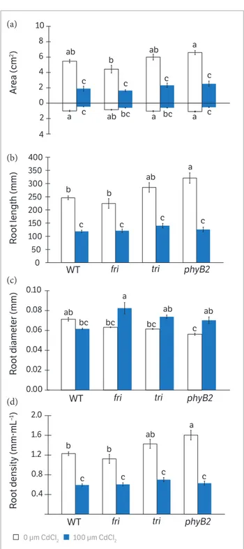
accumulation of Cd was observed in both shoots and roots. This accumulation was more pronounced in roots and was not affected by the genotype (Figure 1). In addition, the translocation index was similar for all genotypes (Figure 1b). The presence of cadmium in the nutrient solution decreased leaf and root area and the length of roots in all genotypes (Figure 2a,b). Although Cd accumulated mainly in roots, we observed an increase in root diameter in fri
and phyB2, whereas WT and tri did not show differences between the control and Cd treatments (Figure 2c). In fact, exposure to Cd inhibited root length, area and density in all genotypes (Figure 2).
After treatment with CdCl2, WT, fri and tri exhibited lower shoot fresh weight, while no significant change in root biomass was detected (Figure 3a). However, only WT and
tri suffered reduction of shoot dry weight after Cd exposure (Figure 3b). Plants cultivated in CdCl2 did not exhibit a reduction in the root dry weight, but phyB2, showed an increase in dry weight after exposure to CdCl2 (Figure 3b). In addition, Cd exposure caused a strong height reduction in Figure 1. (a) Cadmium measurements in 21 days-old tomato mutants
and WT grown with 100 µM·CdCl2 in the coordinate axis, values above
and below the 0 corresponds to the cadmium shoot and root content, respectively; (b) Cd translocation index. Data are means ± SEM (n = 3). The same lowercase letters on the bars of each panel indicate non-significant differences at p ≤ 0.05 by Tukey’s test.
0 10 20 30 40
1 2 3 4 5 6 7 8 9 10 11 12 13 180.000 140.000 100.000 60.000 20.000 20.000 60.000
1 2 3 4 5 6 7 8 9 10 11 12 13
C
d C
ont
ent (µg.
g
–1 dry WT)
C
d tr
ansloc
ation index
WT fri tri phyB2
WT fri tri phyB2
a a a a a a a a a
a a a
4 2 0 2 4 6 8 10
1 2 3 4 5 6 7 8 9 10 11 12 13
Ar e a (cm 2) 0 50 100 150 200 250 300 350 400
1 2 3 4 5 6 7 8 9 10 11 12 13
R
oot length (mm)
0.00 0.02 0.04 0.06 0.08 0.10
1 2 3 4 5 6 7 8 9 10 11 12 13
R oot diamet er (mm) b b ab ab ab a a a c c c c b b b a a a a ab ab
ab bc bc
c c c c c c ab ab
bc bc bc c
c c c c
0.4 0.8 1.2 1.6 2.0
1 2 3 4 5 6 7 8 9 10 11 12 13
R
oot density (mm·mL
–1)
WT fri tri phyB2
WT fri tri phyB2
WT fri tri phyB2
0 µm CdCl2 100 µm CdCl2
Figure 2. Leaf and root measurements in 21 days-old tomato mutants
and WT grown without or with 100 µM·CdCl2. (a) in the coordinate axis,
values above and below the 0 corresponds to the leaf and root area, respectively; (b) Root length; (c) diameter and (d) and density. Control plants of each genotype did not receive Cd. Data are means ± SE (n = 3). Different lowercase letters on the bars of each painel indicate significant differences at p ≤ 0.05 by Tukey’s test.
WT, fri and tri, while plant height was unaltered by high Cd in phyB2 (Figure 3c). However, in the absence of Cd, phyB2
and fri had similar heights as WT, whereas tri was taller.
(a) (a)
(b)
(b)
(c)
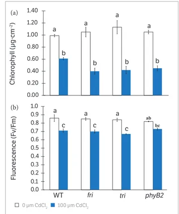
The supply of CdCl2 significantly decreased chlorophyll content in all genotypes, especially in the phytochrome mutants (Figure 4a). Additionally, there was a clear reduction of chlorophyll fluorescence in WT, fri and tri, while no significant change was observed in Cd-treated phyB2 plants (Figure 4b).
All genotypes showed leaf blades with unseriated adaxial and abaxial epidermis, palisade parenchyma presenting one layer of cells, spongy parenchyma thicker than the palisade parenchyma and evident intercellular spaces. The vascular bundles were trapezoidal and surrounded by parenchyma cells in both the adaxial and abaxial sides of the leaf (Figures 5a-f).
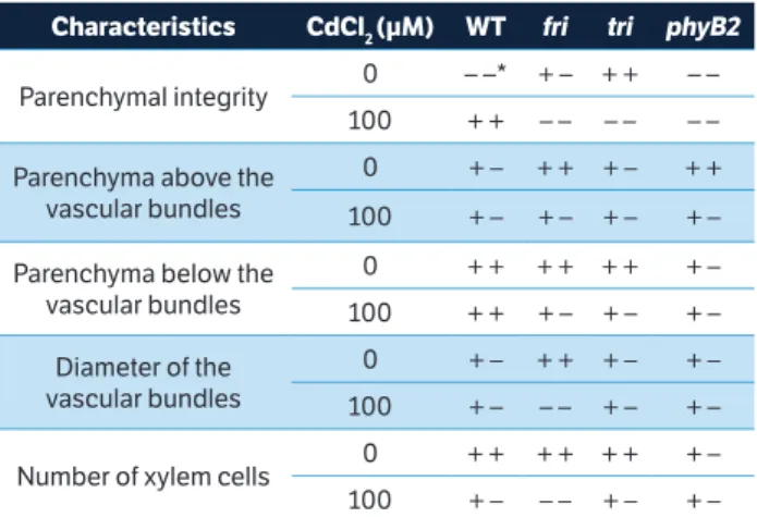
The fri and phyB2 plants (without the addition of CdCl2) had a large amount of parenchyma cells above the vascular bundles, which was not observed in these same genotypes when exposed to Cd, indicating a reduction in the number of cells of this tissue in this area. After the other genotypes were analyzed, we observed that there was no difference in the parenchymal cell density both above and below the vascular bundles. The tri genotype usually had a prominent apical cell above the parenchyma of the vascular bundles (Figure 5).
When we looked at the amount of parenchyma cells located below the vascular bundles in the midrib of the analyzed leaves, we also noticed a reduction in the number of these cells in the fri and tri genotypes supplied with Cd. The vascular bundles of the fri genotype showed a smaller diameter in plants undergoing Cd stress, while only small differences could be observed in the diameter of vascular bundles for the remaining genotypes (Table 1). There was a decrease in the number of xylem cells in all genotypes except
phyB2 in Cd stress conditions. The significant decrease in the number of xylem cells in fri plants, as well as the large
0.00 0.20 0.40 0.60 0.80 1.00 1.20 1.40 Chlor oph yll (µg·cm –2) ab bc 0.0 0.1 0.2 0.3 0.4 0.5 0.6 0.7 0.8 0.9 1.0 Fluor esc enc e (F v /F m)
WT fri tri phyB2
a a
a
b
b b b
a
a
a
a
c c c
0 µm CdCl2 100 µm CdCl2
–0.2 –0.1 0.0 0.1 0.2 0.3 0.4
1 2 3 4 5 6 7 8 9 10 11 12 13
0.010 0.005 0.000 0.005 0.010 0.015
1 2 3 4 5 6 7 8 9 10 11 12 13
a
a a ab a ab a b a
ab
b b
a
b b ab
0 20 40 60 80 100 120 140 Height (mm) Dry w eight (mg) F resh w eight (mg) a bc bc bc cd d b cd
WT fri tri phyB2
a a b d b ab cd cd bc bcd bc ab
ab ab ab
0 µm CdCl2 100 µm CdCl2
Figure 3. Leaf and root measurements in 21 days-old tomato mutants
and WT grown without or with 100 µM·CdCl2. (a) in the coordinate
axis, values above and below the 0 corresponds to the shoot and root fresh weight, respectively; (b) values above and below the 0 corresponds to the shoot and root dry weight, respectively and (c) shoot height. Control plants of each genotype did not receive Cd. Data are means ± SE (n = 3). Different lowercase letters on the bars of each panel indicate significant differences at p ≤ 0.05 by Tukey’s test.
Figure 4. Chlorophyll and fluorescence measurements in 21
days-old tomato mutants and WT grown without or with 100 µM CdCl2.
(a) chlorophyll content and (b) fluorescence. Control plants of each genotype did not receive Cd. Data are means ± SE (n = 3). Different lowercase letters on the bars of each panel indicate significant differences at p ≤ 0.05 by Tukey’s test.
(a)
(b)
(c)
(a)
number of tiny cells of xylem in tri and fri before Cd contact, were clearly noted (Table 1).
It is increasingly evident that phytochromes play a key role in abiotic stress responses (Zilli et al. 2008; Carvalho et al. 2011a; Xie et al. 2011). In this study, we used the phyA (fri), phyB1 (tri), and phyB2 (phyB2) mutants of tomato to evaluate whether some of these phytochromes were involved in plant responses to Cd stress conditions. D’Amico-Damião et al. (2015) reported that phytochrome B
Figure 5. Anatomical analysis in 21 days-old tomato mutants and WT grown without or with 100 µM·CdCl2. (a) WT without Cd; (b) WT with Cd;
(c) fri without Cd; (d) fri with Cd;(e) tri without Cd; (f) tri with Cd; (g) phyB2 without Cd and (h) phyB2 with Cd. Scales = 30 µm.
mutants grow better under drought conditions. Thus, better plant development of these genotypes may be expected in Cd conditions.
However, all plants except phyB2 exhibited reduced shoot fresh and dry weight and reduced height in the presence of Cd (Figures 2a-c). Although this observation could suggest that phyB2 was tolerant to Cd stress, these values were similar to those of the WT plants in the same conditions, suggesting that phyB2 plants grow slower in non-stressful conditions. (a)
(e) (c)
(g)
(b)
(f) (d)
On the other hand, Cd induced a larger root diameter in all photomorphogenic mutants (Figure 1c), especially fri,
which had a larger root diameter than WT, but there was no increase in root dry weight (Figure 2b). These results can be explained by the impairment of root elongation and phytochrome involvement in root development. In fact, it has been observed that phytochromes triggered the expression of ELONGATION HYPOCOTYLS 5 (HY5) and, in turn, the HY5 protein acted as a transcription factor and promoted root growth in response to light (Lee et al. 2016). In addition, it was verified that plants treated with Cd exhibited an enlargement of the roots and a reduction in root elongation (Fusconi et al. 2007; Maksimovic et al. 2007). Changes in root development in response to Cd exposure could be due to negative effects on the root cell microtubule cytoskeleton, which plays a crucial role during root development (Fusconi et al. 2007). However, more studies are needed to clarify these issues.
Additionally, tri was taller than WT in both control and stressful conditions, but similar shoot dry weight was verified, indicating no difference in biomass accumulation (Figure 2). Indeed, it was demonstrated that phyB1 plays a key role in stem elongation and shade avoidance responses (Schrager-Lavelle et al. 2016). Thus, tri, a phyB1-deficient mutant, exhibited a pronounced elongation of the stem due to impaired light perception (Figure 2c).
The Cd accumulation and translocation index were similar in all genotypes (Figure 1). Despite this similarity,
fri plants showed a reduced number of xylem cells and a reduced diameter of the vascular bundles compared to WT plants after Cd exposure (Table 1). The same result Table 1. Anatomical comparison among 21 days-old tomato mutants
and WT grown without or with 100 µM·CdCl2. *Signs ++, +- and --
indicating, respectively, greater, middle and less integrity, diameter or amount of cells from 21 days-old tomato mutants and WT grown
without or with 100 µM·CdCl2.
Characteristics CdCl2 (µM) WT fri tri phyB2
Parenchymal integrity 0 – –* + – + + – –
100 + + – – – – – –
Parenchyma above the vascular bundles
0 + – + + + – + +
100 + – + – + – + –
Parenchyma below the vascular bundles
0 + + + + + + + –
100 + + + – + – + –
Diameter of the vascular bundles
0 + – + + + – + –
100 + – – – + – + –
Number of xylem cells 0 + + + + + + + –
100 + – – – + – + –
was observed by Auge et al. (2012), whose hand-cut stem cross-sections showed a lower xylem vessel number and transversal area in fri plants under high evaporative demand. This reduction in xylem caused a reduction in the water supply to the leaves (Auge et al. 2012) and was expected to affect the transport of Cd from the roots to the shoot. In fact, Cd enters into the root system through the symplast or apoplast and eventually reaches the xylem, where it can be translocated to shoots (Lux et al. 2011). Furthermore, Cd translocation in the roots is impaired by the barriers in the exo- and endoderm (Redjala et al. 2011). Therefore, the destabilization of the root tissue facilitates the diffusion of Cd and its loading in the xylem, thus affecting Cd transport from the roots to the shoot (Lux et al. 2011). In addition, Cd accumulation in the shoot causes severe damage to plant physiological processes such as photosynthesis, water relations, mineral metabolism and leaf morphology (Lopez-Chuken and Young 2010; Gill et al. 2012; Asgher et al. 2015). In our study, we observed Cd in the shoots of all genotypes (Figure 1), and there was also a clear disruption in the xylem and parenchymal tissues (Figure 5 and Table 1), as well as a reduction in chlorophyll content and fluorescence, independent of genotype (Figure 4), which were most likely due to instability in the chloroplast membrane due to the Cd accumulation.
In fact, our results could indicate the control of these responses by phytochromes. Clear anatomical differences were observed between the tested phytochrome mutants. For instance, fri
and phyB2 exhibited more parenchyma cells above the vascu-lar bundles, while tri had a prominent apical cell in the paren-chyma above the vascular bundles. However, under Cd stress conditions, both parenchymal integrity and parenchyma cells above or below the vascular bundles were severely reduced in
fri compared to WT plants (Figure 5 and Table 1).
CONCLUSION
Despite mounting evidence that phytochromes are involved in plant responses to several abiotic stresses (Carvalho et al. 2011b; Auge et al. 2012), none of the three phytochrome mutants assessed in the present study exhibited higher tolerance to Cd. Thus, taken together, these results indicate that phytochromes do not integrate the pathway of Cd stress response in tomato plants.
Ali, B., Deng, X., Hu, X., Gill, R. A., Ali, S., Wang, S. and Zhou, W.
(2015). Deteriorative effects of cadmium stress on antioxidant
system and cellular structure in germinating seeds of Brassica
napus L. Journal of Agricultural Science and Technology, 17, 63-74.
Asgher, M., Khan, M. I., Anjum, N. A. and Khan, N. A. (2015).
Minimising toxicity of cadmium in plants — role of plant growth
regulators. Protoplasma, 252, 399-413. https://doi.org/10.1007/
s00709-014-0710-4.
Auge, G. A., Rugnone, M. L., Cortés, L. E., González, C. V., Zarlavsky, G.,
Boccalandro, H. E. and Sánchez, R. A. (2012). Phytochrome A increase
tolerance to high evaporative demand. Physiologia Plantarum, 151,
228-235. https://doi.org/10.1111/j.1399-3054.2012.01625.x.
Bae, G. and Choi, G. (2008). Decoding of light signals by plant
phytochromes and their interacting proteins. Annual Review
of Plant Biology, 59, 281-311. https://doi.org/10.1146/annurev.
arplant.59.032607.092859.
Balestrasse, K. B., Yannarelli, G. G., Noriega, G. O., Batlle, A. and
Tomaro, M. L. (2008b). Hemeoxygenase and catalase gene
expression in nodules and roots of soybean plants subjected to
cadmium stress. Biometals, 21, 433-441. https://doi.org/10.1007/
s10534-008-9132-0.
Balestrasse, K. B., Zilli, C. G. and Tomaro, M. L. (2008a). Signal
transduction pathways and haem oxygenase induction in soybean
leaves subjected to salt stress. Redox Report, 13, 255-262. https://
doi.org/10.1179/135100008x308966.
Boccalandro, H. E., Mazza, C. A., Mazzella, M. A., Casal, J. J.
and Ballaré, C. L. (2001). Ultraviolet B radiation enhances a
phytochrome-B mediated photomorphogenic response in
Arabidopsis. Plant Physiology, 126, 780-788. https://doi.org/10.1104/
pp.126.2.780.
REFERENCES
Carvalho, R. F., Aidar, S. T., Azevedo, R. A., Dodd, I. C. and Peres,
L. E. P. (2011b). Enhanced transpiration rate in the high pigment 1
tomato mutant and its physiological significance. Plant Biology,
13, 546-550. https://doi.org/10.1111/j.1438-8677.2010.00438.x.
Carvalho, R. F., Campos, M. L. and Azevedo, R. A. (2011a). The role of
phytochrome in stress tolerance. Journal of Integrative Plant Biology,
53, 920-929. https://doi.org/10.1111/j.1744-7909.2011.01081.x.
Cui, W. T., Fu, G. Q., Wu, H. H. and Shen, W. B. (2011).
Cadmium-induced heme oxygenase-1 gene expression is associated with the
depletion of glutathione in the roots of Medicago sativa. Biometals,
24, 93-103. https://doi.org/10.1007/s10534-010-9377-2.
D’Amico-Damião, V., Cruz, F. J. R., Gavassi, M. A., Santos, D. M.
M., Melo, H. C. and Carvalho, R. F. (2015). Photomorphogenic
modulation of water stress in tomato (Solanum lycopersicumL.):
the role of phytochromes A, B1, and B2. Journal of Horticultural
Science and Biotechnology, 90, 25-30. https://doi.org/10.1080/
14620316.2015.11513149.
Essen, L. O., Maillliet, J. and Hughes, J. (2008). The structure of
a complete phytochrome sensory module in the Pr ground state.
Proceedings of the National Academy of Science of the U.S.A.,
105, 14709-14714. https://doi.org/10.1073/pnas.0806477105.
Franklin, K. A. (2016). Photomorphogenesis: plants feel blue in the
shade. Current Biology, 26, 1275-1276. https://doi.org/10.1016/j.
cub.2016.10.039.
Franklin, K. A. and Quail, P. H. (2010). Phytochrome functions in
Arabidopsis development. Journal of Experimental Botany, 61,
11-24. https://doi.org/10.1093/jxb/erp304.
Fusconi, A., Gallo, C. and Camusso, W. (2007). Effects of cadmium
proliferation and microtubule pattern as suitable markers for
assessment of stress pollution. Mutation Research, 632, 9-19.
https://doi.org/10.1016/j.mrgentox.2007.03.012.
Gill, S. S., Khan, N. A. and Tuteja, N. (2012). Cadmium at high dose
perturbs growth, photosynthesis and nitrogen metabolism while
at low dose it up regulates sulfur assimilation and antioxidant
machinery in garden cress (Lepidium sativum L.). Plant Science,
182, 112-120. https://doi.org/10.1016/j.plantsci.2011.04.018.
Gratão, P. L., Monteiro, C. C., Carvalho, R. F., Tezotto, T., Piotto, F. A.,
Peres L. E. P. and Azevedo, R. A. (2012). Biochemical dissection
of diageotropica and Never ripe tomato mutants to Cd-stressful conditions. Plant Physiology and Biochemistry, 56, 79-96. https://
doi.org/10.1016/j.plaphy.2012.04.009.
Gratão, P. L., Monteiro, C. C., Rossi, M. L., Martinelli, A. P., Peres, L.
E. P., Medici, L. O., Lea, P. J. and Azevedo, R. A. (2009). Differential
ultrastructural changes in tomato hormonal mutants exposed to
cadmium. Environmental and Experimental Botany, 67, 387-394.
https://doi.org/10.1016/j.envexpbot.2009.06.017.
Gratão, P. L., Monteiro, C. C., Tezotto, T., Carvalho, R. F., Alves,
L. R., Peters, L. P. and Azevedo, R. A. (2015). Cadmium stress
antioxidant responses and root-to-shoot communication in grafted
tomato plants. Biometals, 28, 803-816. https://doi.org/10.1007/
s10534-015-9867-3.
Hiltbrunner, A., Viczian, A., Bury, E., Tscheuschler A., Kircher, S., Toth,
R., Honsberger, A., Nagy, F., Fankhauser, C. and Schafer, E. (2005).
Nuclear accumulation of the phytochrome A photoreceptor requires
FHY1. Current Biology,15, 2125-2130. https://doi.org/10.1016/j.
cub.2005.10.042.
Jin, Q. J., Zhu, K. K., Xie, Y. J. and Shen, W. B. (2013). Heme oxygenase-1
is involved in ascorbic acid-induced alleviation of cadmium toxicity
in root tissues of Medicago sativa. Plant and Soil, 366, 605-616.
https://doi.org/10.1007/s11104-012-1451-9.
Johansen, D. A. (1940). Plant microtechnique. New York:
McGraw-Hill Book Co. Inc.
Kim, H., Kim, Y., Park, J. and Kim, J. (2002). Light signalling mediated
by phytochrome plays animportant role in cold-induced gene
expression through the C-repeat/dehydration responsive element
(C/DRE) in Arabidopsis thaliana. The Plant Journal, 29, 693-704.
https://doi.org/10.1046/j.1365-313x.2002.01249.x.
Lee, H-J., Ha, J-H., Kim, S-G., Choi, H-K., Kim, Z. H., Han, Y-J., Kim,
J-I., Oh, Y., Fragoso, V., Shin, K., Hyeon, T., Choi, H-G., Oh, K-H.,
Baldwin, I. T. and Park, C-M. (2016). Stem-piped light activates
phytochrome B to trigger light responses in Arabidopsis thaliana
roots. Science Signaling, 9, 1-8. https://doi.org/10.1126/scisignal.
aaf6530.
Liu, J., Zhang, F., Zhou, J., Chen, F., Wang, B. and Xie X. (2012).
Phytochrome B control of total leaf area and stomatal density
affects drought tolerance in rice. Plant Molecular Biology, 78,
289-300. https://doi.org/10.1007/s11103-011-9860-3.
Lopez-Chuken, U. J. and Young, S. D. (2010). Modelling
sulphate-enhanced cadmium uptake by Zea mays from nutrient solution
under conditions of constant free Cd 2+ ion activity. Journal of
Environmental Sciences, 22, 1080-1085. https://doi.org/10.1016/
s1001-0742(09)60220-5.
Lux, A., Martinka, M., Vaculik, M. and White, P. J. (2011). Root
responses to cadmium in the rhizosphere: a review. Journal of
Experimental Botany, 62, 21-37. https://doi.org/10.1093/jxb/erq281.
Maksimovic, I., Kastori, R., Krstic, L. and Lukovic, J. (2007). Steady
presence of cadmium and nickel affects root anatomy, accumulation
and distribution of essential ions in maize seedlings. Biologia
Plantarum, 51, 589-592. https://doi.org/10.1007/s10535-007-0129-2.
Malavolta, E., Vitti, G. C. and Oliveira, S. A. (2011). Avaliação do estado
nutricional de plantas: princípios e aplicações, 2 ed. Piracicaba:
Associação Brasileira para Pesquisa de Potassa e do Fosfato.
Nagajyoti, P. C., Lee, K. D. and Sreekanth, T. V. M. (2010). Heavy metals,
occurrence and toxicity for plants: a review. Environmental Chemistry
Letters, 8, 199-216. https://doi.org/10.1007/s10311-010-0297-8.
Neff, M. M., Fankhauser, C. and Chory, J. (2000). Light: an indicator
of time and place. Genes and Development, 14, 257-271.
Nishihama, R., Ishizaki, K., Hosaka, M., Matsuda, Y., Kubota, A. and
Kohchi, T. (2015). Phytochrome-mediated regulation of cell division
and growth during regeneration and sporeling development in
the liverwort Marchantia polymorpha. Journal of Plant Research.
https://doi.org/10.1007/s10265-015-0724-9.
O’Brien, T. P., Feder, N. and Mccully, M. M. E. (1964). Polychromatic
staining of plant cell wall by toluidine blue. Protoplasma,59,
367-373. https://doi.org/10.1007/bf01248568.
Plusquin, M., Mulder, K., Belleghem, F. V., DeGheselle, O., Pirotte, N.,
Willems, M., Cuypers, A., Salvenmoser, W., Laudurner, P., Artois, T.
and Smeets, K. (2015). Toxic effects of cadmium on flatworm stem
cell dynamics: a transcriptomic and ultrastructural elucidation of
underlying mechanisms. Environmental Toxicology, 31, 1217-1228.
Quail, P. H. (2002a). Photosensory perception and signalling in
plant cells: new paradigms? Current Opinion in Cell Biology, 14,
180-188. https://doi.org/10.1016/s0955-0674(02)00309-5.
Quail, P. H. (2002b). Phytochrome photosensory signalling
networks. Nature Reviews Molecular Cell Biology, 3, 85-93. https://
doi.org/10.1038/nrm728.
Redjala, T., Zelko, I., Sterckeman, T., Legué, V. and Lux, A. (2011).
Relationship between root structure and root cadmium uptake
in maize. Environmental and Experimental Botany, 71, 241-248.
https://doi.org/10.1016/j.envexpbot.2010.12.010.
Rossato, L. V., Nicoloso, F. T., Farias, J. G., Cargnelluti, D, Tabaldi,
L. A., Antes, F. G., Dressler, V. L., Morsch, V. M. and Schetinger, M.
R. C. (2011). Effects of lead on the growth, lead accumulation and
physiological responses of Plucheas agittalis. Ecotoxicology, 21,
111-123. https://doi.org/10.1007/s10646-011-0771-5.
Saijo, Y., Hata, S., Kyozuka, J., Shimamoto, K. and Izui, K. (2000).
Over-expression of a single Ca2+-dependent protein kinase confers both
cold and salt/drought tolerance on rice plants. The Plant Journal,
23, 319-327. https://doi.org/10.1046/j.1365-313x.2000.00787.x.
Schrager-Lavelle, A., Herrera, L. A., and Maloof, J. N. (2016). Tomato
phyE Is required for shade avoidance in the absence of phyB1 and
phyB2. Frontiers in Plant Science,7, 1275. https://doi.org/10.3389/
fpls.2016.01275.
Shen, Q., Jiang, M., Li, H., Che, L. L. and Yang, Z. M. (2011). Expression
of a Brassica napus heme oxygenase confers plant tolerance to
mercury toxicity. Plant Cell and Environment, 34, 752-763. https://
doi.org/10.1111/j.1365-3040.2011.02279.x.
Tsukaya, H., Kozuka, T. and Kim, G. T. (2002). Genetic control of
petiole length in Arabidopsis thaliana. Plant Cell Physiology, 43,
1221-1228. https://doi.org/10.1093/pcp/pcf147.
Usman, A. R. A., Lee, S. S., Awad, Y. M., Lim, K. J., Yang, J. E. and
Ok, Y. S. (2012). Soil pollution assessment and identification of
hyperaccumulating plants in chromated copper arsenate (CCA)
contaminated sites. Korea. Chemosphere, 87, 872-878. https://doi.
org/10.1016/j.chemosphere.2012.01.028.
Vaculik, M., Pavlovic, A. and Lux, A. (2015). Silicon alleviates
cadmium toxicity by enhanced photosynthetic rate and
modified bundles heath’s cell chloroplast ultrastructure in
maize. Ecotoxicology and Environmental Safety, 120, 66-73.
https://doi.org/10.1016/j.ecoenv.2015.05.026.
Verkleij, J. A. C., Golan-Goldhirsh, A., Antosiewisz, D. M.,
Schwitzguebel, J. P. and Schroder, P. (2009). Dualities in plant
tolerance to pollutants and their uptake and translocation to
the upper plant parts. Environmental and Experimental Botany,
67, 10-22. https://doi.org/10.1016/j.envexpbot.2009.05.009.
Wit, M., Galvão, V. C. and Fankhauser, C. (2016). Light-mediated
hormonal regulation of plant growth and development. Annual
Review of Plant Biology, 67, 513-537. https://doi.org/10.1146/
annurev-arplant-043015-112252.
Xie, Y. J., Cui, W. T., Yuan, X. X., Shen, W. B. and Yang, Q.
(2011). Heme oxygenase-1 is associated with wheat salinity
acclimation by modulating reactive oxygen species homeostasis.
Journal of Integrative Plant Biology, 53, 653-670. https://doi.
org/10.1111/j.1744-7909.2011.01052.x.
Yang, X., Kuk, J. and Moffat, K. (2008). Crystal structure
o f Ps e u d o m o n a s a e r u g i n o s a b a c t e r i o p h y t o c h ro m e :
photoconversion and signal transduction. Proceedings of
the National Academy of Sciences of the U.S.A., 105,
14715-14720. https://doi.org/10.1073/pnas.0806718105.
Zao, Y., Zhou, J. and Ding, D. (2014). Phytochrome B-mediated
activation of lipoxygenase modulates an excess red light-induced
defence response in Arabidopsis. Journal of Experimental
Botany, 65, 4907-4918. https://doi.org/10.1093/jxb/eru247.
Zilli, C. G., Balestrasse, K. B., Yannarelli, G. G., Polizio, A. H.,
Santa-Cruz, D. M. and Tomaro, M. L. (2008). Hemeoxygenase
up-regulation under salt stress protects nitrogen metabolism
i n n o d u l e s o f s o y b e a n p l a n t s . E n v i r o n m e n t a l a n d
Experimental Botany, 64, 83-89. https://doi.org/10.1016/j.