Abstract published online: March 30, 2005 Full paper published online: August 31, 2006
Original paper. ISSN 1678-9199.
ANTIBODY AND CYTOKINE SERUM LEVELS IN PATIENTS SUBJECTED TO ANTI-RABIES PROPHYLAXIS WITH SERUM-VACCINATION
AYRES J. A. (1), BARRAVIERA B. (2), CALVI S. A. (2), CARVALHO N. R. (3), PERAÇOLI M. T. S. (4)
(1) Department of Nursing, Botucatu School of Medicine, São Paulo State University, UNESP, Botucatu, São Paulo State, Brazil; (2) Department of Tropical Diseases and Imaging Diagnosis, Botucatu School of Medicine, São Paulo State University, UNESP, Botucatu, São Paulo State, Brazil; (3) University Hospital, Botucatu School of Medicine, São Paulo State University, UNESP, Botucatu, São Paulo State, Brazil; (4) Department of Microbiology and Immunology, Institute of Biosciences, São Paulo State University, UNESP, Botucatu, São Paulo State, Brazil.
ABSTRACT: Rabies is considered a fatal disease once clinical symptoms have
developed. The aim of this study was to evaluate epidemiological aspects and immune response in patients attacked by domestic and wild animals and subjected to post-exposure rabies treatment with equine serum and associated vaccine. Thirty-three patients were evaluated; they were between 13 and 65 years old, 75.8% were male and 24.2% female, and from the Botucatu neighborhood. Twenty healthy control individuals with the same age range were also studied. Specific antibodies to equine immunoglobulins and IFN-γ, IL-2, IL-4, and IL-10 production were evaluated by ELISA. IgM, IgE, IgG and subclasses, and rabies virus antibodies serum levels were determined by nephelometry and seroneutralization methods, respectively. No anaphylactic or serum sickness allergic reactions were observed in patients after treatment. Anti-equine IgG levels were significantly higher than those of IgM after 14 and 28 days of treatment. Protective antibodies to rabies virus > 0.5 UI/ml were detected in 84.6% and 75% of patients at days 14 and 28, respectively. IFN-γ, IL-2 and IL-10 levels in patients before and 48h after treatment were significantly higher than in controls suggesting that both Th1 and Th2 cells were activated in the patients. Serum IgM levels were higher at day 14, and IgG2 and IgE levels were higher at day 28 of treatment. These results suggest that post-exposure rabies treatment in humans induces significant alterations in patient immune response characterized by increased levels of cytokines, serum levels of specific rabies virus antibodies, and the equine serum components employed in the treatment.
KEY WORDS: rabies, anti-rabies prophylaxis, immunoglobulins, cytokines. CORRESPONDENCE TO:
JAIRO A. AYRES, Departamento de Enfermagem, Faculdade de Medicina de Botucatu, UNESP, Distrito de Rubião Júnior, 18618-000, Botucatu, SP, Brasil. Email:
INTRODUCTION
Rabies is a zoonotic, viral disease. The causative agent, Lyssavirus of the family
Rhabdoviridae, is responsible for acute encephalitis. In developed countries, rabies is
spread from wild to domestic animals and consequently to humans; once clinical
signs appear, it is fatal. (10).
From an epidemiological point of view, rabies remains a very important public health
problem in several developing countries from the African, Asiatic (64), and South
American continents (15, 28, 43, 45, 46, 63, 64). It is estimated that around 300
people die from the disease and approximately 300,000 are treated every year with
post exposure serum vaccination (2).
Oceania and Antarctica are free of the disease (2, 37, 50), and it has been
eradicated in Japan, England, the Oceania islands, some Pacific islands, and other
island countries. In countries such as France, Germany, Spain, Canada, and the
United States, it has been controlled by efficient surveillance systems (39, 44).
In Brazil, despite great advances in rabies control, there are still incidences of the
disease in the North, Northeast, Central West, and some southern states (47). The
southern region is considered a controlled area (45). Reports of the disease have
been decreasing in Brazil since the 1980’s, but case frequency is still variable. In
2000, twenty-six cases were recorded and confirmed, four of which were in the
Central West, nine in the North, and 13 in the Northeast (15). The highest incidences
occur in poorer areas, and in areas where there are no effective control programs
and there is higher contact between man and animals.
The Word Health Organization (WHO) recommends that all people highly exposed to
animals with suspected or confirmed rabies diagnosis must receive anti-rabies
prophylactic treatment with serum vaccination (60). This treatment is intended to
protect the host against disease development using the following strategy: the
anti-rabies serum specific immunoglobulin provides a long period of protection, offering
antibodies for immediate virus neutralization, while the vaccine stimulates
endogenous antibody production to give an efficient immunologic memory (14, 61).
Equine anti-rabies serum is a purified immunoglobulin solution with antigenic
properties which induce IgE antibodies formation by a sensitization process and may
cause anaphylaxis on subsequent serum exposure and form immune complexes
which can trigger serum sickness (5, 9, 12, 25, 27, 38, 54). Kaliner & Beleval (26)
Heterologous proteins trigger a complex immunological process in the receptor
organism which in turn triggers hypersensitivity mechanisms (51).
Pre and post-exposure prophylaxis regimens are well established today by the
simultaneous use of vaccine and hyperimmune serum. Serotherapy should be used
with caution because it may trigger major reactions in the receptor, although with
relatively low frequency and severity. Administration is therefore recommended in
hospitals prepared to deal with adverse reactions.
Since both heterologous anti-rabies serum and rabies vaccine have immunogenic
determinants, they can induce cellular and humoral immune response in patients
subjected to serum vaccination treatment. This study evaluated the specific
antibodies produced against equine immunoglobulin and rabies virus, and the serum
immunoglobulin and cytokine levels in patients subjected to post-exposure rabies
prophylactic treatment after domestic animal aggression.
PATIENTS AND METHODS
Patients
There were 33 patients, 25 males and 8 females, with ages ranging from 13 to 65
years old (mean: 40.6 years old); they were admitted to the Tropical Diseases Clinic
at the University Hospital of the Botucatu School of Medicine, São Paulo State
University, UNESP, between January 2000 and October 2001. They reported
exposure to animals with suspected rabies: 12 had been exposed to biological
material from bovine with suspected rabies; three had contact with wild animals, two
with simian and one with a bat; and 15 had been victims of aggression from domestic
animals, canine (seven), feline (five), and swine (three). A group of 20 healthy blood
donors, gender and age matched to patients, was included as controls. This study
was approved by the Ethics Committee of the Botucatu School of Medicine, and
informed consent was obtained from all patients, controls, or their parents.
Treatment
Patients exposed or attacked by rabies-suspected animals were considered at risk of
contracting the rabies virus. Thus, as per World Health Organization
vaccination (45) consisting of passive immunization with 40 UI/kg equine
heterologous serum by intramuscular (IM) route. Active immunization was
administered with human anti-rabies vaccine prepared from the rabies virus strain
WISTAR PM/WI 38-1503-3m, cultured in Vero cells (Pasteur Mérieux); it was also
administered by IM route in a five-dose schedule: the first dose on the day of virus
exposure and the others at days 3, 7, 14, 21, and 28 after exposure. The patients
were questioned about the time elapsed between the accident and medical attention,
and about any previous administration of anti-rabies serum.
Human sera
Blood samples (6 ml) were collected without anticoagulant in 10-ml plastic tubes
before serum vaccination treatment and then after 48 h, 14 and 28 days of treatment.
Blood was centrifuged at 2500 rpm for 10 min; the serum was allowed to separate
and was then stored at -20°C until determination of specific antibodies to virus,
equine immunoglobulin, human serum immunoglobulins and cytokines.
Anti-equine
I
gG and IgM immunoglobulin determinationSerum levels of anti-equine immunoglobulin antibodies in patients subjected to
serum-vaccination treatment were determined by enzyme-immunoassay (ELISA).
Linbro Titertek (Flow Laboratories, Inc., Mclean, VA, USA) 96-well plates were
coated overnight at 4°C with 100 µl of 1:8000 dilution F(ab)2 fraction of equine
anti-rabies in 0.05 M carbonate buffer, pH 9.6. The plates were then washed with
phosphate buffered saline (PBS, pH 7.4) containing 0.05% Tween 20 (T-PBS). At
this and all other steps, wells were washed six times with 200 µl T-PBS. The blocking
solution containing 2% bovine albumin (Sigma Chemical Co., St Louis, MO, USA)
and 0.05% gelatin (Sigma) in 200 µl PBS was added and plates were incubated for
60 min at 37°C and washed six times with T-PBS. Sera of patients or controls (100
µl) were added in duplicate to the wells and incubated for 60 minutes at 37°C. After
washing, 100 µl of 1:1000 rabbit IgG, anti-human IgG, or 1:2000 anti-human IgM
conjugated with peroxidase (Sigma Chemical Co., USA) was added to each well, and
the plates were incubated for 60 min at 37°C. Wells were then washed with T-PBS
and 100 µl of an enzimatic substrate containing 0.4 mg/ml and 2 µl/ml of hydrogen
room temperature in the dark for 15 min, the reaction was stopped by adding 50 µl of
2 M sulphuric acid, and absorbance values were measured at 492 nm using an
ELISA reader (Multiskan Spectrophotometer, EFLAB, Helsinki, Finland). Results
were expressed as optical density (OD). Reaction cutoff was established as the
highest serum OD value obtained from 13 healthy individuals tested at 1:200
dilutions. The OD of each patient serum dilution was compared with reaction cutoff.
OD values above 0.141 for IgG and above 0.183 for IgM were considered as
positive.
Anti-rabies virus antibodies determination
Neutralizing antibodies were measured by Simplified Fluorescent Inhibition Microtest
[SFIMT] (20). Briefly, the test sera and positive and negative standard sera were
distributed into a series of two-fold dilutions in minimum Eagle medium (MEM)
containing 10% bovine fetal serum beginning from 1/5 in 100 µl per well of Linbro
Titertek (Flow Laboratories) 96-well plates; 50 µl of a previously tittered optimal viral
suspension (PV virus) was added to each well to infect 80% of the cells; this was
followed by an incubation period of 60 min at 37°C. Then, 50 µl BHK21 cells (clone
13), growing in MEM with 10% bovine fetal serum (106 cells/ml), was added to each
well. The microplates were incubated at 37°C for 24 h in a humidified CO2
atmosphere. Virus infection rate was determined in each test. After cell fixation with
80% cold acetone for 15 min, cells were stained by an anti-rabies nucleocapsid
fluorescent conjugate (Sanofi cod 72114). The plates were washed with PBS
followed by distilled water. Then, 50 µl of 0.5 M glycerine buffered carbonate, pH 9.6,
was added to each well. The plates were examined in an epi-fluorescence inverted
microscope to evaluate the effect of the virus on the cell culture. Results expressed
the dilution corresponding to the well with 50% decrease of infection. Comparison of
test sera results with those of standard serum were used to obtain titer IU values.
Immunoglobulin determination
The IgM, IgG and isotypes, and IgG1, IgG2, IgG3 and IgG4 levels were determined
in serum of patients subjected to serum vaccination therapy evaluated before and
after 14 and 28 days of treatment. IgM and IgG were determined by nephelometry
the manufacturer’s instructions. Immunoglobulin concentrations were measured with
a BN 100 nephelometer (Dade Behring, Marburg, Germany) and ARRAY 360
equipment (Beckman Coulter, Inc., Los Angeles, CA, USA). Immunoglobulin isotype
results were expressed in mg/100ml.
IgE determination
IgE serum levels were evaluated by an Elecsys Enzymun-test (Roche, diagnostic
Corp., Indianopolis, IN, USA) with a ruthenium-labeled anti-IgE monoclonal antibody.
Reactions were developed according to the manufacturer’s instructions, and IgE
concentrations were obtained in an ELECSYS 2010 apparatus (Roche, Diagnostic
Corp., Indianopolis, IN, USA).
Enzyme-linked immunosorbent assay for cytokines
Cytokines IL-2, IL-4, IL-10, and IFN-γ concentrations were determined in patient
serum obtained before and after treatment (48 h later) by ELISA. Antibody matched
pairs and respective standards were purchased from R&D Systems (Minneapolis,
MN, USA) and used according to the manufacturer’s instructions. Assay sensitivity
limit was 10 pg/ml for all cytokines tested.
Statistical analysis
Data were statistically analyzed using InStat software (Graph Pad, Software, version
3.05, San Diego, CA, 2000). Serum levels of anti-equine immunoglobulin antibodies
and anti-rabies virus found in patients before and after treatment and in the control
group were compared by one-way analysis of variance (ANOVA), followed by
multiple comparisons by the Tukey-Kramer method, and concentrations of IgE, IgM,
and IgG isotypes were analyzed by the paired t-test. Differences in the cytokine
responses between patients and controls were determined by Kruskal-Wallis
non-parametric analysis of variance, followed by Dunn Test multiple comparison.
Significance level was set at p<0.05 in all analyses.
RESULTS
We studied the epidemiological characterization of 33 patients receiving rabies
high exposure to bovine biological material followed by canine and feline attack,
animals of major epidemiological importance.
Bite was the major route (54.6%), followed by animal manipulation (42.4%), allowing
contact with biological materials (blood and saliva), and bite followed by scratching
(3%). Absence of wound was seen in 42.4%, multiple lesions in 33.3%, and single
lesion in 24.2% of cases. Lesions were mainly in the upper limbs (81.2%), inferior
limbs (9.4%), and both (6.2%); there were only 3.1% in the trunk.
Post-exposure serum vaccination was indicated for all patients in the study, using a
five-dose regimen of Vero cell culture produced vaccine in combination with the
equine anti-rabies serum containing 40 UI/kg purified equine immunoglobulin as per
the Pasteur Institute Manual (45). No adverse reactions were seen in either vaccine
or anti-rabies serum inoculation sites. Also, patients did not develop either
anaphylactic or serum sickness hypersensitivity reactions.
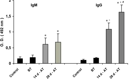
Anti-equine immunoglobulins antibodies were detected in the serum samples of
patients subjected to serum-vaccination treatment and in the control group. Figure 1
shows that patients developed anti-equine IgG and IgM immunoglobulin antibodies
after treatment. Both IgM and IgG antibody levels detected at 14 and 28 days of
treatment were significantly higher than before treatment or in healthy controls. IgG
serum levels were significantly higher than IgM at both 14 and 28 days after
treatment. At day 28, IgG levels were significantly higher than at day 14, while no
significant differences in IgM levels were observed in these periods.
The analysis of neutralizing anti-rabies virus antibodies in patient sera before and
after vaccination showed that patients did not have antibody protection before
vaccine administration. After treatment, patients had clearly developed high levels of
specific antibodies against rabies virus at both 14 and 28 days of vaccine regimen.
Antibody levels in patient serum at these times were not significantly different. Based
on WHO criteria (62) proposing a 0.5-UI/ml limit for establishing serum conversion
after rabies vaccination, 11/13 patients (84.6%) at 14 days and 15/20 patients (75%)
at 28 days treatment were positive for serum conversion.
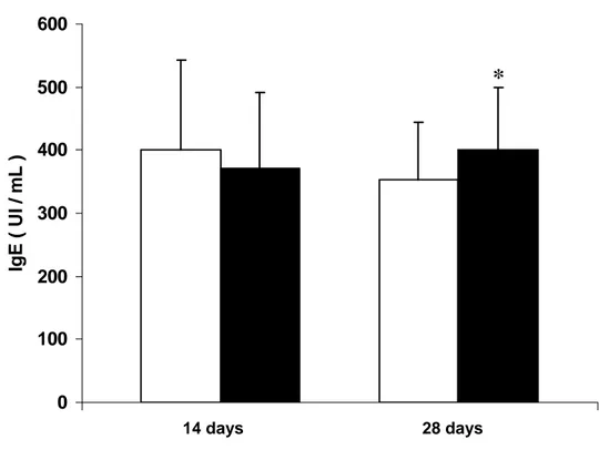
Figures 2 and 3 show serum concentrations for total IgM and IgG and IgG1, IgG2,
IgG3 and IgG4 subclasses in patients before and after 14 and 28 days of treatment
regimen, respectively. Figure 4 shows IgE levels. Immunoglobulin levels after
significantly higher levels of IgM at 14 days treatment (Figure 2) and IgG2 and IgE at
28 days treatment (Figures 3 and 4).
significantly higher levels of IgM at 14 days treatment (Figure 2) and IgG2 and IgE at
28 days treatment (Figures 3 and 4).
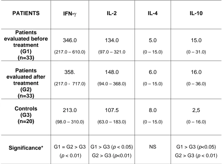
Table 1 shows IFN-γ, IL-2, IL-4, and IL-10 in sera of both equine-immunoglobulin and
rabies-vaccine treated and untreated patients compared to levels in healthy controls
not subjected to serum vaccination treatment. IFN-γ, IL-2, and IL-10 levels were
significantly higher in patient groups both before and after treatment compared to
those in the control group. No significant difference was observed between groups
for IL-4 levels.
0 0,5 1 1,5 2 Cont
rol BT
14 d -
AT
28 d
AT
Cont
rol BT
14 d
AT
28 d -
AT
IgM IgG * + #+ #
O . D . ( 492 n m ) * + * *
Figure 1: Anti-equine IgM and IgG immunoglobulin antibodies in sera of patients
evaluated before (BT) and after (AT) treatment with rabies serum vaccination.
Results are expressed as mean +
Figure 1: Anti-equine IgM and IgG immunoglobulin antibodies in sera of patients
evaluated before (BT) and after (AT) treatment with rabies serum vaccination.
Results are expressed as mean + SD of optical density (OD)
* (p<0.05) versus control, BT;
+
(p<0.05) versus IgM;
0 200 400 600 800 1000 1200 1400
m
g
/
10
0 m
L
*
IgG IgG1 IgG2 IgG3 IgG4 IgM
Figure 2: IgG and subclass serum levels in patients evaluated before and after 14
days of treatment with rabies serum vaccination. Results are expressed as mean +
SD of mg/100 ml.
*(p<0.05) paired t test
0 200 400 600 800 1000 1200 1400
m
g
/
10
0 m
L
*
IgG IgG1 IgG2 IgG3 IgG4 IgM
Figure 3: IgG and subclass serum levels in patients evaluated before and after 28
days of treatment with rabies serum vaccination. Results are expressed as mean +
SD of mg/100 ml.
Figure 4.
0 100 200 300 400 500 600
Ig
E
(
U
I / mL )
28 days 14 days
*
Figure 4: IgE serum levels in patients evaluated before and after 14 and 28 days of
treatment with rabies serum vaccination. Results are expressed as mean + SD of
UI/ml.
Table 1: Serum levels of Interferon-gamma (IFN-γ), Interleukin-2 (2), 4 and
IL-10 detected in 33 patients evaluated before and after 48 h of treatment with equine
immunoglobulin and in 20 healthy controls.
PATIENTS IFN-γ IL-2 IL-4 IL-10
Patients evaluated before treatment (G1) (n=33) 346.0
(217.0 – 610.0)
134.0
(97.0 – 321.0
5.0
(0 – 15.0)
15.0
(0 – 31.0)
Patients evaluated after treatment (G2) (n=33) 358.
(217.0 - 717.0)
148.0
(94.0 – 368.0)
6.0
(0 – 15.0)
16.0
(0 – 36.0)
Controls (G3) (n=20)
213.0
(98.0 – 310.0)
107.5
(63.0 – 183.0)
8.0
(0 – 15.0)
2,5
(0 – 16.0)
Significance* G1 = G2 > G3 (p < 0.01)
G 5)
G2 >
1 > G3 (p < 0.0
G3 (p<0.01)
NS G1 > G3 (p<0.05)
G2 > G3 (p < 0.01)
Results are expressed as median values and range (in parentheses).
* Kruskal-Wallis test
ISCUSSION
prevention using serum and vaccine is undoubtedly the only
accination
D
Human rabies
prevention method available today (4, 21, 23, 30) and has satisfactorily contributed to
decreasing the number of rabies cases in Brazil since the 1980’s (44, 48).
Patients evaluated at both 14 and 28 days after anti-rabies serum v
treatment showed significantly higher anti-equine IgM and IgG immonulogulin levels
than pre-treatment or healthy individuals. These results show the body’s capacity to
produce antibodies against equine antigens persistent in the patients’ circulation,
significantly higher than IgM in both studied groups. Although there was no significant
difference between IgG results at days 14 and 28 after treatment, there was a
tendency to higher values in patients evaluated at day 28. The fact that equine
antibody half-life is approximately 21 days in serum and that they can be detected up
to 44 days after treatment corroborates this hypothesis (7,8,13, 55, 58).
Although patients in this study produced IgM and IgG antibodies specific to
anti-rabies serum proteins, they did not develop clinical signs compatible with
anaphylactic reactions or serum sickness. Treatment therefore characterized the
sensitization process but not the formation of sufficient immune complexes to cause
serum sickness. This suggests that equine immunoglobulin stimulates the immune
system into forming specific antibodies, but without causing adverse effects; this is
probably due to the serum purification process during production. There has been a
low incidence or absence of adverse reactions in patients treated with purified equine
immunoglobulin after exposure to rabies (31). In the past, the use of non-purified
equine heterologous serum induced serum sickness in approximately 15% of
children and 40% of adults (26). Pre-exposure to equine proteins found in animal
hair, serum, and skin peeling products can induce prior sensitization making patients
prone to serum sickness after treatment with immunobiological agents (3, 9, 35).
Kaliner & Beleval (26) reported that adverse reactions in patients treated with
heterologous serum depend on their prior exposure to equine immune serum,
produced and purified by different methods, and volume of serum administered.
Equine heterologous serum purification makes it more efficient and safe in treating
patients who require serotherapy (64, 65).
We evaluated anti-rabies immunization efficiency during a vaccine regimen at days
14 and 28. We detected seroconversion in 84.6% of patients at day 14 and 75% at
day 28 without significant difference between groups for neutralizing antibody levels.
Literature reports that the production of neutralizing antibodies in protective titers
(>0.5 UI/ml) occurs in all patients subjected to serum vaccination from the 14th day of
active immunization (16, 24, 31, 52, 58). Therefore, we can consider that the serum
vaccination regimen used in this study did not attain the desired protection level for
all patients throughout the study period. The lack of serum conversion could be
associated with simultaneous administration of the vaccine and anti-rabies serum.
Non-adherence of some patients to active immunization after hospital treatment
IgM, IgE, and IgG immunoglobulin class and subclass values in patients treated with
anti-rabies serum vaccination were higher than pre-treatment values. Patient IgM
levels were significantly higher at day 14, and IgG2 and IgE were higher at day 28
than in pre-treatment, demonstrating antigenic stimulation which could be associated
with the response to equine immunoglobulin antigens and viral antigens. Therefore,
equine immunoglobulin serotherapies for post-exposure rabies treatment and
transplant rejection have shown IgM and IgG antibody production specific to the
equine protein present in the immunobiological agent (11, 22, 35, 36, 59).
A comparative study on the production of IgG subclasses specific to different
vaccination regimens using vaccines prepared in human diploid cells and obtained in
nerve tissue, type Fuenzalida & Palácios, showed that the IgG1 subclass was the
main immunoglobulin produced regardless of vaccine regimen and vaccine used.
However, individuals vaccinated by intramuscular route had higher IgG2 positivity
than those vaccinated via intradermal route (18).
The significantly higher IgG levels at 28 days of treatment with serovaccination could
have been due to immunization with vaccine prepared in cell cultures. Some authors
have reported IgE specificity in a small number of immunized individuals with
vaccines prepared in cell cultures and directed against beta-propiolactone and
human albumin, components used in the preparation of vaccines which are
responsible for inducing allergic reactions in vaccinated individuals (18, 42, 53).
Therefore, results suggest that both the immunological agent and the vaccine used to
treat patients show many immunogenic epitopes capable of inducing specific immune
responses detectable by ELISA and seroneutralization. Polyclonal activation induced
by these antigens seems to have been sufficient to increase total post-treatment
immunoglobulin levels.
Analysis of serum cytokine production in patients subjected to anti-rabies serum
vaccination showed that IFN-γ, IL-2, and IL-10 levels were significantly increased
before treatment. This can be attributed to the physical and psychological stress to
which the patient was subjected, starting with the attack and lasting until medical
assistance, which varied from 4 to 18 hours. Literature shows that human
psychological stress can suppress or activate immune response functions and
interfere with cytokine synthesis (6, 29, 32, 56). Significantly elevated levels of TNF-α, IL-6, IFN-γ, and IL-10 were seen in individuals subjected to psychological stress
resulting in decreased IFN-γ production and increased IL-10 synthesis (33), while
stress-induced anxiety state is related to Th1 pattern response with increased IFN-γ
and less IL-4 and IL-10 production. These results indicate that changes in the
production of pro-inflammatory cytokines, TNF-α, IL-6 and IFN-γ, and IL-4 and IL-10,
considered negative immunoregulatory cytokines, play a major role in homeostasis
regulation due to psychological stress (32).
Cytokine levels after 48 h of serovaccination treatment showed persistently high IFN-γ, IL-2, and IL-10 values. These suggest that, in addition to stress effects, immune
system stimulation by virus-exogenous antigens and administered equine
immunoglobulin could be responsible for the sustained production of these cytokines.
Previous studies have shown that anti-rabies vaccination induces IFN-γ production
(46) in mice with correlation between cytokine production and degree of resistance to
the virus (19). T CD4+ lymphocyte clones specific for G & N proteins of viral
nucleocapsid, producers of IFN-γ and IL-2, have been detected in individuals
immunized against rabies (17, 67). Also, lymphocytes in vaccinated individuals
specifically respond to antigens from different virus samples with IL-2 production,
suggesting that this cytokine can be considered a good parameter for studying cell
immunity and T memory cell induction by anti-rabies vaccination (41).
Evidence from literature on Th1, IFN-γ, and IL-2 cytokine production by T cells from
vaccinated individuals supports our hypothesis that high levels of these cytokines in
patient sera may result from stimulation by the vaccine virus. Anti-virus neutralizing
antibody production, equine anti-globulin IgM and IgG synthesis, and higher IgG2
serum levels in patients evaluated at day 28 suggest IFN-γ and IL-2 participation in
inducing the immune response to serum vaccination. Therefore, IFN-γ induces
protective antibody response which participates in phagocytosis and elimination of
microorganisms by macrophages, which are also activated by this cytokine, while
IL-2 acts predominantly on cell immune response stimulation (1).
However, maintenance of high IL-10 levels in patients evaluated after treatment
suggests a cytokine modulatory role on the immune response. IL-10 is mainly
produced by human Th2 cells (57), macrophages, and B cells (40). This cytokine
stimulates B lymphocyte proliferation aiding humoral response while it has an
inhibitory effect on IFN-γ production (1). Elevated IL-10 levels can be produced by
considered a superantigen capable of inducing B and T cell polyclonal activation
(49). The fact that viral superantigen preferentially activates Th2 cells (34) seems to
explain the increased production of antibodies in serovaccination-treated patients in
our study.
As high IL-10 levels have been detected 48 h after serum vaccination treatment, their
production may be mainly related to neuroendocrine circuit homeostasis
maintenance, caused by psychological and physical stress in patients after attack,
and B cell polyclonal activation induced by the superantigen action of virus
components, then involved in immune response regulation triggered by serum
vaccination treatment.
Together, the results of this study show that anti-rabies serum vaccination induces
significant alterations in the immune response of treated patients, demonstrated by
the production of cytokines and virus specific antibodies and equine
anti-immunoglobulin.
REFERENCES
1 ABBAS AK, LICTMAN AH, POBER JS. Cytokines. In:__________. Cellular and
molecular immunology. Eds. Cellular and molecular immunology.
Philadelphia: Saunders Company, 2000: 253-76.
2 ACHA PN., ARAMBULO PV. Rabies in the tropicos: history and current status. In:
KUWERT E, MÉRIUX C, KOPROWISKI H, BÖGEL K. EDS. Rabies in the
Tropics. New York: Spinger – Verlag, 1985: 343-59.
3 AMEMIYA H.,HASHIGAWI, N., PUTNAM CW.,STARZL TE. Cross reactivity
studies of horse, goat and rabbit anti-lymphocyte globulin. Clin. Exp.
Immunol., 1970, 6, 279 – 89.
4 ATANASUI P., FUENZALIDA E. El suero antirrábico. Salud Publica Méx., 1974,
16, 465-8.
5 ATKINSON P., KALINER MA. Anafilaxia. Clin. Med. Am. Norte., 1992, 4, 855-70.
6 BACHEN EA., MANUCK SB., MARSLAND AL., COHEN S., MALKOFF SB.,
MULDOON MF., RABIN BS. Lymphocyte subset and cellular immune
responses to a brief experimental stressor. Psychosom. Med., 1992, 54,
673-9.
7 BAER GM., CLEARY WF. A model in mice for the pathogenesis and treatment of
8 BARRAVIERA B., KANENO R., SARTORI A., PEREIRA SILVA MF., PERAÇOLI
MTS. Use of an ELISA assay to evaluate venom, antivenom, IgG and IgM
human antibody levels in serum and cerebrospinal fluid from patients bitten
by Crotalus durissus terrificus in Brazil. J. Venom. Anim. Toxins., 1996, 2,
14-26.
9 BARRAVIERA B., PERAÇOLI MTS. Soroterapia heteróloga. In: BARRAVIERA B.
Ed. Venenos animais - uma visão integrada. Rio Janeiro: EPUC, 1994:
361-72.
10 BHANGANADA, M., WILDE, H., SAKOPLSATAYDORN, P., OONSOMBAT, P.
Dog-bite injuries at a Bangkok teaching hospital. Acta. Trop., 1993, 55,
249-255.
11 BIELORY L., KEMENY DM., RICHARDS D., LESSOF MH. IgG subclass antibody
production in human serum sickness. J. Allergy. Clin. Immunol., 1990, 85,
573-7.
12 BOGHNER BS., LIGHTENTEIN LM. Anaphylaxis. N. Engl. J. Med., 1991, 324,
1785-90.
13 BRASIL. Ministério da Saúde. Fundação Nacional de Saúde. Manual dos centros
de referência de imunobiológicos especiais. Brasília: Ministério da Saúde;
2001:120.
14 BRASIL. Ministério da Saúde. Fundação Nacional de Saúde. Raiva humana no
Brasil em 1982. Bol. Epidemiol., 1984, 16, 1-4.
15 BRASIL. Ministério da Saúde. Raiva humana: distribuição de casos confirmados
por unidade federativa. Brasil, 1980-2000. Disponível em:
http://www.funasa.gov.br/epi/epioo.htm. Access: 25 Jul. 2002.
16 BRIGGS DJ., BANZHOFF A., NICOLAY U., SIRIKWIN S., DUMAVIBHAT B.,
TONGSWAS S., WASI C. Antibody response of patients after postexposure
rabies vaccination with small intradermal doses of purified chick embryo cell
vaccine on purified Vero cell rabies vaccine. Bull. World Health Organ., 2000,
78, 69-83.
17 CELIS E., MILLER R., WIKTOR T., DIETZSCHOLD B., KOPROWSKI H. Isolation
and characterization of human T cell lines and clones reactive to rabies virus:
antigen specificity and production of interferon-gamma. J. Immunol.,
18 CHAVES LB. Resposta imune humoral na imunização anti-rábica humana.
Comparação de títulos de anticorpos neutralizantes, de isotipos de
imunoglobulinas e de avidez de IgG na vacinação intramuscular e
intradérmica. São Paulo: Universidade Federal de São Paulo, Escola
Paulista de Medicina, 1997. 111p. [Master’s Dissertation].
19 CHUTIVONSGSE S., WILDE H., FISHBEIN DB., BAER GM., HEMACHUDHA T.
One-year study of the 2-1-1 intramuscular postexposure rabies vaccine
regimen in 100 severely exposed Thai patients using rabies immune globulin
and Vero cell rabies vaccine. Vaccine., 1991, 9, 573-6.
20 CONSALES C., MENDONÇA R., LUCHIARI M., VASSÃO R., PEREIRA CA.
Macrophage activity in rabies virus infection of genetically selected high and
low antibody responder lines of mice. Res. Virol., 1990, 141,57-67.
21 FAVORETTO SR., CARRIERI ML., TINO MS., ZANETTI CR., PEREIRA OAC.
Simplified fluorescent inhibition microtest for the titration of rabies neutralizing
antibodies. Rev. Inst. Med. Trop. São Paulo., 1993, 35, 171-5.
22 GARDNER SD. Rabies. Practitioner., 1983; 227: 333-7.
23 HARKIS GD., WEIRZBICKI AS. Isotype and antigen analysis of immune
complexes isolated from the sera of heart transplant recipients. J. Clin. Lab.
Immunol., 1984.,13: 155-60.
24 HELMICK CG., JOHNSTONE C., SUMMER J., WINKLER WG., FAGER S. A
clinical study of Merieux human rabies immune globulin. J. Biol.
Stand.,1982, 10, 357-67.
25 IMBELONI LE., MANHÃES WL. Reações anafiláticas e anafilactóides. Rev. Bras.
Anestesiol., 1987, 37, 261-70.
26 KALINER JS., BELEVAL GS. Incidence of reactions following administration of
antirabies serum. J. Am. Med. Assoc., 1965, 193, 109-12.
27 KAPLAN A. Anfilaxia. In: WYNGAARDEN JB., SMITH LH., BENNET JC. Eds.
Cecil tratado de medicina interna. Rio de Janeiro: Guanabara Koogan, 1993:
1494-6.
28 KHAWPLOD P., WILDE H., CHOMCHEY P., BENJAVONGKULCHAI M.,
YENMUANG W., CHAIYABUTR N., SITPRIJVA V. What is an acceptable
delay in rabies immune globulin administration when vaccine alone had been
29 KNAPP PH., LEVY EM. GIORGI RG., BLACK PH., FOX BH., HEEREN TC.
Short-term immunological effects of induced emotion. Psychosom. Med., 1992, 54,
133-48.
30 KOPROWISKI H. The long shadow of Pasteur. Bol. Cent. Panamerican Fiebre
Aftosa, 1977, 25, 5-12.
31 LANG J., ATTANATH P., QUIAMBAO B., SINGHASIVANON V.,
CHANTHAVANICH P., MONTALBAN C., LUTSCH C, PEPIN-COVATTA S.,
LE MENES V., MIRANDA M., SABCHAREON A. Evaluation of the safety,
immunogenicity, and pharmacokinetic profile of a new, highly purified,
heat-treated equine rabies immunoglobulin, administered either alone or in
association with a purified, Vero-Cell rabies vaccine. Acta. Trop., 1998, 70,
317- 33.
32 MAES M., SONG C., LIN A., DE JONGH R., VAN GASTEL A., KENIS G.,
BOSMANS E., DE MEESTER I., BENOY I., NEELS H., DEMEDTS P.,
JANCA A., SCHARPE S., SMITH RS. The effects of psychological stress on
humans: increased production of pro-inflammatory cytokines and a Th1-like
response in stress-induced anxiety. Cytokine, 1998, 10, 313-8.
33 MARSHALL GDJR., AGARWAL SK., LLOYD C., COHEN L., HENNINGER EM.,
MORRIS CJ. Cytokine dysregulation associated with exam stress in healthy
medical students. Brain Behav. Immunol., 1998, 12, 297-307.
34 MARTINEZ- ARENDS A., ASTOUL E., LAFAGE M., LAFON M. Activation of
human tonsil lymphocytes by rabies virus nucleocapsid superantigen. Clin.
Immunol. Immunopathol. 1995, 77, 177-84.
35 PIN MATHIEU C., RENOULT E., KENNEL DE MARCH A., BENE MC., KESSLER
M., FAURE GC. Serum anti-rabbit and anti-horse IgG, IgA, and IgM in kidney
transplant recipients. Nephrol. Dial. Transplant., 1997, 12, 2133-9.
36 NIBLACK G., JOHNSON K., WILLIAMS T., GREEN W., RICHIE R., MACDONELL
R. Antibody formation following administration of antilymphocyte serum.
Transplant. Proc., 1987,19, 1896-7.
37 NICHOLSON KG. Modern vaccines. Rabies. Lancet, 1990, 335, 1201-5.
38 NISHIOKA AS., SILVEIRA PVP. A clinical and epidemiologic study of 292 cases
of lance head viper bite in a Brazilian teaching hospital. Am. J. Trop. Med.
39 NOAH DL., DRENZEK CL., SMITH JS., KREBS JW., ORCIARI L., SHADDOCK
J., SANDERLIM D., WHITFIELD S., FEKADU M., OLSON JG.,
RUPPRECHT CE., CHILDS JE. Epidemiology of rabies in the United States,
1980 to 1996. Ann. Intern. Med., 1998, 128, 922-30.
40 O’GARRA A., CHANG R., GO N., HASTINGS R., HAUGHTON G., HOWARD M.
Ly-1 B (B-1) cells are the main source of B cell-derived interleukin 10. Eur. J.
Immunol., 1992, 22, 711-7.
41 PERRIN P., JOFFRET ML., ZANETTI C., BOURHY H., GONTIER C., FRITZEL
C., LECLERC C., SUREAU P. Rabies-specific production of interleukin-2 by
peripheral blood lymphocytes from human rabies vaccinees. Vaccine., 1991,
9, 549-58.
42 PLOTKIN AS., RUPPRECHT CE., KOPROWSKI H. Rabies vaccine In: PLOTKIN
AS., ORENSTEIN WA. Eds. Vaccines. Philadelphia: Saunders Company.,
1999. 743-66.
43 QUIMONEZ JM., STEELE RW. Hydrophobic horse sense. Clin. Pediatr., 1998;
37: 483-4.
44 RECHMANN MLAB. Raiva. Bol. Inf. Control. Zoonoses urbanas, 1988, 11, 86-94.
45 SÃO PAULO. Educação e promoção da saúde no programa de controle da
Raiva. São Paulo: Instituto Pasteur, 2000. (Manual Técnico do Instituto
Pasteur, 27)
46 SÃO PAULO. Profilaxia da raiva humana. São Paulo: Instituto Pasteur; 1999.
(Manual Técnico do Instituto Pasteur, 4).
47 SCHIJNS VECJ., CLAASSEN IJM., VERMEULEN AA., HORZINECK MC.,
OSTERHAUS ADME. Modulation of antiviral immune responses by
exogenous cytokines: effects of tumour necrosis factor-α interleukin-1α,
interleukin 2 and interferon-γ on the immunogenicity of an inactivated rabies
vaccine. J. Gen. Virol., 1994, 75, 55-63.
48 SCHNEIDER MC., ALMEIDA GA., SOUZA ML., MORARES NB., DIAZ RC.
Controle da raiva no Brasil de 1980-1990. Rev. Saúde Pública., 1996, 30,
196- 203.
49 SCHNEIDER MC. Estudo de avaliação sobre área de risco para raiva no Brasil.
50 SCOTT-ALGARA D., LAFON M., VUILLIER F., PIALOUX G., DAUGUET C.,
DIGHIERO G. Viral superantigen-induced hyporesponsiveness of T cells and
polyclonal B cell activaton in HIV-1 infection. Eur. J. Immunol, 1994, 24,
2595-601.
51 SIKES RH. Rabies. In: HUBBERT WT., MCCULLOCH WF.,
SCHUMERRENBERGER PR, Eds. Diseases transmitted from animals to
man. Springfield: Charles Thomas, 1975: 871-96.
52 SISA MAAR., ANDRADE CEO. Reações adversas à soroterapia heteróloga. Rev.
Bras. Alerg. Imunopatol., 1987, 10, 56-7.
53 SUNTHARASAMAI P., WARRELL MJ., WARRELL DA., VIRAVAN C.,
LOOAREESUWAN S., SUPANARANOND W., CHANTHAVANICH P.,
SUPAPOCHANA A., TEPSUMETHANON W., POURADIER-DUTEIL X. New
purified Vero-cell vaccine prevents rabies in patients bitten by rabid animals.
Lancet., 1986, 19, 129-31.
54 SWANSON MC., ROSANOFF E. GURWITH M., DEITCH M.,
SCHNURRENBERGER P., REED CE. IgE and IgG antibodies to
beta-propiolactone human serum albumin associated with urticarial reactions to
rabies vaccine. J. Infect. Dis., 1987,155, 909-13.
55 TANTAWICHIEN T., BENJAVONGKULCHAI M., WILDE H., JAIJAROENSUP W,
SIAKASEM A., CHAREONWAI S., YOUNTONG C., SITPRIJA V. Value of
skin testing for predicting reactions to equine rabies immune globulin. Clin.
Infect. Dis., 1995, 21, 660-2.
56 THESKSTON RDG., FAN HW., WARREL DA., DIAS DA SILVA WD., WARD AS.,
HIGASHI HG. Butantan Institute Antivenom Study Group. Use for enzyme
immunoassays to compare the effect and assess the dosage regimes of
three Brazilian Bothrops antivenoms. Am. J. Trop. Med. Hyg., 1992, 47,
593-604.
57 UCHAKIN PN., TOBIN B., CUBBAGE M., MARSHALL JR G., SAMS C. Immune
responsiveness following academic stress in first year medical students. J.
58 VIEIRA P., DE WAAL-MALEFYT R., DANG MN., JOHNSON KE., KASTELEIN R.,
FIORENTINO DF., DEVRIES JE, RONCAROLO NG., MOSMAN TR.,
MOORE KW. Isolation and expression of human cytokine synthesis inhibitory
factor cDNA clones: homology to Epstein-Barr virus open reading frame
BCRFI. Proc. Natl. Acad. Sci. USA. 1991, 88, 1172-6.
59 VODOPIJA I., SUREAU P., SMERDEL S., LAFON M., BAKLAIC Z., LJUBICIC M.,
SVJETLICIC M. Interaction of rabies vaccine with human rabies
immunoglobulin and reliability of a 2-1-1 schedule application for
postexposure treatment. Vaccine, 1988, 6, 283-6.
60 WALLER M., PIERCE JC., HUME DM., MALLORY J., MILLINGTON GA.
Anti-horse globulin antibodies in human sera. Clin. Exp. Immunol., 1970, 6,
645-53.
61 WHO. Comité de Expertos da la OMS sobre Rabia. Ginebra: Organization
Mundial da La Salud, 1984. (Informe, 7)
62 WHO. Consultation on intradermal application of human rabies vaccines. Wkly.
Epidemiol. Rec., 1975, 47, 336-7.
63 WHO. Rabies – Fact Sheet Nº 99-2001 Available at: http:// www.who.int/inf-fs/en/fact099html. Access: May 13, 2002.
64 WILDE H., CHOMCHEY P., PRAKONGSRI S., PUNYARATABANDHU P. Safety
of equine rabies immune globulin. Lancet, 1987, 28, 1275.
65 WILDE H., CHOMCHEY P., PRAKANGSRI S., PUYARATABANDHU P.,
CHUTIVONGSE S. Adverse effects of equine rabies immune globulin.
Vaccine., 1989, 7, 10-1.
66 WILDE H., CHOMECHEY P., PUYARATABANDHU P., PHANUPAK P.,
CHUTIVONGSE S. Purified equine rabies immune globulin: safety and
affordable alternative to human rabies immune globulin. Bull. World Health
Organ., 1989, 67, 731-6.
67 XIANG ZQ., SPITALNIK S., TRAN M., WUNNER WH., CHENG J., ERTL HC.
Vaccination with a plasmid vector carrying the rabies virus glycoprotein gene
induces protective immunity against rabies virus. Virology., 1994,199,