(1) Setor de Fonoaudiologia da Associação de Assistência à Criança Deiciente – AACD - São Paulo (SP), Brasil. Conlict of interest: non-existent
The effect of the neuromuscular electrical stimulation
on the suprahyoid muscle activity during swallowing
in subjects with dysphagia
O efeito da eletroestimulação neuromuscular na contração da musculatura
supra-hióidea durante a deglutição de indivíduos com disfagia
Maíra Barbosa Lobo(1) Natasha De Luccia(1) Andréa Castor Nogueira(1) Carolina Castelli Silvério(1)
Received on: December 30, 2015 Accepted on: June 15, 2016
Mailing address: Carolina Castelli Silvério
Avenida Professor Ascendino Reis, 724 Vila Clementino – São Paulo, SP CEP: 04027-000
E-mail: [email protected]
ABSTRACT
Purpose: the purpose of this study is to verify the effect of Neuromuscular Electrical Stimulation on the
suprahyoid muscle activity during swallowing, in post stroke subjects, with oropharyngeal dysphagia.
Methods: participated eight post-stroke subjects with dysphagia, male and female, referred to speech
--language therapy to early swallowing rehabilitation. Before the irst rehabilitation session, the patient´s muscular electrical activity of suprahyoid muscle was measured using Surface Electromyography Biofeedback. Patients were randomly divided into two groups: Experimental Group (n=4): patients who received both traditional speech-language therapy and the application of Neuromuscular Electrical Stimulation. Control Group (n=4): patients submitted to traditional speech-language therapy. After eight sessions, all patients measured the suprahyoid activity with the Surface Electromyography Biofeedback. The pre and post treatment results were compared in both groups. Results: comparing the two groups
average of time and amplitude of the muscle contraction during swallowing of saliva and two swallows of paste, no statistically signiicant differences were observed in the pre and post treatment values.
Conclusion: the use of Neuromuscular Electrical Stimulation on the parameters and methodology used
did not prove eficient in promoting greater contraction of the suprahyoid muscles during the swallowing, in post-stroke individuals with oropharyngeal dysphagia. The data obtained may be due to the methodo
-logy used in this study relative to the protocol application and the way to measure the results.
Keywords: Transcutaneous Electric Nerve Stimulation; Deglutition Disorders; Stroke; Speech Therapy;
Electromyography
RESUMO
Objetivo: veriicar o efeito da Eletroestimulação Neuromuscular na contração da musculatura supra
--hióidea durante a deglutição em indivíduos pós-Acidente Vascular Cerebral com disfagia orofarín -gea. Métodos: participaram da pesquisa oito indivíduos pós-Acidente Vascular Cerebral com disfagia,
de ambos os sexos, encaminhados para terapia fonoaudiológica com objetivo de trabalhar a função da deglutição, em início de processo terapêutico. Anteriormente ao início da primeira sessão, foi realizada a mensuração do tempo e amplitude da atividade elétrica muscular através da Eletromiograia de Superfície de Biofeedback. Os pacientes foram divididos aleatoriamente em dois grupos: Grupo Experimental (n=4): pacientes que receberam a fonoterapia tradicional e aplicação da Eletroestimulação Neuromuscular; Grupo Controle (n=4): pacientes que receberam a fonoterapia tradicional. Após oito sessões, todos os pacientes passaram novamente pela Eletromiograia de Superfície de Biofeedback para veriicar a ativi
-dade elétrica da musculatura supra-hióidea. Os registros dos exames pré e pós intervenção foram com -parados nos dois grupos. Resultados: comparando-se as médias das variáveis de amplitude e tempo da
atividade elétrica muscular durante a deglutição de saliva e nas duas deglutições de pastoso, não foram observadas diferenças estatisticamente signiicantes entre os grupos estudados, na comparação dos valores pré e pós intervenção. Conclusões: o uso da Eletroestimulação Neuromuscular nos parâmetros
e na metodologia empregada não mostrou-se eiciente em promover maior contração da musculatura supra-hióidea durante a deglutição em indivíduos pós-Acidente Vascular Cerebral com disfagia orofarín
-gea. Os dados encontrados podem ser decorrentes da metodologia utilizada nesta pesquisa com relação ao protocolo de aplicação da técnica e à forma de mensuração dos resultados.
Descritores: Estimulação Elétrica Transcutânea; Transtornos da Deglutição; Acidente Vascular Cerebral;
INTRODUCTION
The deglutition is responsible for leading the food to the stomach, thus assuring the nutritional aspect, and for protecting the lung of the aspiration of the food and/or saliva. Deglutition disorders lead to the presence of oropharyngeal dysphagia, which can bring clinical problems, such as malnutrition, dehydration and pulmonary complications. In post-stroke patients the main risk factor for the occurrence of aspiration pneumonia is the presence of dysphagia1. According to studies, 51% of post-stroke patients present changes in swallowing function in acute phase2.
In the presence of oropharyngeal dysphagia, several rehabilitation techniques have beem used to minimize the clinical impact, as compensation (changes in consistencies, cervical postures and eating utensils) and active exercises that act in the muscles of tongue, pharynx, larynx, supra and infra-hyoid. Since the 90´s decade, Neuromuscular Electrical Stimulation (NMES) has been used internationally in the rehabilitation of oropharyngeal dysphagia. In Brazil, its use for this purpose in a more evident way a few years ago1.
Studies have investigated the use of NMES in the rehabilitation of oropharyngeal dysphagia in
poststroke individuals, showing beneits in swallowing
biomechanic3.
The use of NMES for dysphagia aims establish or restore the minimum conditions of contraction, muscle functionality, proprioception and kinesthesis linked directly and indirectly to laryngeal mobility in swallowing and its phases. Electrical stimulation in the muscle has immediate effects, such as muscle contraction and muscle disorders. Over the long term there is a muscle
strengthening and structural changes in muscle ibers4. The scientiic literature about the therapeutic effec -tiveness of NMES in dysphagia is still controversial, lying since studies5-7 that does not correlate improvement of biomechanic of swallowing as a result of its use, as well
as studies that veriied this biomechanic optimization
and, consequently of dysphagia1,3,8-16.
Due to clinical problems of dysphagia and the need to use techniques that can accelerate the process of rehabilitation of swallowing, it is necessary to verify quantitatively and objectively the effects of NMES on suprahyoid muscles and consequently in swallowing for patients with neurological impairment.
The choice of suprahyoid muscles (mylohyoid muscles, genius-hyoid, digastric and style-hyoid) to evaluate the effect of NMES in dysphagia intervention occurs for the importance of the contraction of these muscles in the swallowing. This contraction promotes the elevation, forward and stabilization of the hyoid bone during swallowing, protecting the lower airway from aspiration of saliva and/or food.
Thus, this study aimed to verify the effect of NMES in the contraction of suprahyoid muscles during swallowing in post-stroke patients with oropharyngeal dysphagia.
METHODS
This study was approved by the Ethics Committee
of the Associação de Assistência à Criança Deiciente
(AACD), protocol number 783,444. A Term of Consent was given to the patient or his responsable.
Participated in this study eight post-stroke individuals in chronic phase, both sexes, with oropha-ryngeal dysphagia referred for speech therapy with the purpose of work the swallowing function in a reference center of physical rehabilitation.
The diagnosis of oropharyngeal dysphagia was carried out by a specialized speech therapist, during clinical evaluation of swallowing at the start of subject´s rehabilitation at the center. In addition to the diagnosis
of dysphagia, the patient should speciically present deicit in the hyoid elevation during swallowing,
showing weakness in the suprahyoid muscles during
this function. The deicit in the hyoid elevation was also veriied during the clinical evaluation of swallowing.
It is known that the location of brain injury in post-stroke patients can bring different cognitive and senso-rimotor symptomatology, therefore uniform the brain injury location would promote greater homogeneity of the group of patients investigated. However, even in hospitals and rehabilitation centers of reference, there is great diversity with regard to this location, making it hard the formation of groups of patients for studies, since the number of patients would be reduced. In this way, it was decided in this study to seek the homoge-neity of the patients studied by pathophysiology of swallowing presented, as this function is the object
of study and will suffer the inluence of therapeutic
Patient Gender Age Injury Injury time Group
1 Female 34 CVAi brainstem 3m experimental
2 Masculino 70 CVAi right 8y experimental
3 Female 81 CVAi right 4y controle
4 Female 63 CVAi right 2a controle
5 Female 57 CVAh cerebellar 3a experimental
6 Female 53 CVAi right 2a controle
7 Female 84 CVAh cerebellar 1a2m controle
8 Female 57 CVAi right 4a6m experimental
Legenda: CVAi = ischemic cerebro vascular accident; CVAh hemorrhagic cerebro vascular accident; y=years; m=months.
Figure 1. Data according to gender, age (years), type and time of brain injury and the research group of patients in this study
Aiming to prevent clinical problems resulting from dysphagia, like the occurrence of aspiration pneumonia, all the patients in this study received speech orientation with respect to consistency, volumes, utensils and posture to eat, to reduce or eliminate clinical signs of tracheal aspiration, and promote nutritional gain.
The patients in this study were divided randomly into two groups:
• Experimental group: patients who received NMES concomitant to the traditional speech therapy;
• Control Group: patients receiving only traditional therapy.
The randomization process was conducted by lot, being initially chosen patients who were part of the experimental group. The exclusion criteria were the
presence of: tracheostomy tubes that could inluence
the dynamics of swallowing; exclusive feeding tube due to the probable impossibility for swallowing training with food in the short-term therapy; cognitive and/ or language changes that hindered the miofunctional active exercises; convulsive syndromes or cardiac pacing, which impede the use of NMES in the cervical region; history of speech therapy with previous use of NMES in suprahyoid region.
In all of the subjects in this study were measure the amplitude and time of the electrical activity of
suprahyoid muscles during swallowing, before the irst
therapy session. This measurement was performed by biofeedback surface electromyography (sEMG-biofeedback), with use of electromyograph “Myotrac
Ininiti” of brand “Thought Technology Ltda”. The
amplitude and time of muscular electrical activity were obtained through adhesive electrodes with a diameter
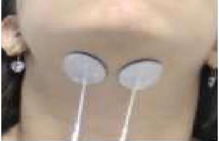
of 2 cm, positioned and ixed in suprahyoid area (photo
1) with the skin previously cleaned with gauze soaked in 70% alcohol. The researchers requested for the patient one swallowing of saliva, and later two swallows of 3
ml of homogeneous paste food, measured in syringe and offered in metal teaspoon. The graphics about the amplitude and time curves obtained by the equipment
used was stored in computer iles for later analysis.
Figure 2. electrode placement illustration in suprahyoid region
Statistical analysis
For statistical analysis we used the MS-Excel
electronic spreadsheet in its MS-Ofice 2010 version
for the data organization, and IBM SPSS (Statistical Package for Social Sciences), in its version 22.0, to obtain the results. In the statistical analysis, we adopted
the signiicance level of 5% (0.05) for the application of
statistical tests.
The test of Wilcoxon Signed Posts was used in order to verify possible differences between the two obser-vation moments for the variables of interest in each group studied. To verify possible differences between both groups studied for the variables of interest was applied the Mann-Whitney test. As we have only four sampling units per group, we can consider that there
are effective trends to ind differences when ‘p’ (signii -cance) is between 5% (0.050) and 10% (0,100).
RESULTS
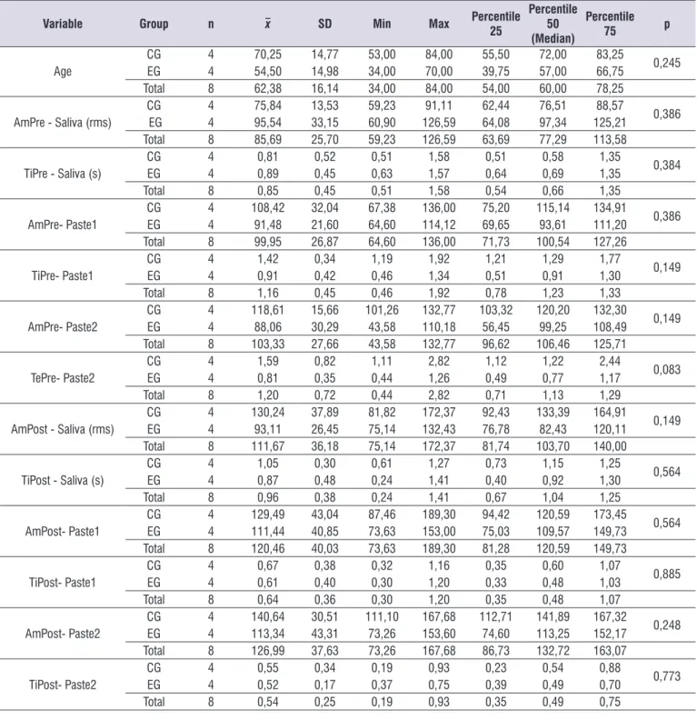
In comparing the mean age presented by the two
groups there was no signiicant difference between
them, showing homogeneity between the groups with respect to age (Table 1). Comparing the average of the variables range and time of muscle electrical activity during swallowing saliva and two swallows of paste, no
statistically signiicant differences between groups was
observed, both in the comparison of pre-intervention values, as in the post intervention values (Table 1). In the comparison between groups is possible to see tendency to real difference (where p<0.100) only in mean comparison in time of the electrical muscle activity during second swallowing of paste food, in the pre-intervention moment time, with highest value for the mean of control group.
In the averages of amplitude and time in muscle electrical activity in the control group (Table 2), there
were no signiicant differences when comparing the pre- and post-intervention. It is veriied that the compar -isons of the amplitude in muscle electrical activity in saliva swallowing, in the second paste swallowing and time of muscle electrical activity in two offers of paste found values with effective tendency to difference (p <0.100).
The average of the range of muscular electrical activity, both in saliva swallowing as the two swallows of paste, were higher in the post-intervention period compared to the pre-intervention period in the control group. However, the average of time muscle electrical activity only were higher in the post-intervention exercises and application of elastic bandage in the
sessions established by the study.
The use of NMES in the experimental group included the application of electric stimulus through two adhesive electrodes 3 cm in diameter applied on clean skin in suprahyoid muscles area. The application of electrical stimulus occurred in combination with the realization of active exercises already described in the traditional speech therapy, and followed the protocol that has been used by speech therapy department in the rehabilitation center in which the research was conducted:
• ive minutes of application of Transcutaneous Nerve Stimulation (TENS), frequency of 30 Hz and pulse 200ms, in order to promote greater proprioception of the patient on the muscles being worked. The intensity is adjusted in accordance with the comfort level of each patient;
• twenty minutes of application of Electrical Stimulation Functional (FES) frequency of 80 Hz
and pulse 250ms, with a time of ive seconds with
the power on (time on), alternating with ten seconds of rest (off current - time off) with order to provide the contraction and muscle strengthening. The intensity is adjusted in accordance with the comfort level of each patient;
• ive minutes new application of TENS current with 30Hz frequency and pulse 200ms, aiming muscle deceleration at the end of the session. The intensity is adjusted in accordance with the comfort level of each patient.
Therapy sessions for both groups occurred weekly for 40 minutes. After eight sessions, all patients measure the EMG biofeedback again to check the amplitude and time of muscle electrical activity during swallowing saliva and two swallowing of paste food, with the same volume and utensils used in the pre intervention measurement. The electrodes and the placement of these were also maintained accordance with the initial measurement. The choice of eight sessions to a new survey of the data was based on criteria of the insti-tution where the research was developed, which is the stipulated time to check progress of any therapeutic process applied.
Table 1. Average of age, range and time of activity electric muscular, pre and post therapeutic intervention according the group studied
Variable Group n x SD Min Max Percentile
25
Percentile 50 (Median)
Percentile
75 p
Age
CG 4 70,25 14,77 53,00 84,00 55,50 72,00 83,25 0,245 EG 4 54,50 14,98 34,00 70,00 39,75 57,00 66,75
Total 8 62,38 16,14 34,00 84,00 54,00 60,00 78,25
AmPre - Saliva (rms) EGCG 44 75,8495,54 13,5333,15 59,2360,90 126,5991,11 62,4464,08 97,3476,51 125,2188,57 0,386
Total 8 85,69 25,70 59,23 126,59 63,69 77,29 113,58
TiPre - Saliva (s)
CG 4 0,81 0,52 0,51 1,58 0,51 0,58 1,35 0,384 EG 4 0,89 0,45 0,63 1,57 0,64 0,69 1,35
Total 8 0,85 0,45 0,51 1,58 0,54 0,66 1,35
AmPre- Paste1 EGCG 44 108,4291,48 32,0421,60 67,3864,60 114,12136,00 75,2069,65 115,1493,61 111,20134,91 0,386
Total 8 99,95 26,87 64,60 136,00 71,73 100,54 127,26
TiPre- Paste1 EGCG 44 1,420,91 0,340,42 1,190,46 1,341,92 1,210,51 0,911,29 1,301,77 0,149
Total 8 1,16 0,45 0,46 1,92 0,78 1,23 1,33
AmPre- Paste2 EGCG 44 118,6188,06 15,6630,29 101,2643,58 110,18132,77 103,3256,45 120,2099,25 108,49132,30 0,149
Total 8 103,33 27,66 43,58 132,77 96,62 106,46 125,71
TePre- Paste2 EGCG 44 1,590,81 0,820,35 1,110,44 1,262,82 1,120,49 0,771,22 1,172,44 0,083
Total 8 1,20 0,72 0,44 2,82 0,71 1,13 1,29
AmPost - Saliva (rms) EGCG 44 130,2493,11 37,8926,45 81,8275,14 132,43172,37 92,4376,78 133,3982,43 120,11164,91 0,149
Total 8 111,67 36,18 75,14 172,37 81,74 103,70 140,00
TiPost - Saliva (s)
CG 4 1,05 0,30 0,61 1,27 0,73 1,15 1,25 0,564 EG 4 0,87 0,48 0,24 1,41 0,40 0,92 1,30
Total 8 0,96 0,38 0,24 1,41 0,67 1,04 1,25
AmPost- Paste1 EGCG 44 129,49111,44 43,0440,85 87,4673,63 153,00189,30 94,4275,03 109,57120,59 149,73173,45 0,564
Total 8 120,46 40,03 73,63 189,30 81,28 120,59 149,73
TiPost- Paste1 EGCG 44 0,670,61 0,380,40 0,320,30 1,201,16 0,350,33 0,480,60 1,031,07 0,885
Total 8 0,64 0,36 0,30 1,20 0,35 0,48 1,07
AmPost- Paste2 EGCG 44 140,64113,34 30,5143,31 111,1073,26 153,60167,68 112,7174,60 113,25141,89 152,17167,32 0,248
Total 8 126,99 37,63 73,26 167,68 86,73 132,72 163,07
TiPost- Paste2 EGCG 44 0,550,52 0,340,17 0,190,37 0,750,93 0,230,39 0,490,54 0,700,88 0,773
Total 8 0,54 0,25 0,19 0,93 0,35 0,49 0,75
Legend: n= sample;x = range; SD=Standard deviation; p= signiicance; Am= amplitude; Ti= time; CG= control group; EG= experimental group; Min= minimum; Max= maximum.
moment for saliva swallowing, in this same group (Table 2).
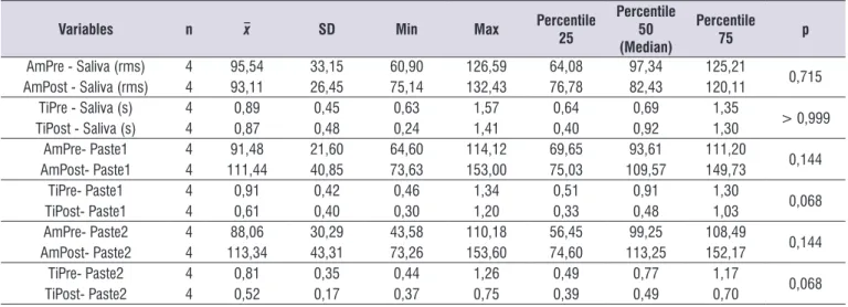
There were no statistical differences in mean values of amplitude and time of electrical muscle activity during swallowing in the pre- and post-intervention the experimental group (Table 3). The comparisons of average in time muscle electrical activity in pre and post intervention in two swallows of paste showed values with effective tendency to difference (p <0.100). In the
experimental group, this trend was not found in average in amplitude and time in muscle electrical activity for saliva swallowing and in the average amplitude of electrical muscle activity in swallowing of paste (Table 3).
In the experimental group, only the averages of the muscular electrical activity amplitude values for the two swallows of paste were higher in post-intervention moment than in the pre-intervention (Table 3).
Table 2. Average of age, range and time of activity electric muscular, pre and post therapeutic intervention, in control group
Variables n x SD Min Max Percentile
25
Percentile 50 (Median)
Percentile
75 p
AmPre - Saliva (rms) 4 75,84 13,53 59,23 91,11 62,44 76,51 88,57 0,068 AmPost - Saliva (rms) 4 130,24 37,89 81,82 172,37 92,43 133,39 164,91
TiPre - Saliva (s) 4 0,81 0,52 0,51 1,58 0,51 0,58 1,35
0,465
TiPost - Saliva (s) 4 1,05 0,30 0,61 1,27 0,73 1,15 1,25
AmPre- Paste1 4 108,42 32,04 67,38 136,00 75,20 115,14 134,91 0,465 AmPost- Paste1 4 129,49 43,04 87,46 189,30 94,42 120,59 173,45
TiPre- Paste1 4 1,42 0,34 1,19 1,92 1,21 1,29 1,77 0,068 TiPost- Paste1 4 0,67 0,38 0,32 1,16 0,35 0,60 1,07
AmPre- Paste2 4 118,61 15,66 101,26 132,77 103,32 120,20 132,30 0,068 AmPost- Paste2 4 140,64 30,51 111,10 167,68 112,71 141,89 167,32
TiPre- Paste2 4 1,59 0,82 1,11 2,82 1,12 1,22 2,44 0,068 TiPost- Paste2 4 0,55 0,34 0,19 0,93 0,23 0,54 0,88
Legend: n= sample; x = range; SD=Standard deviation; p= signiicance; Am= amplitude; Ti= time; Min= minimum; Max= maximum. Wilcoxon Signed Posts Test. p<0,05
Table 3. Average of age, range and time of activity electric muscular, pre and post therapeutic intervention, in experimental group
Variables n x SD Min Max Percentile
25
Percentile 50 (Median)
Percentile
75 p
AmPre - Saliva (rms) 4 95,54 33,15 60,90 126,59 64,08 97,34 125,21 0,715 AmPost - Saliva (rms) 4 93,11 26,45 75,14 132,43 76,78 82,43 120,11
TiPre - Saliva (s) 4 0,89 0,45 0,63 1,57 0,64 0,69 1,35
> 0,999
TiPost - Saliva (s) 4 0,87 0,48 0,24 1,41 0,40 0,92 1,30
AmPre- Paste1 4 91,48 21,60 64,60 114,12 69,65 93,61 111,20 0,144 AmPost- Paste1 4 111,44 40,85 73,63 153,00 75,03 109,57 149,73
TiPre- Paste1 4 0,91 0,42 0,46 1,34 0,51 0,91 1,30 0,068 TiPost- Paste1 4 0,61 0,40 0,30 1,20 0,33 0,48 1,03
AmPre- Paste2 4 88,06 30,29 43,58 110,18 56,45 99,25 108,49 0,144 AmPost- Paste2 4 113,34 43,31 73,26 153,60 74,60 113,25 152,17
TiPre- Paste2 4 0,81 0,35 0,44 1,26 0,49 0,77 1,17 0,068 TiPost- Paste2 4 0,52 0,17 0,37 0,75 0,39 0,49 0,70
DISCUSSION
This study investigated the effect of electrical stimu-lation Neuromuscular (NMES) in the contraction of suprahyoid muscles during swallowing in post-stroke patients with oropharyngeal dysphagia. Both in the group of patients who received intervention of NMES as in the control group were not found values with
statis-tical signiicance that could prove the improvement of
(contraction) muscle electrical activity of these muscles due to therapeutic intervention performed.
According to the data found in this research is not
possible to afirm that the use of NMES associated with
active exercises or the realization only of the latter, was effective in promoting increased contraction of supra-hyoid muscles. Furthermore, neither of the
interven-tions applied showed statistically signiicant for more eficiency when compared to other intervention.
Although the values found, the authors of this study clinically proven improvement in swallowing functionality, by observing the reduction of clinical signs suggestive of tracheal aspiration, and increased
eficiency of swallowing. However, these data are
derived from clinical observation, and present consid-erable subjectivity. In addition, we found that patients in the experimental group showed no new complaints
related to swallowing. These indings corroborate those
from Beom et al.5 who found that NMES not shown quantitative improvement as assessed by videoluo -roscopy in swallowing in patients with dysphagia and reduced laryngeal elevation. However, they found that the patients who received NMES showed qualitative improvement observed through functional scale, compared to the traditional speech therapy. The same authors warn that further studies are needed with more homogeneous control group and high sample, in order to fully establish the effects of NMES in patients with dysphagia.
In this research, in addition to individuals were divided randomly in each group and without the knowledge of the invention to be held, featuring a blind study, there was no statistical difference in age between the control and experimental groups, which features homogeneity between groups. In contrast, the sample with a small number of participants may have been an
impeditive factor for statistical signiicance. In some comparisons of averages was observed a signiicance
trends which may indicate that a larger sample might
give statistical signiicance. However, as these trends
were found in both groups, it did not prove the greater
therapeutic eficacy of NMES compared to isolated
active exercises.
Another factor that in this research could have
contributed to not statistical signiicance refers to the
number and frequency of treatment sessions, being realized eight sessions, once a week. There is no
consensus in the scientiic literature about the number of minimum sessions to produce therapeutic eficiency,
but it can be inferred that the increase in the number of sessions or in the frequency of therapy, could produce different results.
Crary and Carnaby25, in a literature review article, emphasizes the importance of recent publications describing traditional therapy and the contribution of electrostimulation. The authors believe that NMES could play a useful role, as a supporting, to better devel-opment of rehabilitation exercises in dysphagia, but are necessary further studies regarding to its impact on the physiological potential of the swallowing mechanism and its functional results.
The scientiic literature is controversial in relation
to the therapeutic effects of NMES in the rehabilitation of dysphagia. While some studies1,3,8-16 show beneits in the pathophysiology of swallowing in patients with oropharyngeal dysphagia submitted to NMES, others5-7 report the absence of physiological and functional changes resulting from these stimuli. It must be said that even the publications that demonstrate positive results present questionable methodological designs in
scientiic relevance.
The physiological effects that are produced with
the use of NMES are inluenced, among other things,
by the location of the electrodes on the region to be stimulated, by the selection of the frequency of electric stimulus and of the duration of the stimuli during therapy4. Thus, according to the anatomical region in which the electrode was inserted and especially according the format of a NMES application protocol, the result arising from therapeutic intervention may be different. There is no homogeneity among the studies that relate the effect of the NMES in the rehabilitation of dysphagia in relation to the positioning of the electrodes and protocols used, thus the difference of results can be explained by these aspects.
The NMES application protocol in this study priori-tized the needs of muscle preparation by the appli-cation of electric current TENS at the beginning and
end of each session, and stimulation of muscle ibers
type II with the realization of electric current FES at a
responsible for the “explosion” of muscle contraction
and the strength gain, but more susceptible to fatigue26. After the data found in this study, the authors have questioned if the electrical stimulation of the muscle
ibers type I, through application of FES current with a
lower frequency, could result in greater gains in relation
to the therapeutic eficiency. This questioning is due to the fact that the muscle ibers type I are more resistant
to fatigue26 and patients in this study were at the beginning of the rehabilitation process, at which usually the muscle condition is more weakness4.
The veriication of eficiency of therapeutic
techniques in the rehabilitation of oropharyngeal dysphagia requires the use of evolution indicator that can be clinical aspects, as episodes of pneumonia and weight gain, or quantitative and qualitative measures through scales and objective assessment like
video-luoroscopy and electromyography.
In this study, the veriication of NMES eficiency
was performed through the use of biofeedback surface electromyography (sEMG-Biofeedback). The same method of measurement was used in another study27 that evaluated sixty healthy individuals with sEMG-Biofeedback, aiming to check the electrical activity of suprahyoid muscles during swallowing function. The
results of this study showed statistically signiicant
differences in electrical activity of suprahyoid muscles in swallowing requested by verbal command and in those that occurred spontaneously. The authors discussed that the sEMG-Biofeedback is a sensitive instrument to the electrical muscle activity (muscle contraction) during swallowing, but questioned the small sample of individuals to establish normal values.
Crary et al.28 performed a study using the sEMG-Biofeedback as a rehabilitation tool, not evaluation, with results indicating that this type of intervention promotes improvement in swallowing function when associated with therapy. Because this instrument is used primarily in rehabilitation, the authors of this study questioned the sensitivity of this in assessing the electrical muscle activity (muscle contraction) during swallowing as a form of assessment in two different moments.
Cola et al.3, in a literature review to verify the effec-tiveness of the use of NMES in the rehabilitation of oropharyngeal dysphagia concludes that NMES is an effective method in the treatment of dysphagia, with
beneicial changes, such as the return of the diet by
mouth, reduced episodes of tracheal aspiration, among others. The results show that NMES associated with traditional therapy demonstrates higher effectiveness.
Although lacking consensus in the literature about
the eficiency of the use of NMES in the rehabilitation
of oropharyngeal dysphagia and data found in this
research, clinical practice shows therapeutic beneits
with the application of this technique in patients with dysphagia. Thus, further studies are needed with greater uniformity about the location of electrodes, the choice of the evolution indicator and the protocol used for the application of NMES.
CONCLUSION
The use of NMES in parameters and methodology used in this study showed not be effective in promoting greater contraction of suprahyoid muscles during swallowing after stroke in patients with oropharyngeal
dysphagia. The indings may be due to the method -ology used in this study with regard to technical appli-cation protocol and how to measure results.
REFERENCES
1. Guimarães BTL, Furkim AM, Silva RG.
Eletroestimulação neuromuscular na reabilitação da disfagia orofaríngea. Rev Soc Bras Fonoaudiol.2010;15(4):615-21.
2. Gonçalves MIR, César SR. Disfagia neurogênicas: avaliação. In: Ortiz KZ. Distúrbios neurológicos adquiridos: fala e deglutição. 2.ed. Barueri: Manole, 2010. p. 258-81.
3. Cola PC, Dantas RO, Silva RG. Estimulação elétrica neuromuscular na Reabilitação da Disfagia Orofaríngea Neurogênica. Rev Neurocienc. 2011;
in press:1-9.
4. Guimarães BTL, Guimarães MSMA.
Eletroestimulação funcional (EEF) em disfagia orofaríngea. São José dos Campos, SP: Pulso Editorial, 2013.
5. Beom J, Kim SJ, Han TR. Electrical stimulation of the suprahyoid muscles in brain-injured patients with dysphagia: a pilot study. Ann Rehabil Med. 2011;35(3):322-7.
6. Heck FM, Doeltgen SH, Huckabee ML. Effects of submental neuromuscular electrical stimulation on pharyngeal pressure generation. Arch Phys Med Rehabil. 2012;93(11):2000-7.
18. Wheeler KM, Chiara T, Sapienza CM. Surface electromyographic activity of the submental muscles during swallow and expiratory pressure threshold training tasks. Dysphagia. 2007;22(2):108-16.
19. Shaker R, Kern M, Bardan E, Taylor A, Stewart ET, Hoffmann RG et al. Augmentation of deglutitive upper esophageal sphincter opening in the elderly by exercise. Am J Physiol 1997;272(35):G1518-22.
20. Shaker R, Easterling C, Kern M, Nitschke T, Massey B, Daniels S et al. Rehabilitation of swallowing by exercise in tube-fed patients with pharyngeal dysphagia secondary to abnormal UES opening. Gastroenterol. 2002;122(5):1314-21.
21. Easterling C, Grande B, Kern M, Sears K, Shaker R. Attaining and maintaining isometric and isokinetic goals of the Shaker exercise. Dysphagia. 2005;20(2):133-8.
22. Huckabee ML, Steele CM. An analysis of
lingual contribution to submental surface electromyographic measures and pharyngeal pressure during effortful swallow. Arch Phys Med Rehabil.2006;87(8):1067-72.
23. Fujiu M, Logemann JA. Effects of a tongue- holding maneuver on posterior pharyngeal wall movement during deglutition. Am J Speech Lang Pathol. 2006;5(1):23-30.
24. Robbins J, Gangnon RE, Theis SM, Kays SA, Hewitt AL, Hind JA. The effects of lingual exercise on swallowing in older adults. J Am Geriatr Soc. 2005;53(9):1483-9.
25. Crary MA, Carnaby GD. Adoption into clinical practice of two therapies to manage swallowing disorders:exercise-based swallowing rehabilitation and electrical stimulation. Curr Opin Otolaryngol Head Neck Surg. 2014;22(3):172-80.
26. Burkhead LM, Sapienza CM, Rosenbek
JC. Strength-training exercise in dysphagia rehabilitation: principles, procedures, and directions for future research. Dysphagia. 2007;22(3):251-65.
27. O’Kane L, Groher ME, Silva K, Osborn L. Normal
muscular activity during swallowing as measured by surface electromyography. Ann Otol Rhinol Laryngol. 2010;119(6):398-401.
28. Crary MA, Baldwin BO. Surface electromyographic characteristics of swallowing in dysphagia secondary to brainstem stroke. Dysphagia. 1997;12(4):180-7.
8. Permsirivanich W, Tipchatyotin S, Wongchai M, Leelamanit V, Setthawatcharawanich S, Sathirapanya P et al. Comparing the effects of rehabilitation swallowing therapy vs. neuromuscular electrical stimulation therapy among stroke patients with persistent pharyngeal dysphagia: a randomized controlled study. J Med Assoc Thai. 2009;92(2):259-65.
9. Rofes L, Arreola V, López I, Martin A, Sebastián M, Ciurana A et al. Effect of surface sensory and motor electrical stimulation on chronic poststroke oropharyngeal dysfunction. Neurogastroenterol Motil. 2013;25(11):888-e701.
10. Park JW, Oh JC, Lee HJ, Park SJ, Yoon TS, Kwon BS. Effortful swallowing training coupled with electrical stimulation leads to an increase in hyoid elevation during swallowing. Dysphagia. 2009;24(3):296-301.
11. Ludlow CL, Humbert I, Saxon K, Poletto C, Sonies B, Crujido L. Effects of surface electrical stimulation both at rest and during swallowing in chronic pharyngeal dysphagia. Dysphagia. 2007;22(1):1-10.
12. Lim KB, Lee HJ, Lim SS, Choi YI. Neuromuscular electrical and thermal-tactile stimulation for dysphagia caused by stroke: a randomized controlled trial. J Rehabil Med. 2009;41(3):174-8. 13. Shaw GY, Sechtem PR, Searl J, Keller K, Rawi
TA, Dowdy E. Transcutaneous neuromuscular electrical stimulation (VitalStim) curative therapy for severe dysphagia: myth or reality? Ann Otol Rhinol Laryngol. 2007;116(1):36-44.
14. Gallas S, Marie JP, Leroi AM, Verin E. Sensory transcutaneous electrical stimulation improves post-stroke dysphagic patients. Dysphagia. 2010;25(4):291-7.
15. Verina E, Maltete D, Ouahchi Y, Marie JP, Hannequin D, Guegan Massardier E et al. Submental sensitive transcutaneous electrical stimulation (SSTES) at home in neurogenic oropharyngeal dysphagia: a pilot study. Ann Phys Rehabil Med. 2011;54(6):366-75.
16. Toyama K, Matsumoto S, Kurasawa M, Setoguchi H, Noma T, Takenaka K et al. Novel neuromuscular electrical stimulation system for treatment of dysphagia after brain injury. Neurol Med Chir (Tokyo). 2014;54(7):521-8.